Test 2 Content
1/196
There's no tags or description
Looks like no tags are added yet.
Name | Mastery | Learn | Test | Matching | Spaced | Call with Kai |
|---|
No analytics yet
Send a link to your students to track their progress
197 Terms
perimysium
middle layer, wraps fascicles, connective fiber sheath
endomysium
innermost, between fibers
epimysium
lines the outside of skeletal muscle, perimysium bundles with blood vesicles and fascia
myosin
thick filaments, on m line in h zone of sarcomere
actin
thin filaments, in I band, off shoot of z disc on sarcomere
elastic tinin filament
on I band, offshoot of z disc
type 1 muscle fibers
slow twitch, doesn't fatuous, high mitochondria, high blood, high myoglobin, larger, in back and soles
type 2 fibers
fast twitch, fatigues, less mitochondria, in legs and arms
fast twitch types
slow fast and fast fast
tear in rotator cuff muscles
due to sudden stop or shoulder drop
isotonic contraction
same mixture, build up muscle units, at the end of muscle
isometric contraction
same measure, same size when contracting and not moving, start of muscle
eccentric contraction
slowly relaxing, more energy than concentric
concentric contraction
layers, builds on, shorten as contract
trunk and common
large artery that branches
muscle week 4
myotome grows into limb buds, cells fuse to make multinucleated cell
limb buds become
skeletal muscle
z disc
holds actin and elastic filaments together
I bands
has thin filaments, shrinks with contraction
A band
has interlocking myosin and actin
M line
only holds myosin
compartment syndrome
CT covered muscle is inflammed, causing artery cut off and nerve compression
treatment of compartment syndrome
RICE, fasciotomy (cut outer CT)
bursitis
inflammation of bursa from overuse
bible bump
bursa expands near joint, causing ganglion cyst
muscles of facial expression innervation
facial nerve or cranial nerve 7
muscles of mastication innervation
trigeminal nerve or nerve 5
bones in temporal fossa
greater wings of sphenoid, frontal, parietal, sphenoid bone
above muscle origin and insertion
muscle belly, where contractive tissue is
pharyngeal constrictors innervation
vagus, glossopharyngeal, trigeminal
submandibular triangle
anterior digastric, stylohyoid, mandible
larynx
hyoid to thyroid cartilage
larynx elevation when swallowing
mylohyoid, geniohyoid, stylohyoid
muscular triangle
both scms and posterior digastrics
carotid triangle
scm, anterior digastric, omohyoid superior
posterior triangle of neck
scm, clavicle, trapezius
iliocostalis
cervicis, thoracis, lumborum
erector spinae
iliocostalis, longissmis, spinalis
linea alba made of
dense regular ct
inspiration
lower pressure, high volume, diaphragm down
expiration
high pressure, low volume, diaphragm up
coughing
rapid forced expiration by transverse abdominis
forced inspiration
scm, scalenes
forced expiration
internal intercostals, external and internal oblique, transverse abdominis
urogenital triangle
superficial transverse perineal, median raphe, ishiocavernous
anal triangle
superficial transverse perineal, coccyx, median raphe
pectoral girdle
clavicle, scapula, humerus
rotator cuff muscles
stabilize scapula
large serratus anterior
in boxers and body builders
deltopectoral triangle
deltoid, pectorals major, clavicle
axilla
latissimus dorsi, serratus anterior, pectorais major and minor
supraspinatus
prevents inferior dislocation
infraspinatus
prevents sliding forward
teres minor
prevents sliding upwards
subscapularis
prevents sliding backwards
synergy muscles to flex elbow
biceps brachii, brachialis, brachioradialis
common flexor tendon muscles
pronator teres, flexor carpe ulnaris, palmares longus, flexor carpi radialis
anatomical snuff box
ligaments of extensor pollicus longus, adductor pollicus longus, find scaphoid bone
trigger finger
tendonitis causes tendon to get stuck
femoral triangle
adductor longus, sartorius, inguinal ligament, femoral hernia here
piriformis syndrome
compressed sciatic nerve, enlarged muscle, rest and ice treatment
IM injection inferior and posterior to iliac crest
gluteus maximus
hamstrings
biceps femoris, semitendinosus, semimembranosus
flex knee
hamstrings, popliteus muscle
tibialis anterior antagonist
tibialis posterior
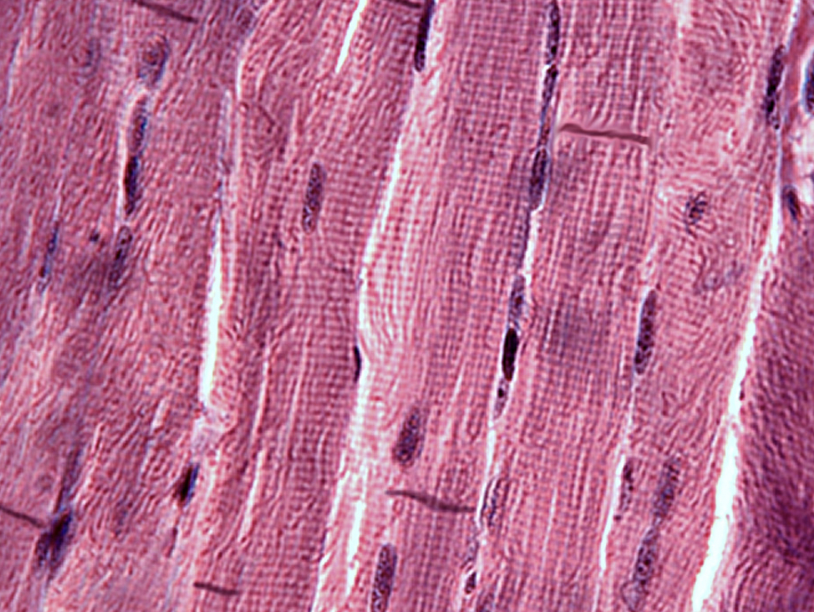
striated skeletal muscle
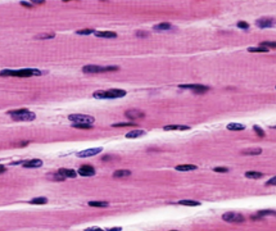
striated cardiac muscle
more mitochondria than skeletal, intercalated discs
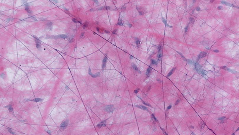
areolar tissue
has elastic (thin) fibers, collagen (thick) fibers, fibroblasts
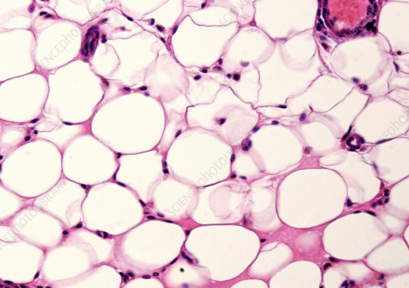
adipose tissue
has nuclear dots and large cells, energy source in areas with high oxygen
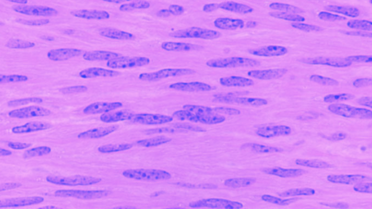
smooth muscle
no stripes and no sarcomeres, 1 nuclei per cell, low mitochondria
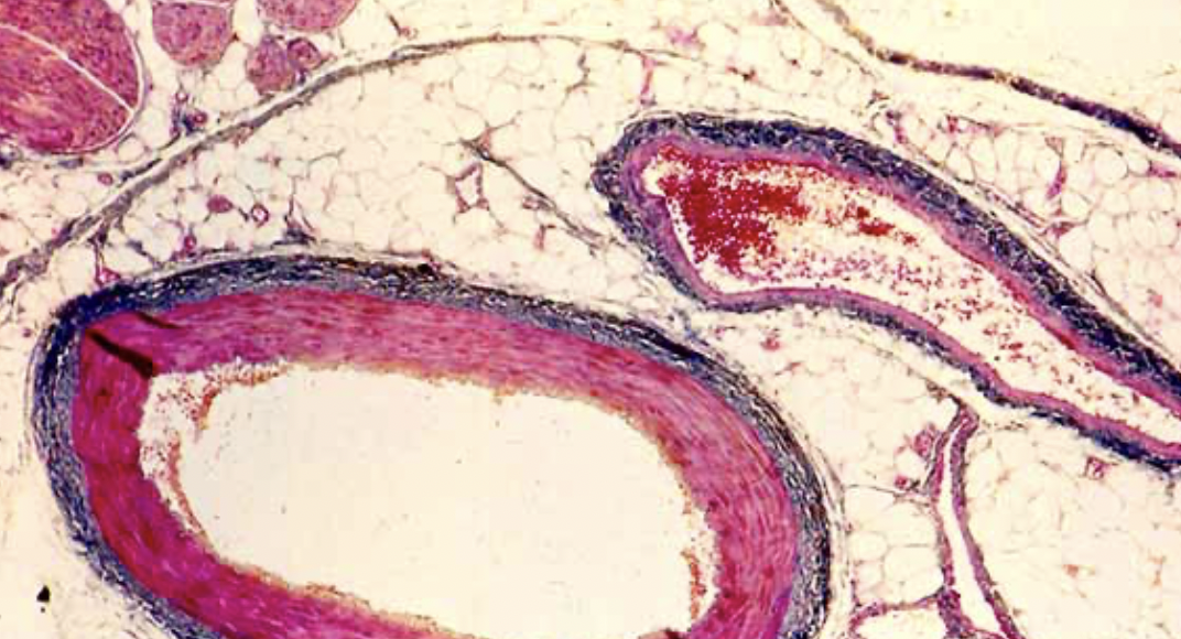
vein and artery
long vein and round artery
intercalated disc junctions
desmosome, gap junction
heart layers (in to out)
endocardium, myocardium, epicardium, pericardium
endocardium tissue
simple squamous epithelial
myocardium
striated cardiac muscle
epicardium
simple squamous epithelium
pericardium
fibrous (outer) and serous (inner) layers, around myocardium
pericardial sac
fluid filled
evolution in neck
pharyngeal gill slits
heart location
in mediastinum, towards left in chest
sa node doesn’t work, av takes over
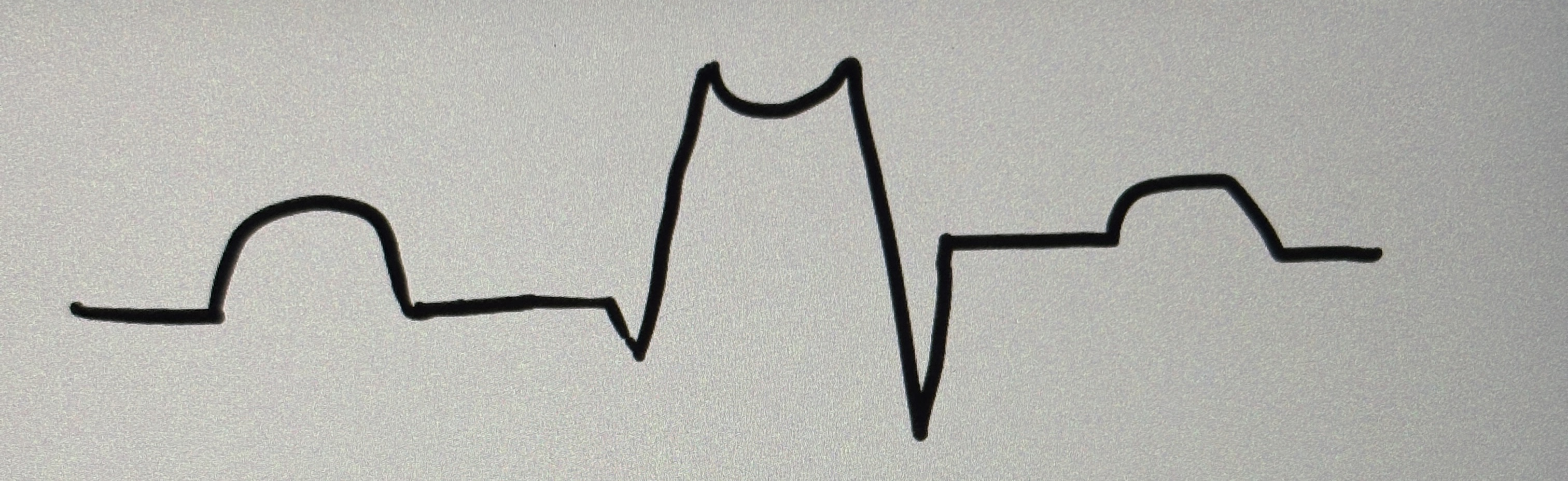
elongated qrs complex due to bundle branches blockage
leads on wrong side or situs inverses
has pulse, but one or more leads came off
positive charge on ekg
left side of heart
negative charge on ekg
right side of heart
SA firing rate
60 second
electrical heart components
sa node, av node, bundle of his, left and right bundle branches, purkinje fibers
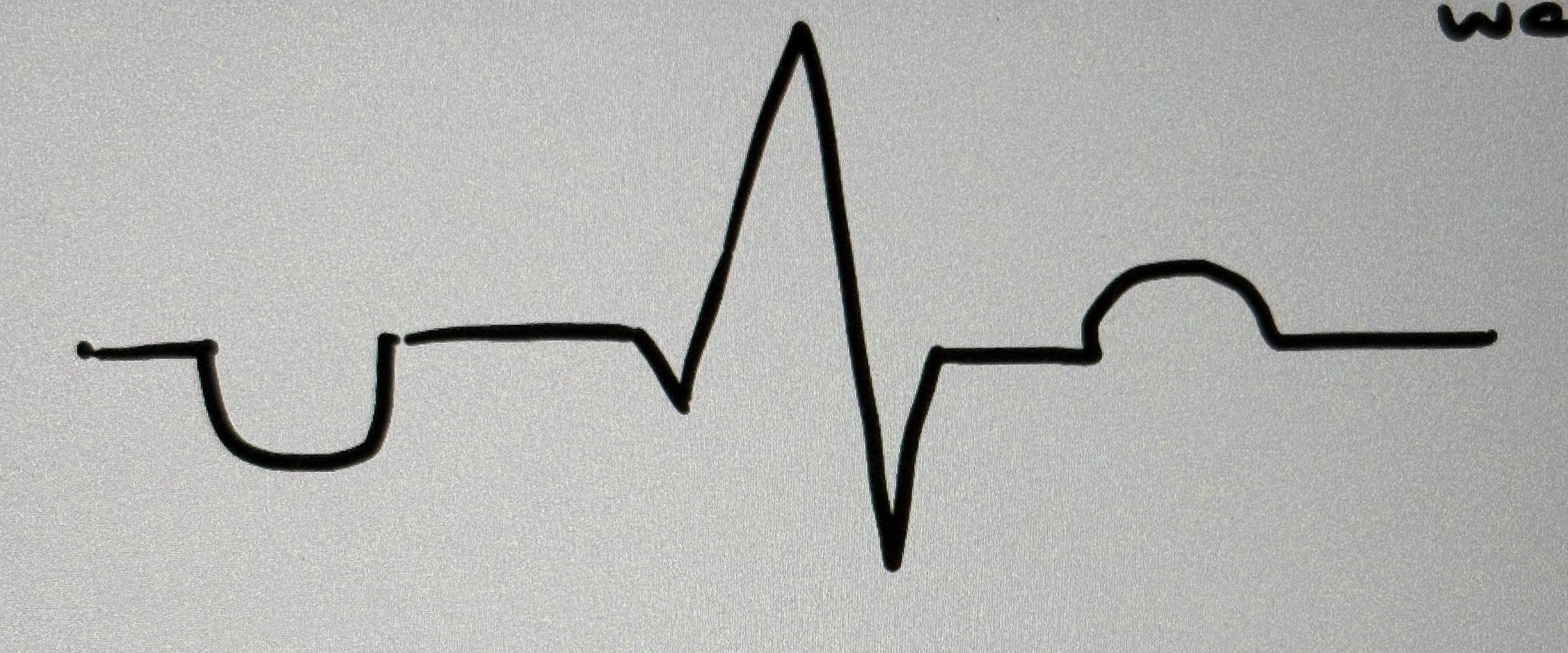
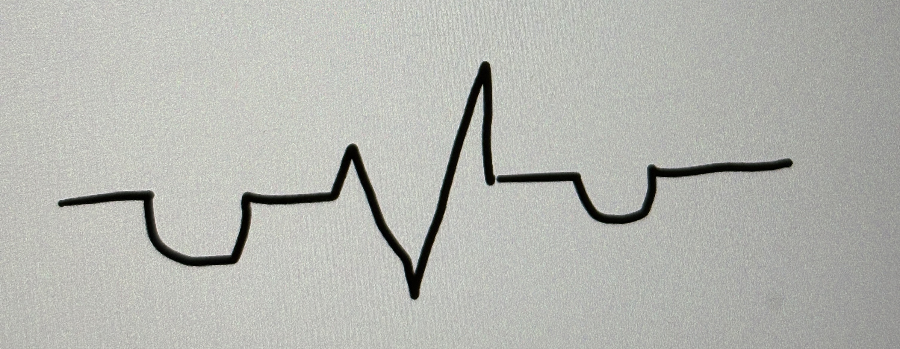
P wave
atrial depolarization, sa node fires
QRS complex
ventricular depolarization, AV node fires at beginning (PR), depolarization of qrs
T wave
ventricular repolarization
between p wave and qrs complex
atrial contraction
brady
slow
tachy
fast
myocardial infarction and rhabdomyolysis causes
high creatine kinase, high troponin, high lactate dehydrogenase, high myoglobin
between endocardium and myocardium
loose irregular ct
between myocardium and epicardium
areolar tissue
fibrous pericardium tissue
outer layer, dense irregular ct
serous pericardium
3 layers, parietal (outer) space in middle, and visceral (inner)
parietal serous pericardium
simple squamous and dense irregular ct