A/P - semester two final
1/72
There's no tags or description
Looks like no tags are added yet.
Name | Mastery | Learn | Test | Matching | Spaced | Call with Kai |
|---|
No analytics yet
Send a link to your students to track their progress
73 Terms
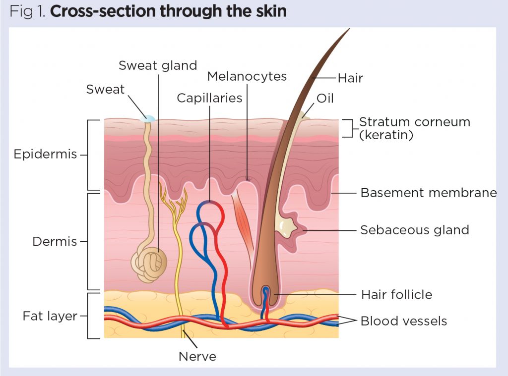
function of arrector pili
causes body hair to stand up straight, temperature regulation
hair follicle
small cavity surrounding the a hair, responsible for hair growth
sebaceous gland
produces sebum, aka oil, helps retain moisture, protect skin, antibacterial
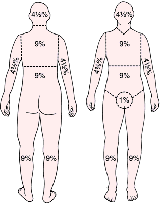
rule of nines
first degree burns
first layer of skin, red and no blistering
second degree burns
first and second layer of skin, red with blistering
third degree burns
all layers of skin, white or purple or black
skin diagram
skeletal system function
support, protection, movement, storage, blood cell production
hematopoiesis
blood cell production, happens in bones
osteoclasts
bone destroying cells
osteoblasts
bone building cells
osteocytes
mature bone cells
bone names and locations
i made a seperate quizlet
https://quizlet.com/853149870/anatomy-bones-and-stuff-flash-cards/
tendons
connective tissue, muscle to bone
ligaments
connective tissue, bone to bone
epimysium
connective tissue, surrounds skeletal muscle
perimysium
connective tissue, surrounds fascicles
endomysium
connective tissue, surrounds fibers
smooth muscle
slow, rhythmic contractions. involuntary, moves things in organs and blood vessels
cardiac muscle
in the heart, striated and branching, rhythmic contractions, involuntary
skeletal muscle
voluntary, strong contractions, striated and multinucleated
“all or nothing” muscle contractions
muscle fibers are either contracted or not contracted, not partially contracted. strength is just a number of fibers contracted. once threshold potential is met, fibers fully contract
sliding filament theory
1.) Calcium removes troponin and tropomyosin.
2.) ATP moves myosin heads which attach to Actin.
3.) Myosin heads pull on actin to shrink sarcomere and contract muscle (power stroke)
4.) myosin heads receive ATP and release from actin (return stroke)
5.) Calcium pumps out of cell and troponin/tropomyosin return blocking myosin from binding.
*AChE removes excess ACh
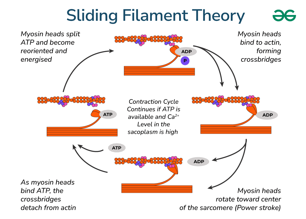
sacromere
functional unit of muscle fibers; made of actin and myosin(which bind together during muscle contraction)
neuromuscular junction
1.) Action Potentional (electrical impulse) travels down axon
2.)ACh released into synaptic cleft
3.) Ach binds to gated ion channels
4.)Na+ flows through channels raising the charge in the muscle cell.
5.) Na+ pump opens further raising the charge.
6.) Ca+ is released initiating muscle contraction in the myofibrils.
7.) K+ pump removes K+ in the cell to resting state
but jayda how do i label the neuromuscular junction diagram
idk how to insert it here but i found a good quizlet (same exact diagram as the study guide)
https://quizlet.com/548749390/neuromuscular-junction-labeling-diagram/
skeletal muscle diagram (fibers and stuff)
surprise! another quizlet! also the same exact diagram
https://quizlet.com/452782672/anatomy-skeletal-muscle-fiber-diagram/
skeletal muscle names and locations
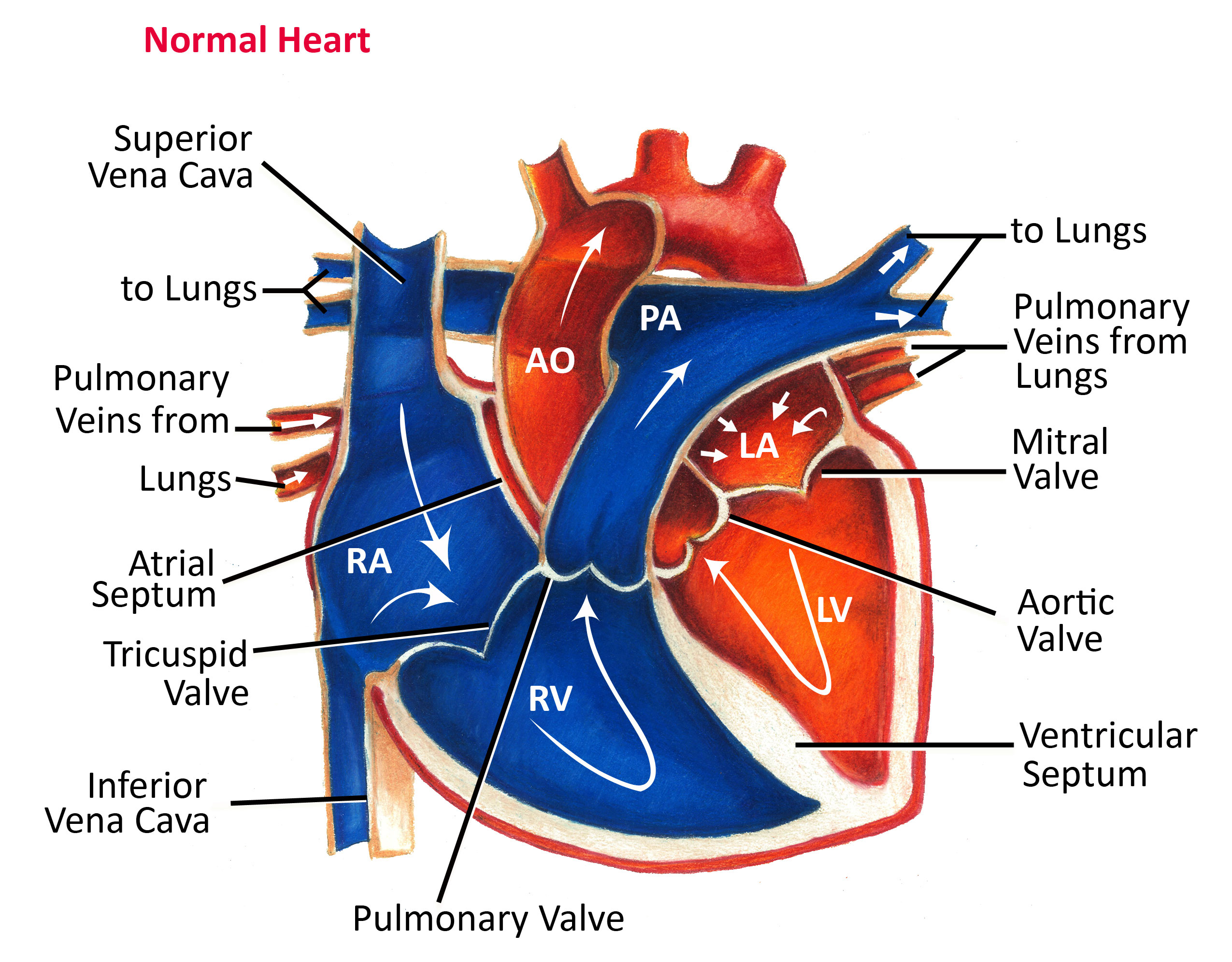
veins
carry blood TOWARDS the heart, unoxygenated usually
capillaries
smallest blood vessels, connect arteries and veins, exchange of nutrients and waste products with tissues
arteries
carry blood AWAY from the heart, oxygenated usually
pericardium
serous membrane surrounding the heart. reduces friction
plasma
clear, liquid part of blood
hemoglobin
proteins in red blood cells that carry oxygen
systole
heart contraction. BP is highest, blood is leaving the heart
diastole
heart relaxation. BP is lowest, blood is entering the heart
blood pressure (BP)
pressure in artery and vein walls
atrioventricular valves
separates atriums and ventricles. bicuspid and tricuspid
semilunar valves
aortic and pulmonary valves
how does blood flow through the heart
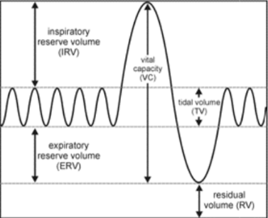
tidal volume (TV)
~500ml, amount of air inhaled and exhaled in a single breath
residual volume (RV)
volume of air left in lungs after max exhalation, needed in order to keep alveoli partially inflated
vital capacity
total amount of air that can be inhaled and exhaled
diffusion
movement of a substance from high to low concentration
pleura
serous membrane that surrounds lungs, protects them and helps them inflate/deflate
respiratory system labeling
this is the best quizlet i could find sorry 💔
https://quizlet.com/905904263/respiratory-system-label-flash-cards/
respiratory graph labeling
whole thing, from top to bottom, is the total lung capacity or TLC
bile
basic (as in not acidic) substance produced by liver, breaks down fats into fatty acids
gastrointestinal (GI) track
part of digestive system where food and liquid passes through
peristalsis
rhythmic, smooth muscle contractions that moves stuff through the GI track
small intestine
contains villi, increases surface area to absorb more nutrients
large intestine
absorbs water and propels feces for expulsion
liver
produces bile and detoxifies blood
mechanical digestion
physical breakdown of food by mouth and stomach
chemical digestion
enzymes breaking down food
kidney
filters bloods, keeps vitamins and electrolytes, turns waste into urine
nephron
filtering unit of kidney. kidneys have millions of them
digestive system labeled
pituitary gland
command center of endocrine system, controls growth and development
thymus
produces immune cells
thyroid gland
produces hormones that regulate metabolism, like thyroxin
pancreas
produces insulin which tells cells to take in sugar from blood
adrenal glands
produces adrenaline (epinephrine), which is a vasoconstrictor and a bronchodilator
ovaries
produce eggs and estrogen
testes
produce sperm and testosterone
endocrine system labeling
ignore the pineal gland but yeah
https://quizlet.com/900023668/endocrine-system-labeling-diagram/
central nervous system (CNS)
brain and spinal cord, body’s processing center
peripheral nervous system (PNS)
feeds info to brain from senses. carries signals for muscle contractions
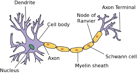
myelin sheath
covers axon and speeds up transmission
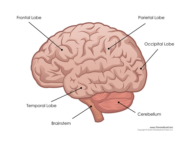
label brain parts
label neuron parts
i was gone when we went over this so sorry if its too detailed or not detailed enough