Comprehensive Orthopedic Fractures and Dislocations: Shoulder to Foot
1/57
There's no tags or description
Looks like no tags are added yet.
Name | Mastery | Learn | Test | Matching | Spaced | Call with Kai |
|---|
No analytics yet
Send a link to your students to track their progress
58 Terms
What does the ABCS approach stand for in orthopedic imaging?
Alignment, Bone, Cartilage (joint space), Soft tissues
What is the most common direction for a glenohumeral dislocation?
Anterior
What radiographic sign is associated with a posterior shoulder dislocation?
Lightbulb appearance of the humeral head
What is a Bankart lesion?
An anterior labral tear, sometimes accompanied by a glenoid fracture
What is a Hill-Sachs lesion?
A compression defect in the posterior humeral head caused by repeated anterior dislocations
What is the significance of a visible posterior fat pad on an elbow radiograph?
It is an abnormal sign indicating potential fracture
What is the most common pediatric elbow fracture?
Supracondylar fracture
What defines a Monteggia fracture-dislocation?
A proximal ulna fracture combined with a radial head dislocation
What is the difference between a Colles and a Smith's fracture?
Colles has dorsal angulation of the distal radius; Smith's has volar angulation
Why is a scaphoid fracture clinically significant?
It has a high risk of avascular necrosis
What is a Bennett's fracture?
An intra-articular fracture at the base of the 1st metacarpal with associated dislocation
What is a Mallet finger?
A distal phalanx avulsion resulting in a drooping fingertip
What constitutes an 'open book' pelvis injury?
Separation of the pubic symphysis and the sacroiliac (SI) joints
What is the clinical threshold for pubic symphysis diastasis?
10 mm separation
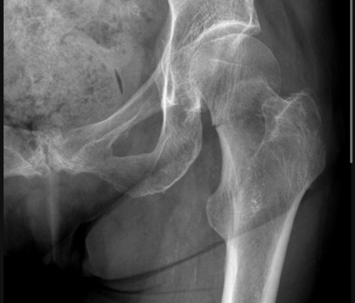
What is Shenton's line used for in hip imaging?
Assessing for hip fractures; it should be a smooth, continuous line
What is lipohaemarthrosis in the knee?
A mixture of fat and blood in the joint, indicating an intra-articular fracture
What is a Segond fracture?
A small lateral tibial avulsion that serves as a marker for an ACL injury
What is a Maisonneuve fracture?
A proximal fibula fracture associated with an ankle injury
How are Weber ankle fractures classified?
A: below the joint, B: at the joint, C: above the joint
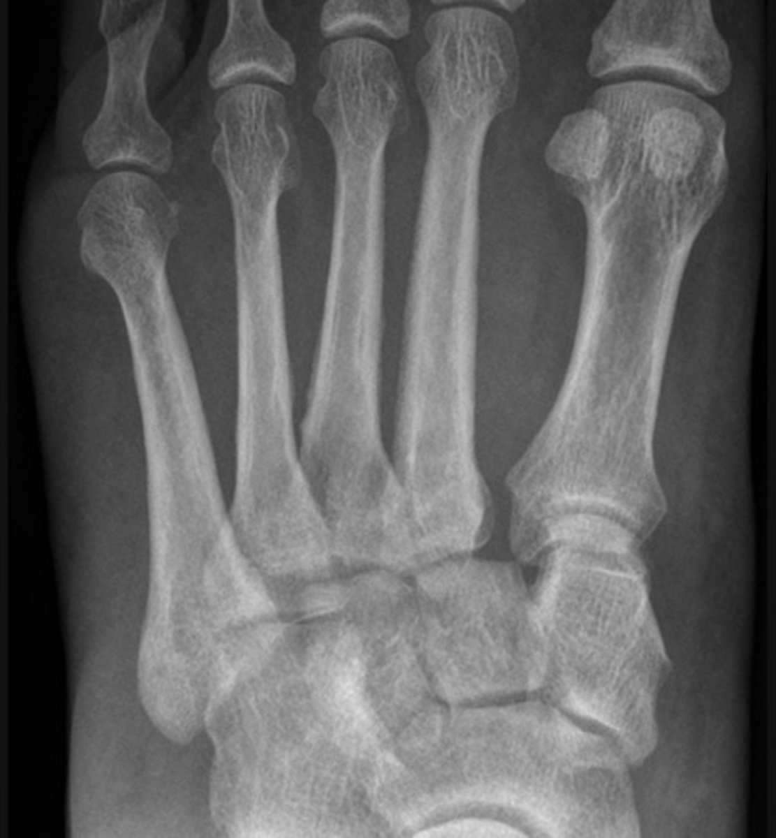
What is a Lisfranc injury?
An injury resulting in midfoot instability
What is the difference between dislocation and subluxation?
Dislocation is complete separation; subluxation is when partial contact remains
What is the mnemonic CRITOL used for in pediatric elbow imaging?
The order of ossification: Capitellum, Radial head, Internal epicondyle, Trochlea, Olecranon, Lateral epicondyle
The distal component of this clavicle fracture is inferiorly displaced - pulled down by the weight of the arm
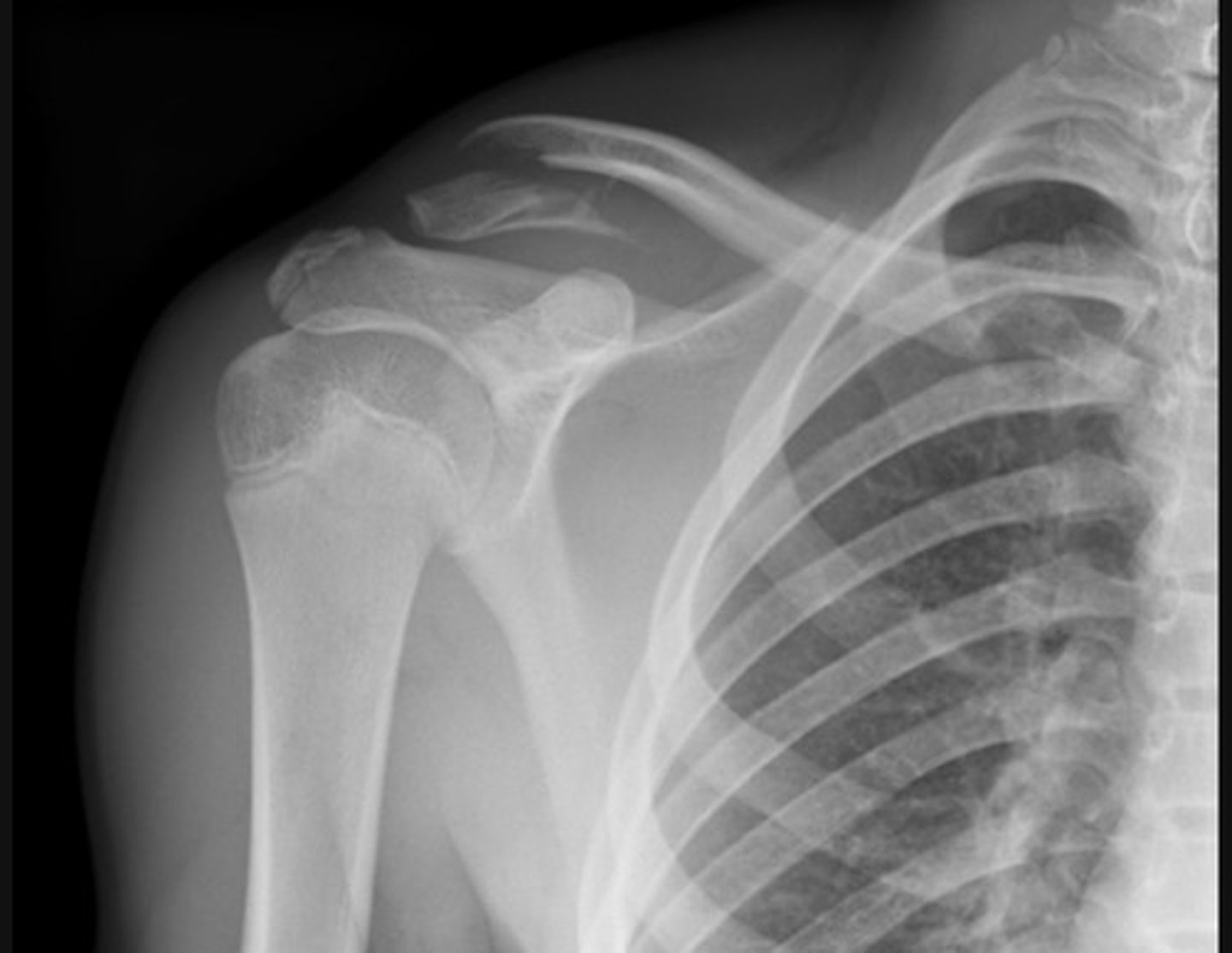
The inferior edge of the acromion is inferiorly displaced and is no longer in line with the inferior edge of the distal clavicle
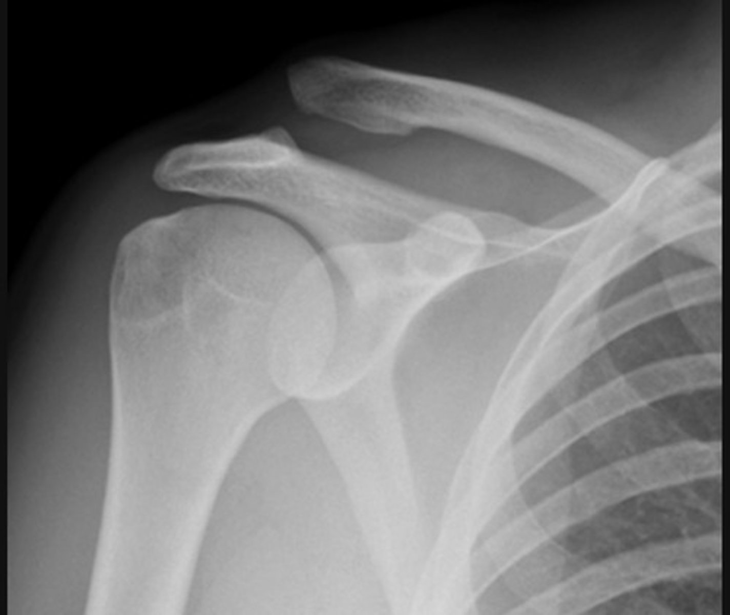
The distal component of this clavicle fracture remains aligned with the proximal clavicle
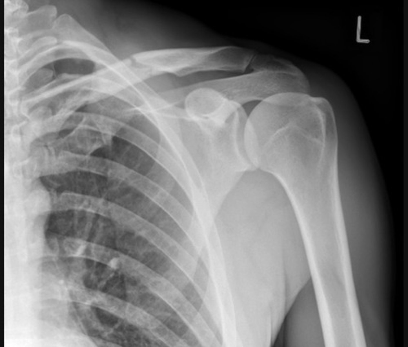
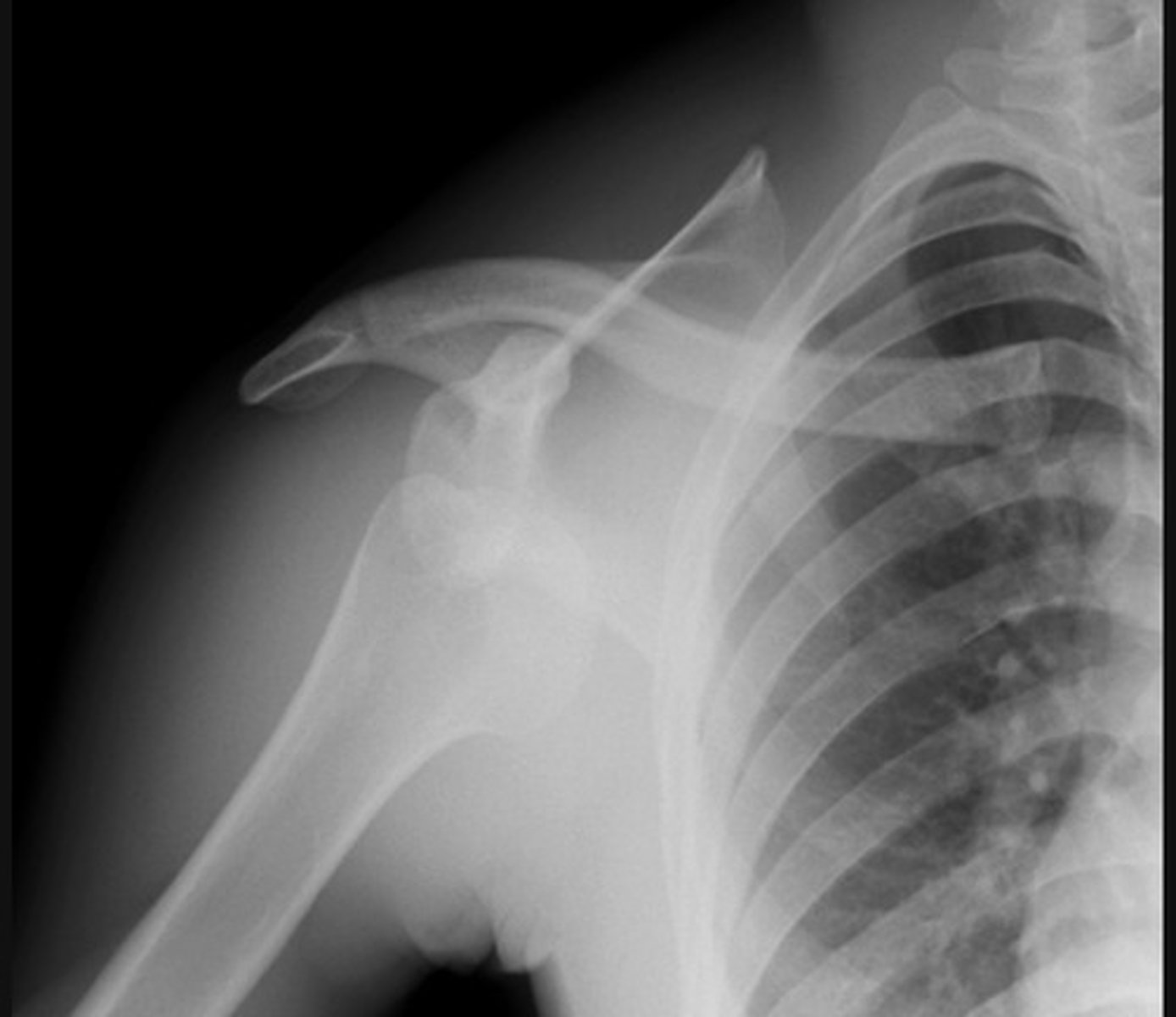
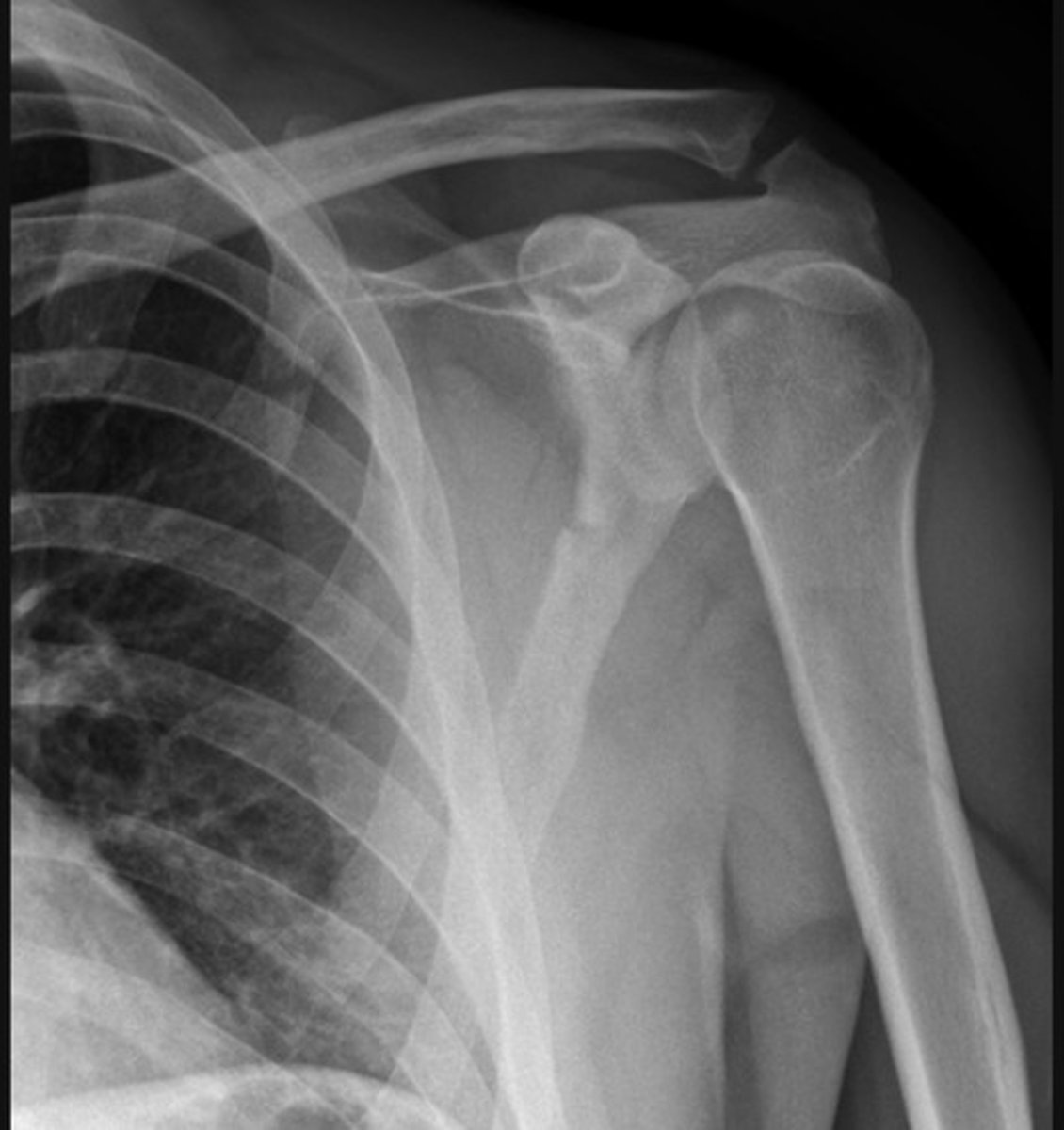
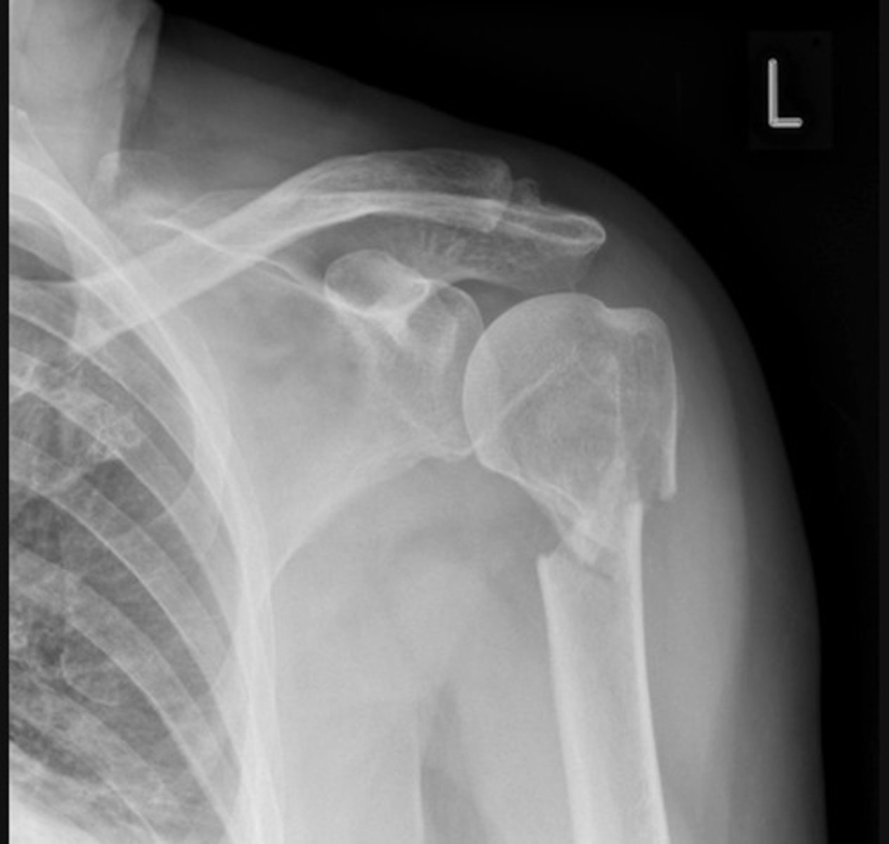
Anterior dislocation. The humeral head is dislocated from the glenoid of the scapula and is now located inferior to the coracoid process of the scapula.
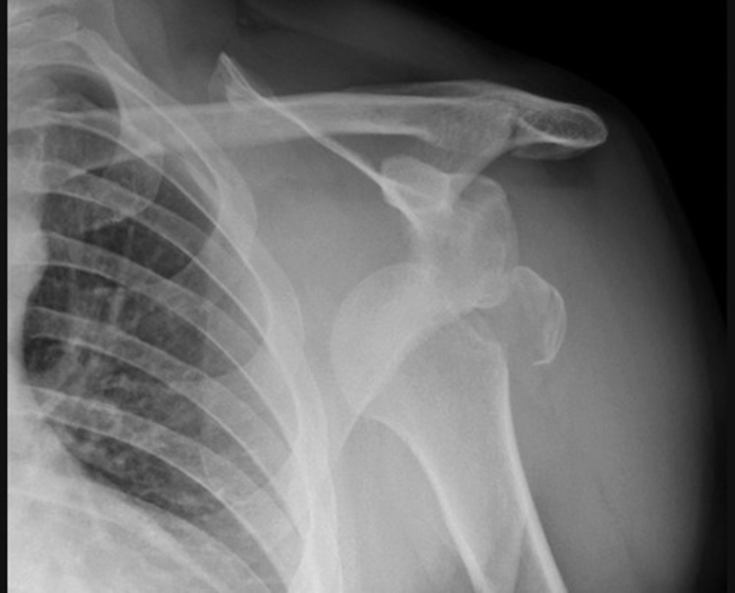
This shoulder anterior dislocation is complicated by a greater tubercle fracture of the humeral head
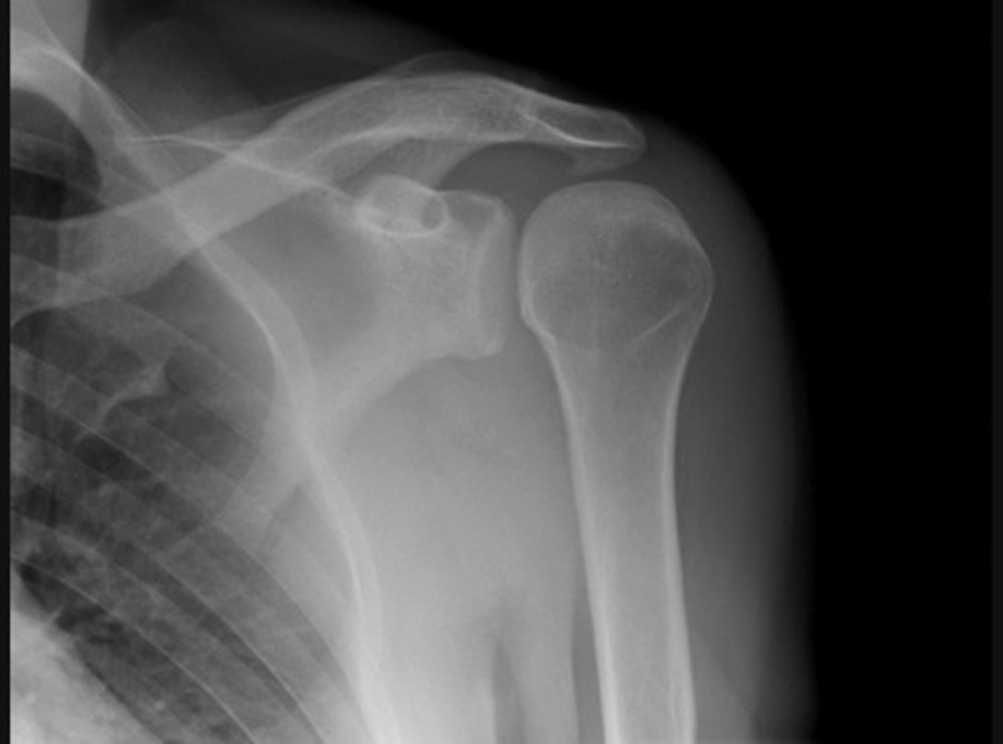
Anterior dislocation and Hill-Sachs lesion

Anterior dislocation and glenoid fracture (bankhart lesion)
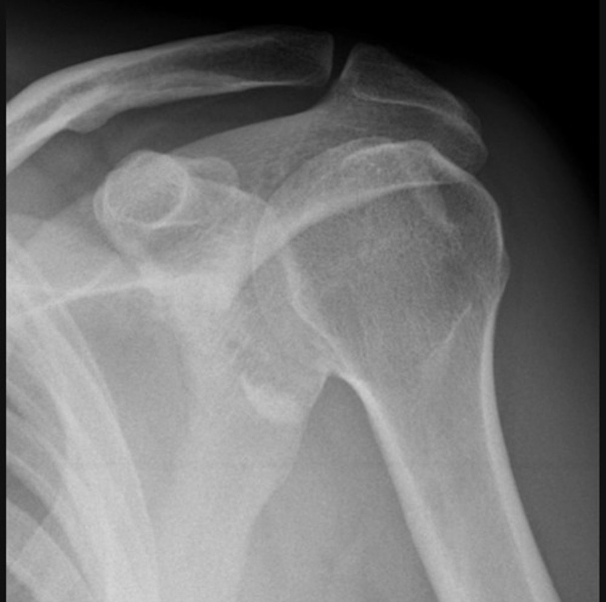
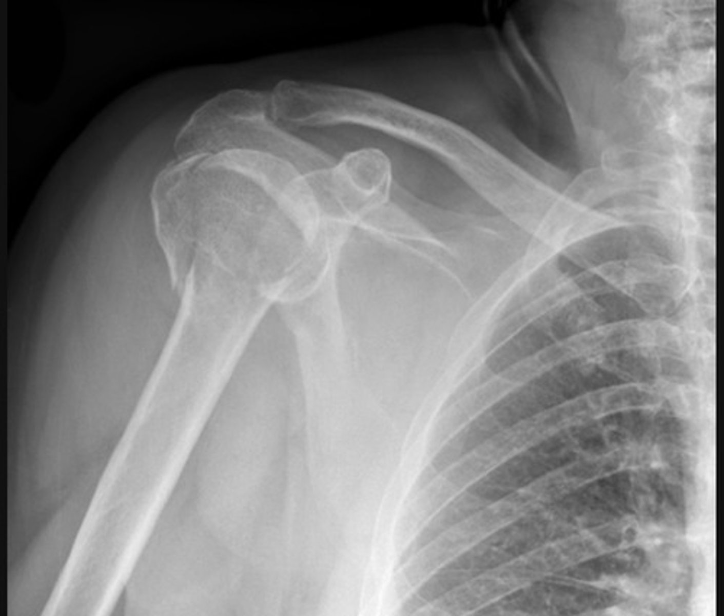
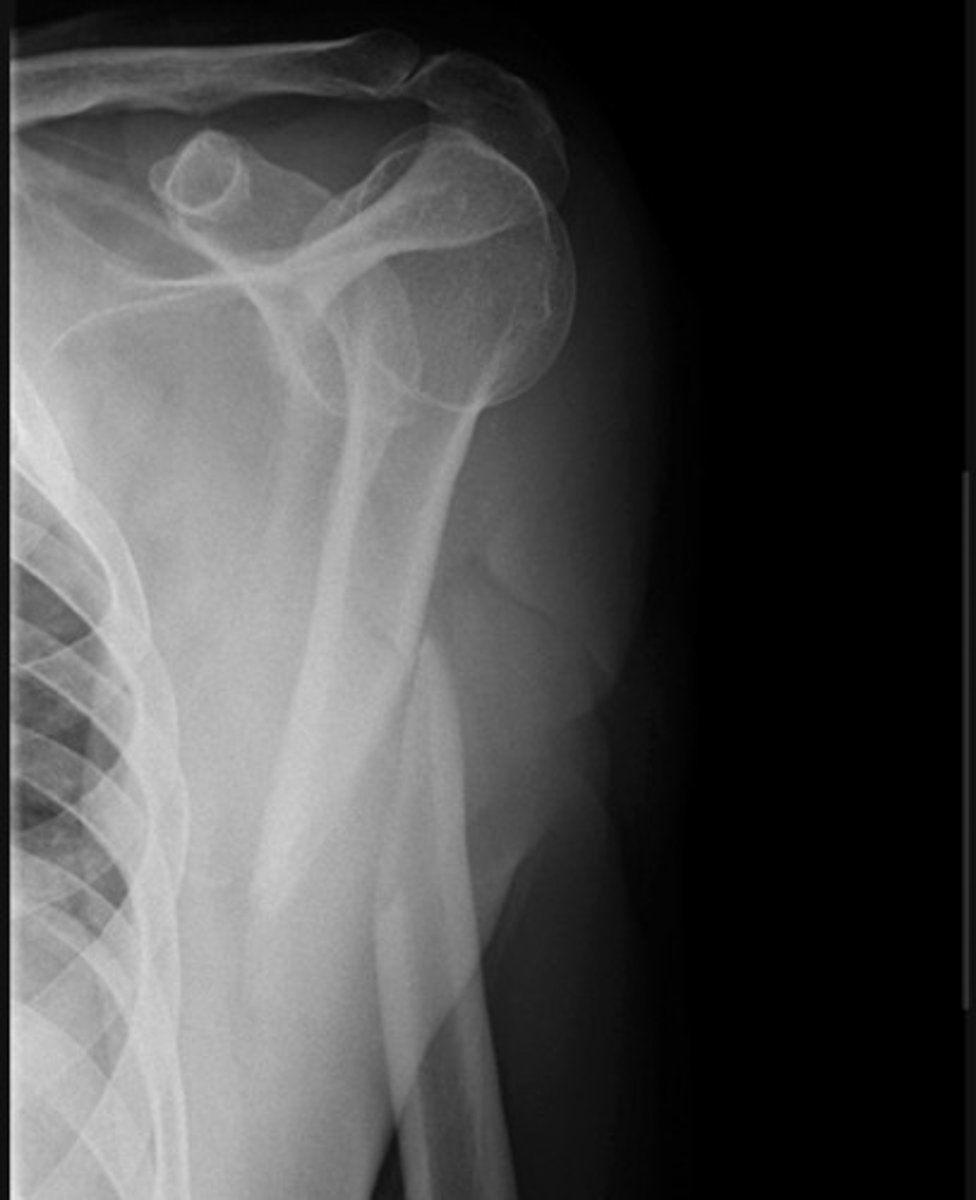
Posterior dislocation, 'lightbulb sign'
Transverse fracture of the scapula body
Surgical neck fracture-
Transverse fracture through the proximal humerus with displacement of the distal component
Comminuted fracture-
A transverse fracture of the surgical neck of humerus is accompanied by a fracture line separating the greater tubercle from the rest of the humeral head
Shaft fracture
Oblique fracture of the humerus shaft
As with many long-bone fractures the distal component is markedly displaced
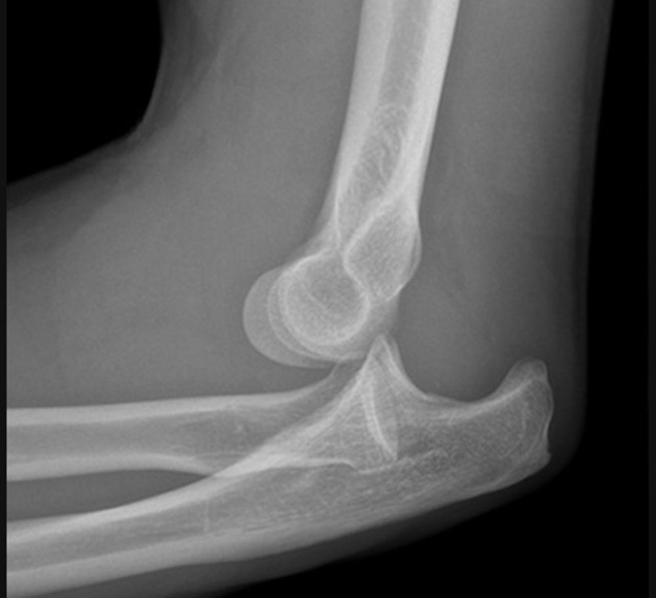
The ulna has dislocated posteriorly from the trochlea of the humerus
The radius has dislocated from the capitulum of the humerus
Visible posterior fat pad.
Supracondylar fracture - Lateral
This is the commonest elbow fracture in children
A joint effusion raises the anterior and posterior fat pads, indicating intra-capsular injury
Less than one third of the capitulum of the humerus lies in front of the anterior humeral line
The distal fracture fragment is pulled posteriorly by the triceps muscle
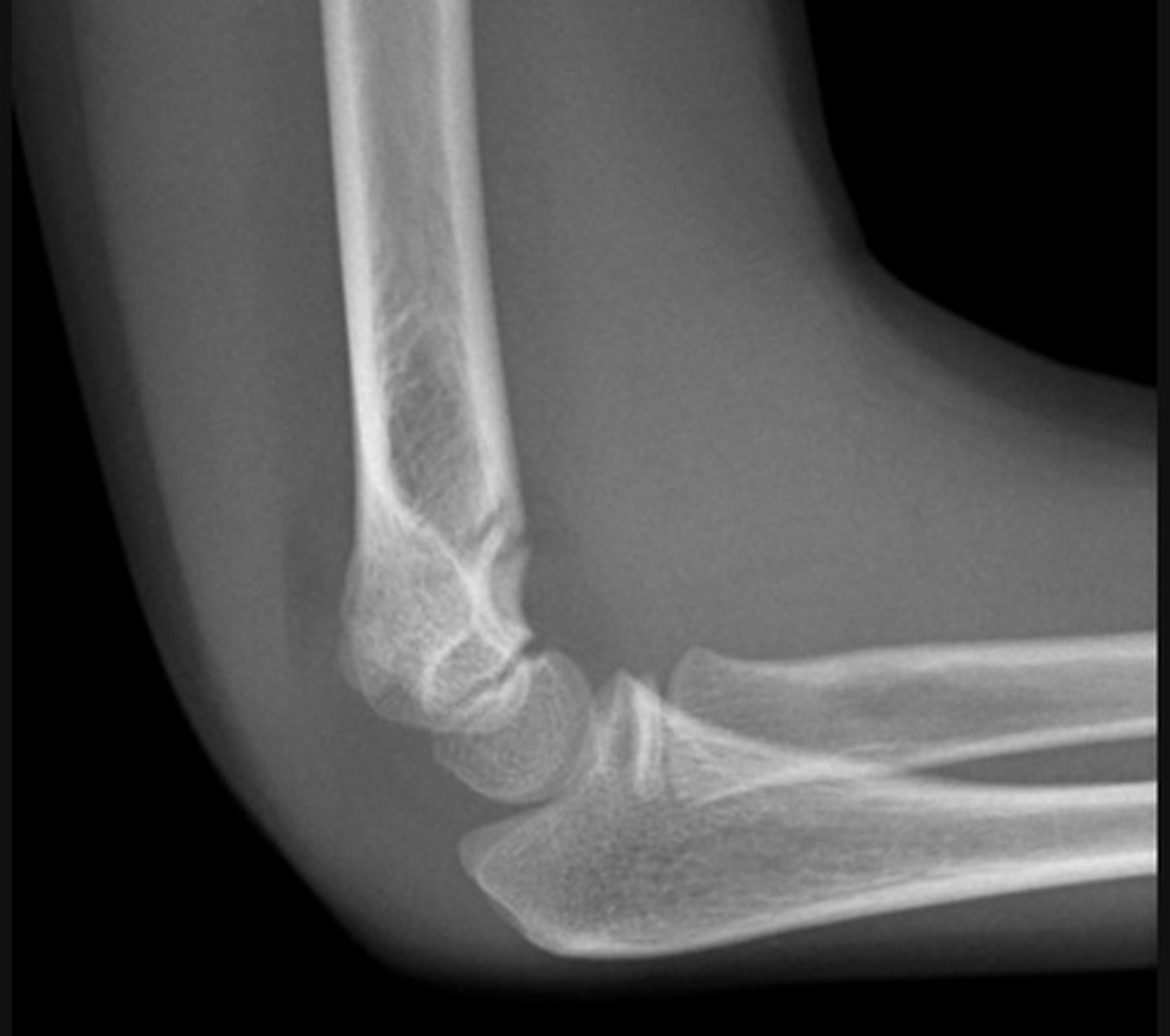
The fracture line is not visible on the lateral view in this case
The effusion - indicated by raised fat pads - is the only visible sign of injury and in the context of trauma should be taken to indicate an undisplaced intra-capsular fracture
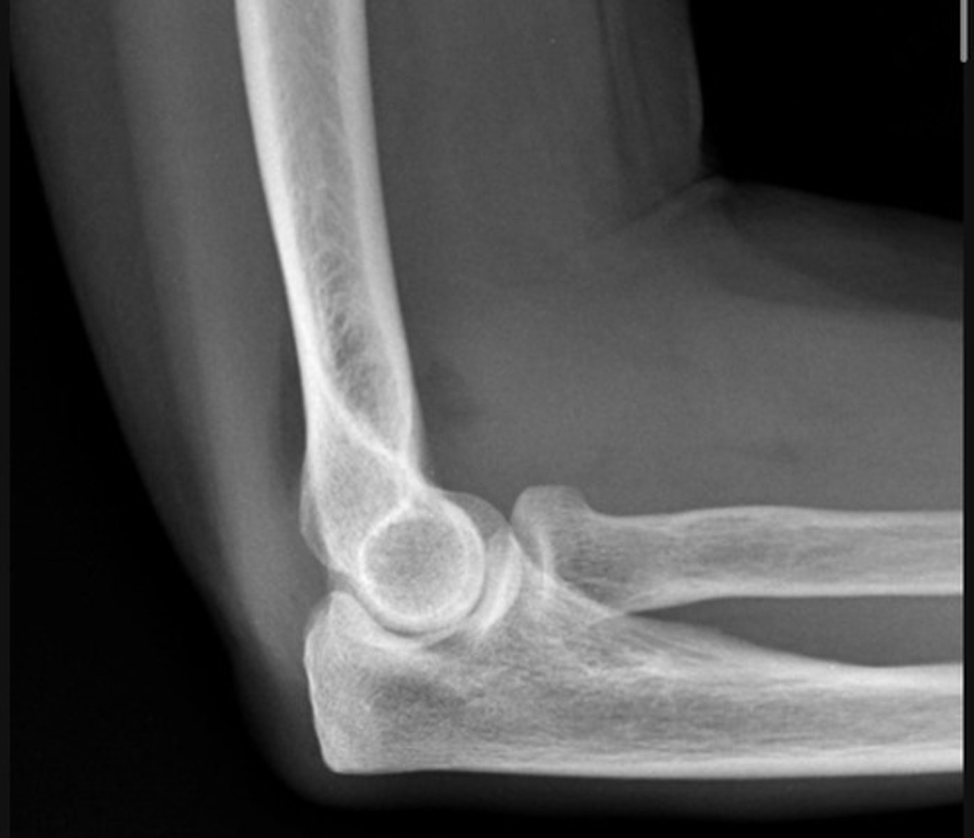
Likely a radial head fracture (adult)
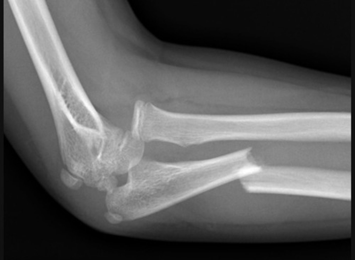
Monteggia fracture-dislocation - Lateral
a fracture of the ulnar shaft with dislocation of the radial head at the elbow
The radiocapitellar line should pass through the middle of the capitulum of the humerus
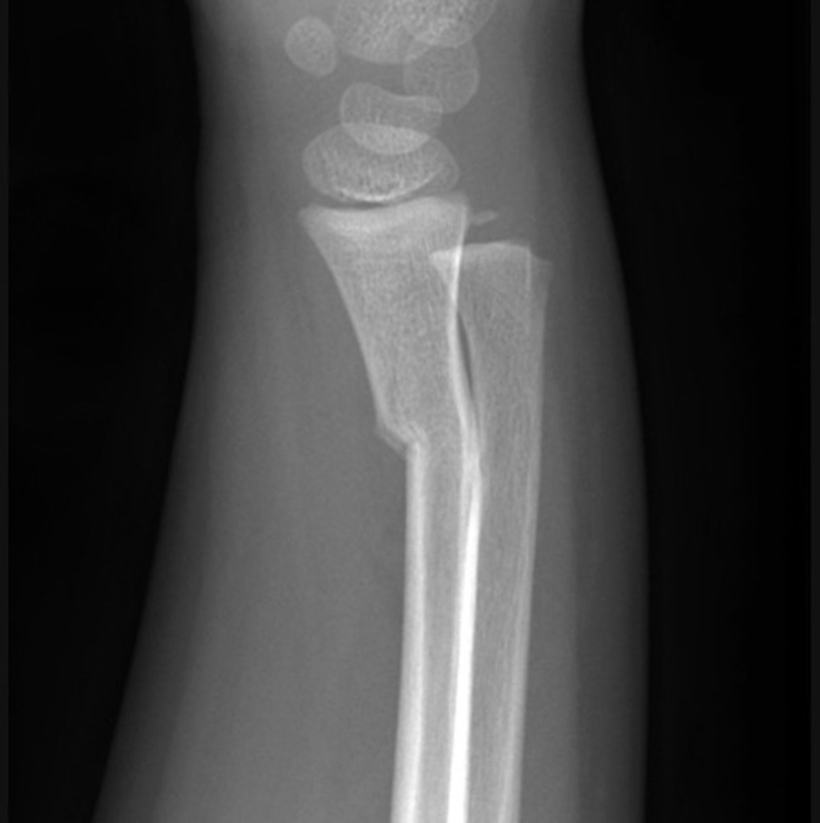
Buckle fracture - Greenstick of the distal radius
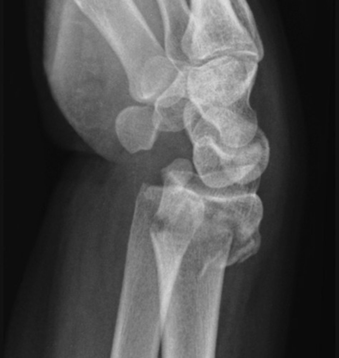
Colles fracture- shortening and dorsal displacement of the distal radius fracture fragment.
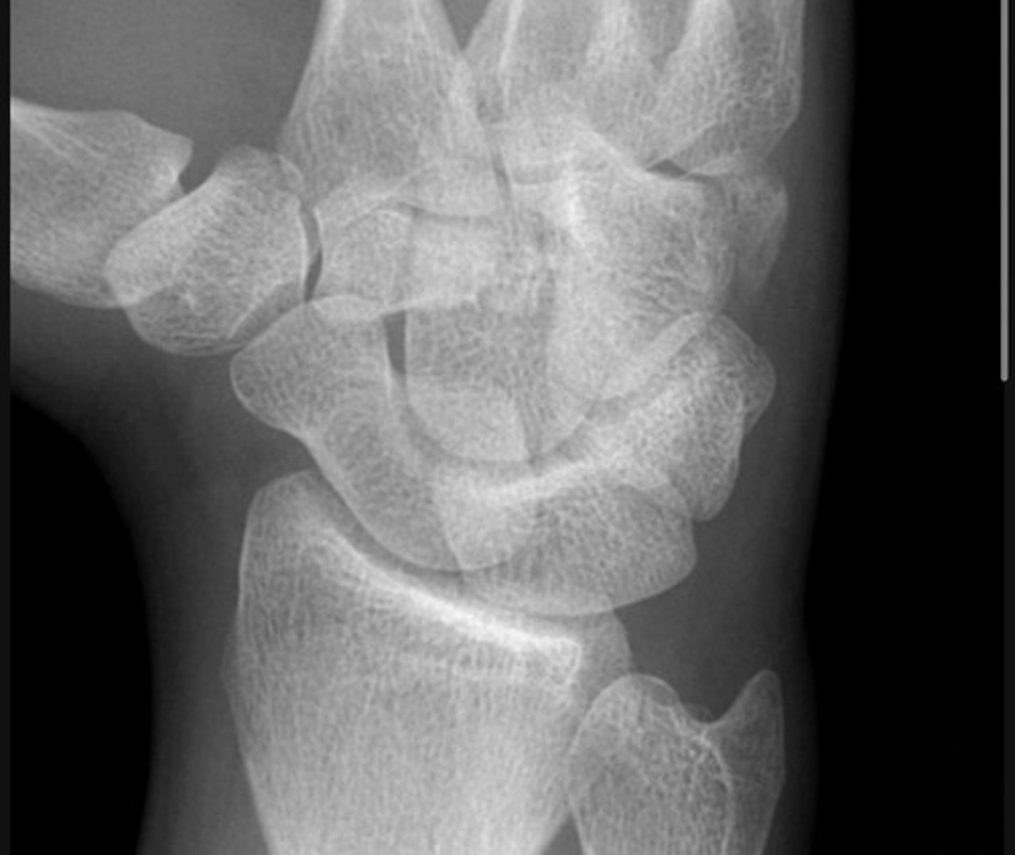
There is a lucent fracture line through the body of the hamate with preserved carpal alignment and no obvious associated dislocation. Findings are consistent with a minimally displaced hamate fracture
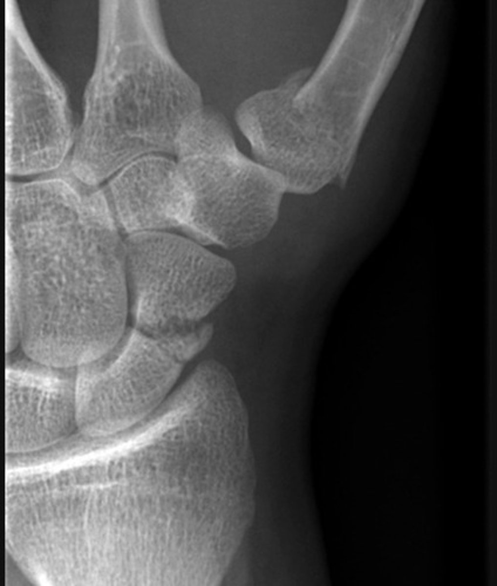
There is a transverse to slightly oblique fracture through the waist of the scaphoid, with no significant displacement or carpal malalignment. Additionally, there is an oblique intra-articular fracture at the base of the first metacarpal, extending into the carpometacarpal joint. The distal metacarpal fragment is displaced proximally and dorsally, consistent with a Bennett's fracture, with associated subluxation at the CMC joint.
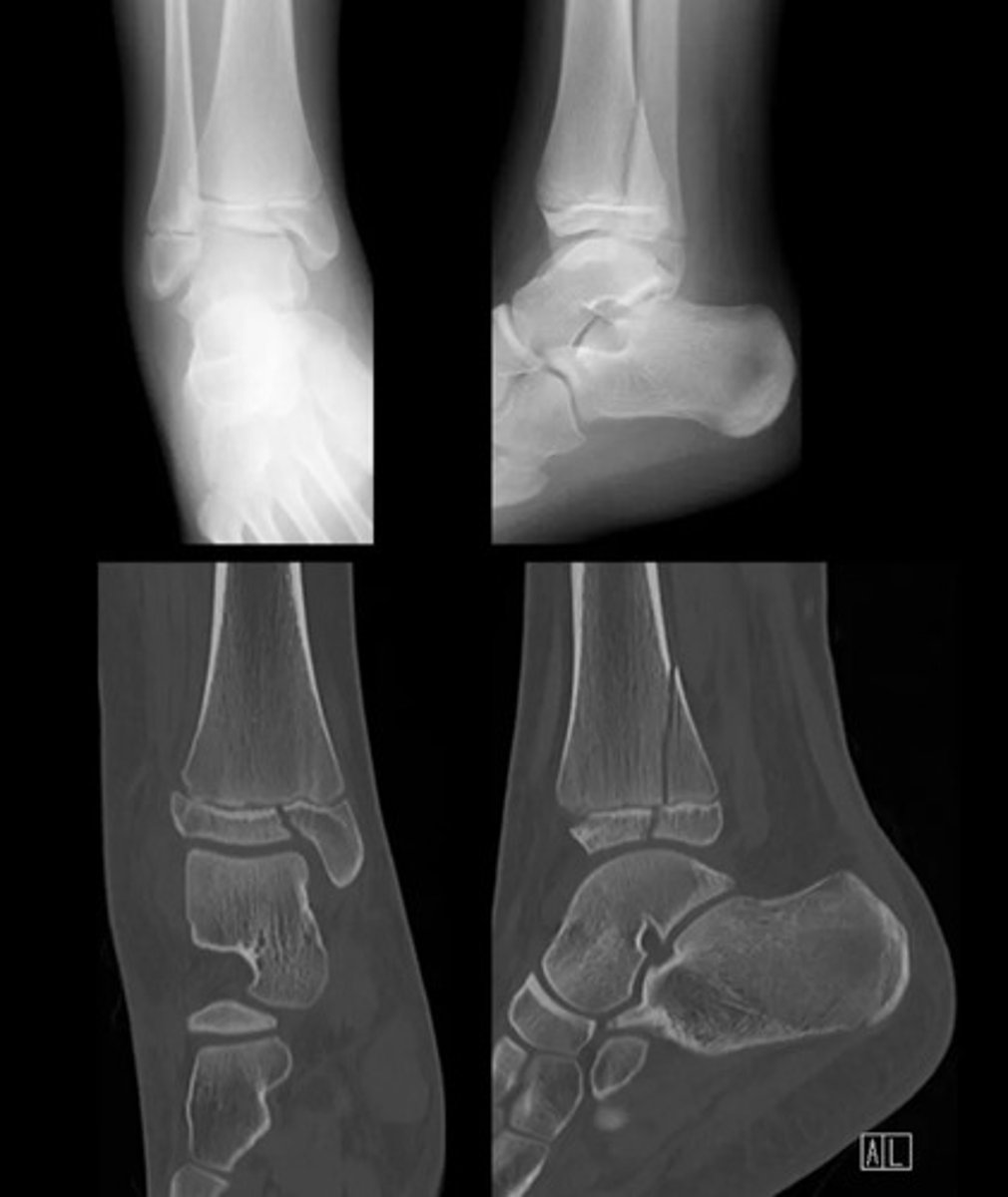
SH type 3
SH 1
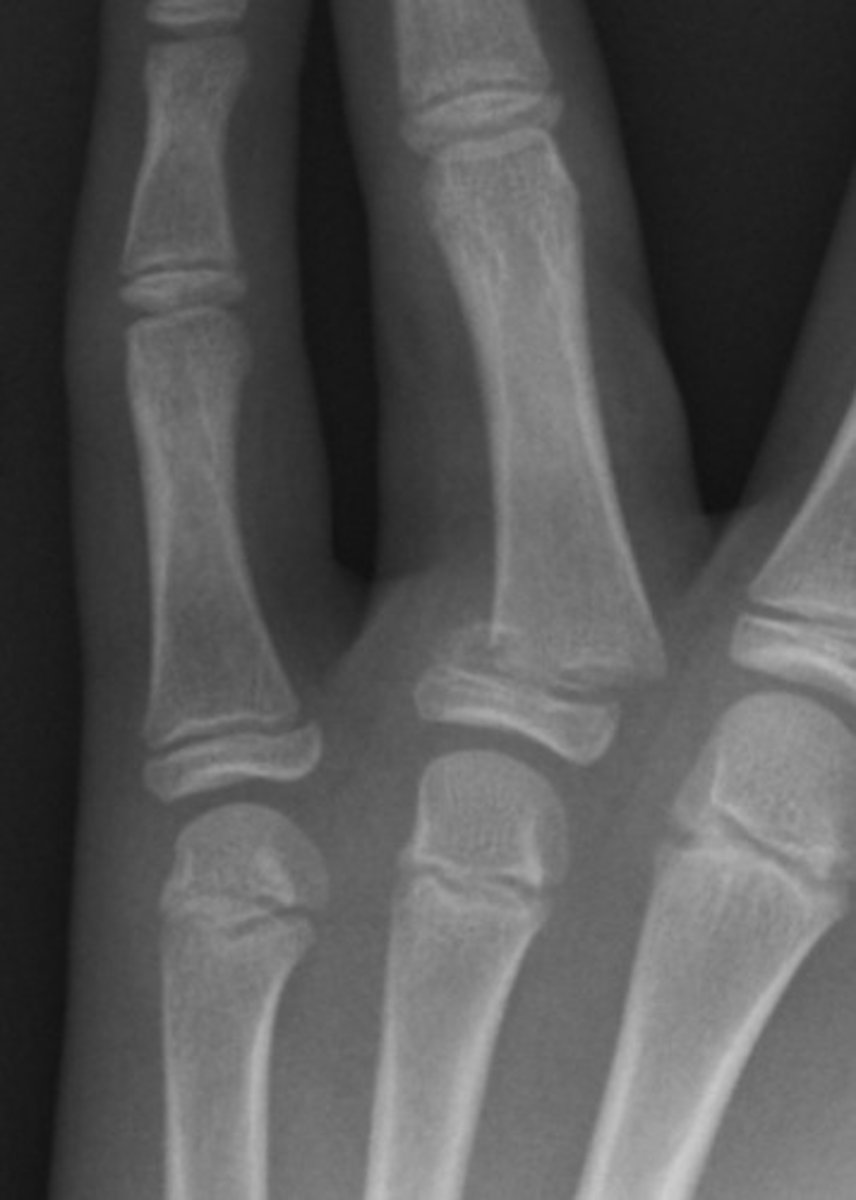
SH 4
Subcapital fracture with complete displacement
Transcervical fracture with minor displacement
Lisfranc injury
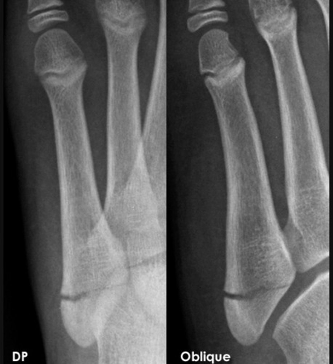
base of 5th metatarsal fracture
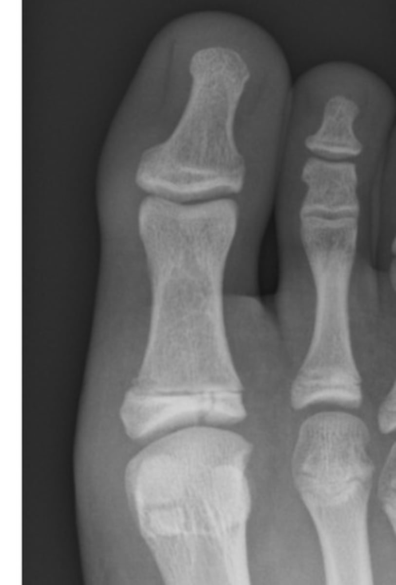
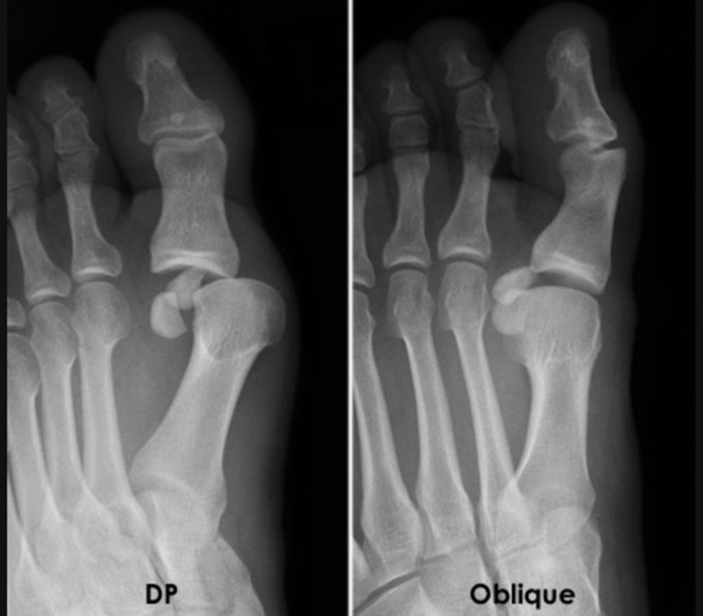
dislocation of the proximal phalanx of the big toe at the MTPJ (metatarsophalangeal joint)
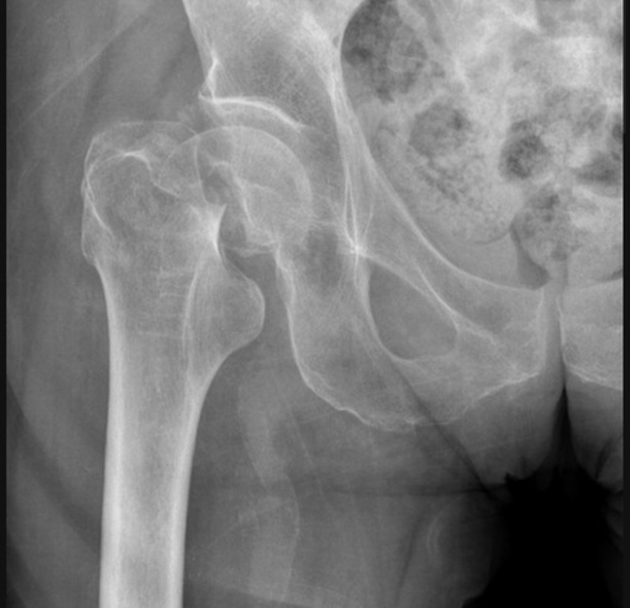
Subtrochanteric fracture
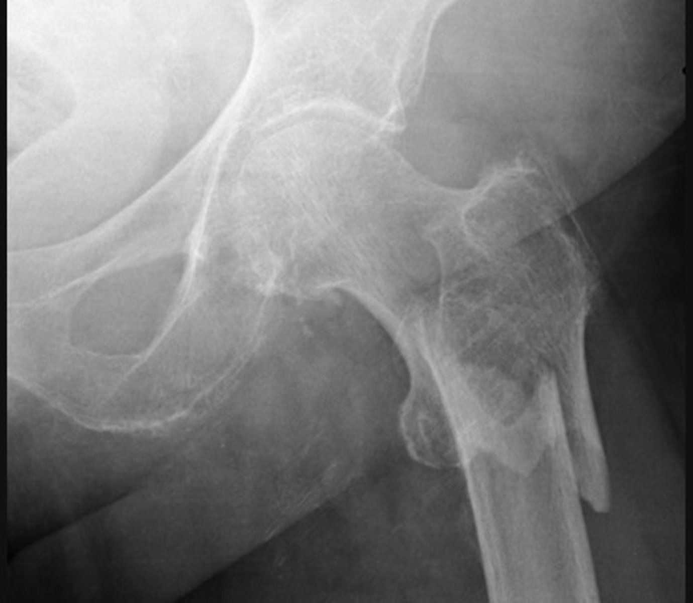
The bone texture is abnormal in this patient with a known malignancy - indicating a pathological fracture
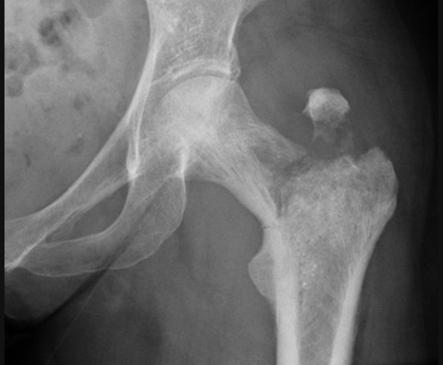
A fracture of the femoral neck is accompanied by avulsion of the greater trochanter
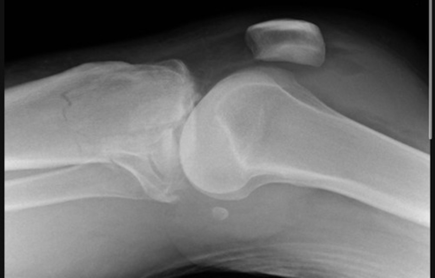
severely comminuted fracture of the tibial plateau and a lipohaemarthrosis
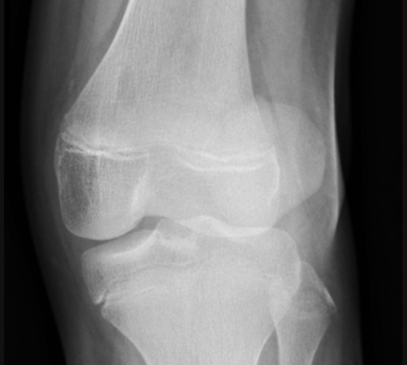
Lateral patella dislocation
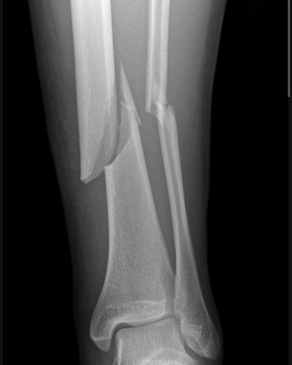
There are complete fractures involving the distal third diaphyses of both the tibia and fibula. The tibial fracture is oblique. The distal tibial fragment is laterally displaced and angulated relative to the proximal shaft. The fibula demonstrates an associated oblique fracture with mild displacement.
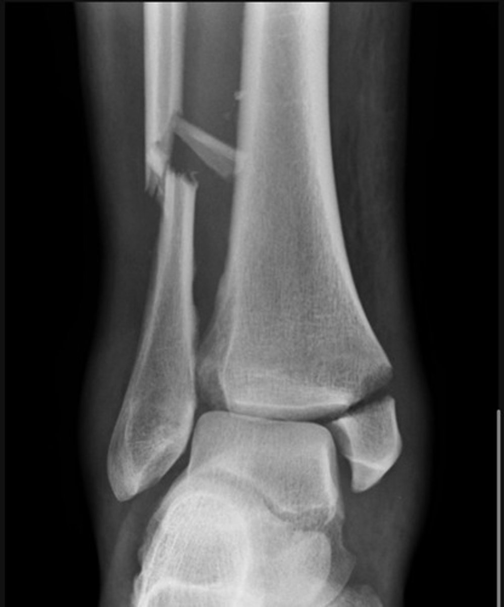
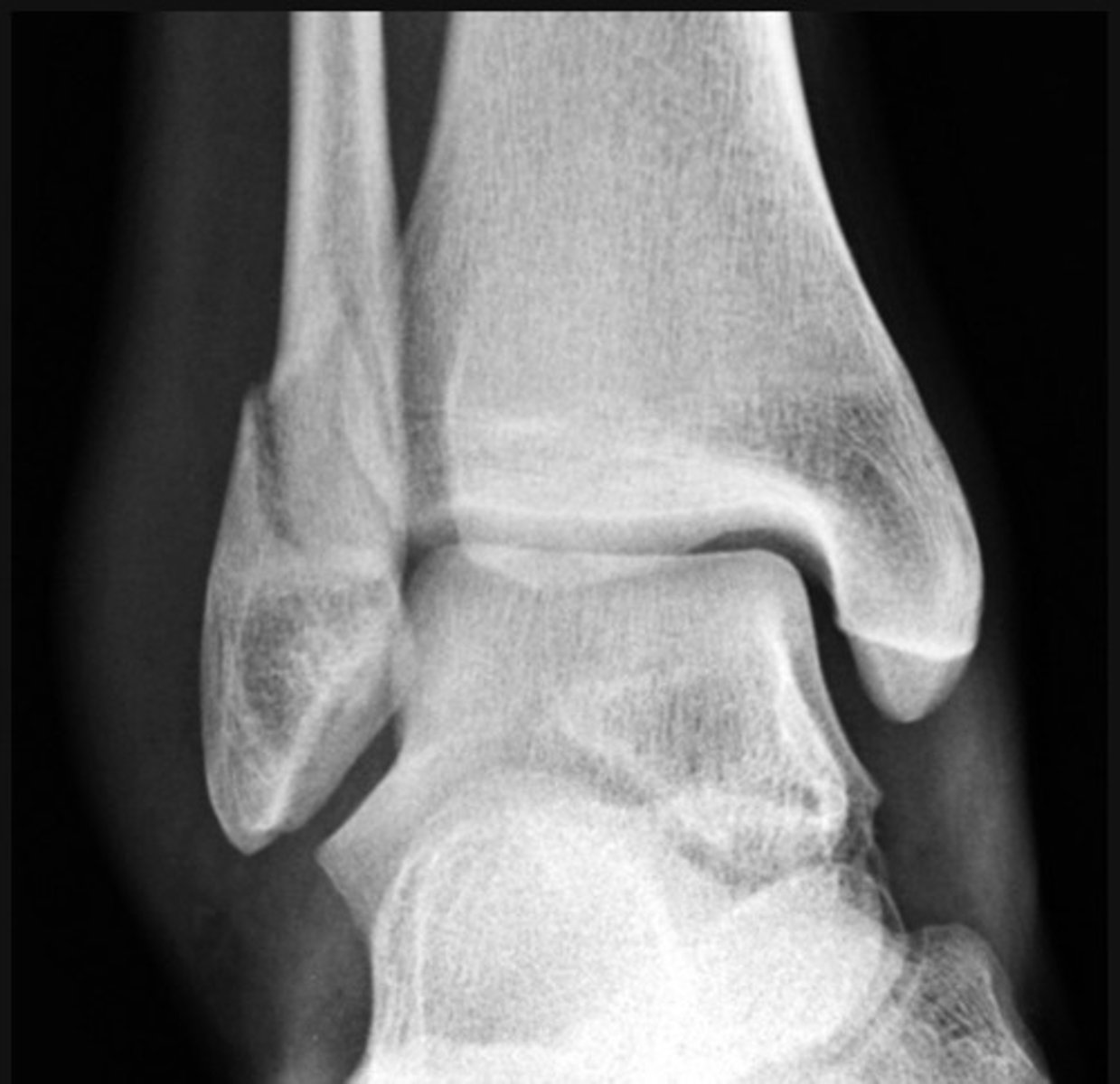
Weber A fracture of the fibula/ lateral malleolus
Weber B feature of the fibula/ lateral malleolus
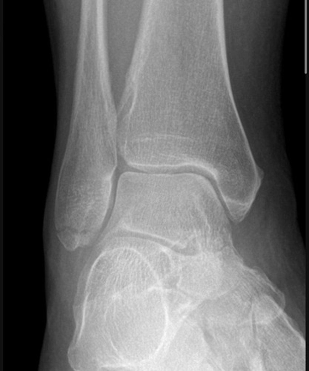
Bimalleolar fracture.
Weber C
There is a transverse medial malleolus fracture
The distal tibiofibular joint is also widened