Week 2: Mechanics of the nervous system
1/75
There's no tags or description
Looks like no tags are added yet.
Name | Mastery | Learn | Test | Matching | Spaced | Call with Kai |
|---|
No analytics yet
Send a link to your students to track their progress
76 Terms
What is the primary focus of the cerebellum?
Movement and coordination
What factors are involved with the spinal cord in the CNS?
Continuous with brain stem
Long conical structure, cone-like
Thickness of adult’s little finger, 1cm in diameter
Mediates transmission of information
between brain & body
What are the three major functions of the spinal cord?
Coordinating reflexes
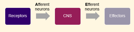
Serving as a conduit for sensory and motor information: messages from brain to body (efferent), messages from body to brain (afferent)
Mediating messages between brain and body
What do the dorsal roots of spinal nerves carry? Where are they located?
Afferent neuron axons for sensory input.
Located in the back of the spinal cord
Afferent neuron axons enter cord in dorsal root and terminate in dorsal horn - sensory input
What do the ventral roots of spinal nerves carry? Where are they located?
Efferent neurons that send messages from the CNS to the body
Located in the front of the spinal cord
Efferent neurons have a cell body in ventral horn and axons leave cord in ventral root - comes out into receptive part of body
What are the functions of the PNS?
Connects CNS to limbs & organs via cranial and spinal nerves
Carries information from environment to CNS
(afferent neurons), processed in CNSCarries messages from CNS to muscles and glands (efferent neurons)
How many pairs of nerves are there in the PNS?
43 pairs of nerves, nerves come in pairs
12 cranial nerve pairs
31 spinal nerve pairs
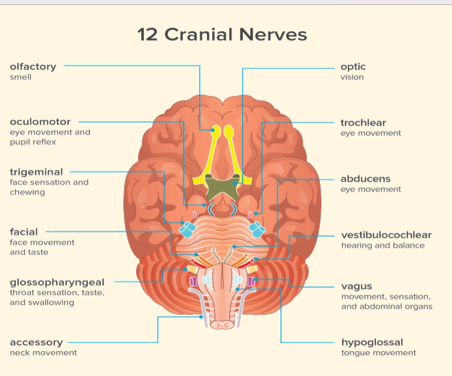
How many pairs of cranial nerves are there?
12 pairs
10 located in brainstem
1 and 2 located in forebrain
How many pairs of spinal nerves are there?
31 pairs
Name all of the 12 cranial nerves
Old
Owls
Often
Take
Tiny
Acorns
For
Very
Grumpy
Village
Angry
Hedgehogs
What is the function of the somatic nervous system?
Voluntary control of body movement
Receives sensory information and controls spinal nerves that innervate skin, joints & muscles
Afferent neurons carry sensory info from skin (sensory neuron)
Efferent neurons control skeletal muscles (motor neuron) - support contractions, muscle fibres pulling across each other
-> Neurons are excitatory
What does the autonomic nervous system control?
Controls involuntary functions and internal environment
Afferent neurons carry sensory info from internal organs to CNS
Efferent neurons control smooth muscle, cardiac muscle & glands (production of hormones)
Neurons are excitatory or inhibitory (e.g. slow down heart rate - parasympathetic)
What are the three sub-divisions of the autonomic nervous system?
Sympathetic Nervous System
Parasympathetic Nervous System
Enteric Nervous System
What is the role of the sympathetic nervous system?
Coordinates the body's fight or flight response
Responses for activities which expand energy
What is the role of the parasympathetic nervous system?
Coordinates rest and relaxation responses
Activities involved with increase in the body’s supply of stored energy
What are the main roles of the enteric nervous system?
The “second brain”
Links digestive system to the brain
Lines your gastrointestinal tract from oesophagus to rectum
Main role is controlling digestion
swallowing
release of enzymes
control of blood to facilitate nutrient absorption
What is the gut-brain axis?
The complex interplay between gut microbiota, the immune system, and the central nervous system
Gut Microbiota (GM) regulates brain function by preserving the CNS immune homeostasis
Prevents neuroinflammation and degredation
What are sensory neurons responsible for?
Part of PNS
Contain sensory receptors for detecting sensory changes
Sends information about these changes to CNS
Cell body in PNS, axon enters CNS (axon terminals located in CNS)
Taste, touch, movement, pressure, temperature
What do motor neurons do?
Part of PNS
Synapses to skeletal muscle to command movement or onto glands (inhibition/activation) to release hormones
Relays signal from CNS to PNS
Dendrites & cell body in CNS, axon enters PNS
What is the role of interneurons?
In CNS
Receives info from sensory neurons
Sends info to motor neurons
Integrate/change signal
-> Integrate - inputs from multiple afferent neurons - average signal
-> Changer - interneurons can provide excitatory or inhibitory signals
What is the primary function of the central nervous system (CNS)?
To receive information from sensory neurons, send information to motor neurons, and integrate/change signals.
What are the two main types of signals that interneurons can provide?
Excitatory and inhibitory signals.
What is the structure of the neuronal membrane?
Made of two layers of lipid molecules
Lipid molecules - attracted to the intracellular and extracellular fluid
Hydrophilic (water attracting) heads
Hydrophobic (water repelling) tails
Barrier: water soluble molecules cannot pass through
Particularly impermeable to ions, stop ions passing through
What drives the movement of ions across the neuronal membrane?
Concentration gradients (via diffusion) and electrical forces (via electrostatic pressure).
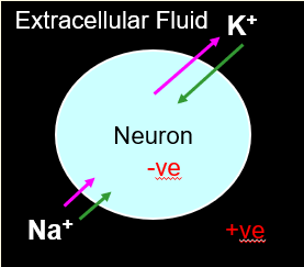
Explain the fluid environment containing ions
Made up of intracellular fluid and extracellular fluid
Cations (+ charged)
Sodium (Na+)
Potassium (K+)
→ predominantly intracellular
Anions (- charged)
Chloride (Cl-) → predominantly extracellular
Organic ions (A-) → only intracellular
What happens to ions under electrostatic pressure?
Charges of opposite sign attract, while charges of the same sign repel.
Outline the process of the electrical polarity of neurons
Neuron is polarised
At rest, neurons are negatively charged compared to extracellular fluid
Negative charge occurs if there are less positive ions and/or more negative ions inside cell
Whilst there is a difference in charge, an electrical force tends to move ions across the membrane
At rest, the resting potential of the inside of the neuron is -70 mV
0 mV in the extracellular of the neuron
What is the resting potential of a neuron?
-70 mV, indicating that the inside of the neuron is negatively charged compared to the extracellular fluid.
Outline the role and function of border guards
Controlled by a gate
Ion channels (leak channels) - always open
Passive ion specific conduits
Selected ions rush down gradients of concentration and electric potential
More K+ gates open compared to Na+
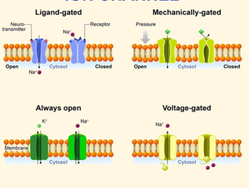
What role do ion channels play in neuronal function?
They allow specific ions to pass through the membrane, contributing to the resting potential and action potentials.
What is the primary ion responsible for maintaining resting potential?
Potassium ions (K+), which are highly concentrated inside the cell.
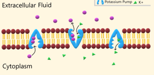
Outline the ion pump process
Cytoplasm has less Na+ than extracellular fluid
3 Na+ out and 2 K+ which maintains this resting potential of -70Mz
Energy consuming
Active transport: against gradient
Maintains and builds gradients
Slower
Outline the process of diffusion of potassium ions
K+ highly concentrated in cell
K+ wants to move out of cell down concentration gradient
At rest, K+ leak channels allows K+ to leave neuron down concentration gradient
Inside cell becomes more negative
Ions will stop moving when opposing forces are at equilibrium: -70Mz
→ This happens in a resting cell
Outline the process of diffusion of chloride ions. Give reference to electrostatic pressure
Generally equally distributed
Cl- highly concentrated outside cell
Cl- wants to move into cell down concentration gradient
Inside of cell is + charged
Cl- also wants to move out of cell due to repel of electric charge
Outline the process of diffusion of sodium ions. Give reference to electrostatic pressure
Na+ is highly concentrated outside cell
Na+ wants to move into cell down concentration gradient
Inside of cell is - charged
Na+ also wants to move into cell due to electric charge attraction
→ Net force for Na+ = move into cell
Outline what is meant by the resting membrane potential
Two forces act on ions
Membrane is a barrier to ion movement
At rest membrane is permeable to K+ so mainly K+ ions move
K+ ion movement stops once opposing forces reach equilibrium
→ unequal distribution of positive and negative ions on the inside and outside of membrane
Resting membrane potential = difference in charge across membrane at rest = -70mV
What occurs during depolarisation of a neuron?
The inside of the neuron becomes more positively charged as sodium ions (Na+) enter the cell.
What is the threshold potential for an action potential to occur?
Approximately -55 mV.
What is meant by action potential?
A brief electrical impulse that provides the basis for conduction of information along an axon
What are the phases of an action potential?
Depolarisation: inside becomes more +
Repolarisation: inside becomes more -
Hyperpolarisation: more - than at rest
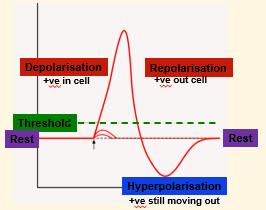
What is meant by the 'all-or-nothing' phenomenon in action potentials?
An action potential occurs only if the threshold is reached; otherwise, it does not occur.
If depolarisation reaches threshold (-55mV), an AP occurs automatically
→ -55mV
What regulates the strength of a neural response?
The rate of neural firing, not the size of a single action potential.
What occurs during depolarisation of a cell?
Stimulus causes a small amount of Na+ to move into the cell
Na+ is + charged → neuron becomes less - (slightly depolarised)
If depolarisation changes charge by +15mV, it activates voltage-gated channels in membrane
Outline the process of voltage-gated channels
Activated by changes in charge of membrane

Outline the process of a voltage-gated action potential
Voltage-gated Na+ channels open. Na+ influx → more +ve
Na+ channels become refractory at peak (neuron is resistant at firing another AP)
Voltage-gated K+ channels open. K+
efflux → less +veOpen K+ channels allow outflow
Overshoot caused by slow closing K+ channels
What is the role of voltage-gated sodium channels during an action potential?
They open in response to depolarisation, allowing Na+ influx, which further depolarises the neuron.
What happens during repolarisation of a neuron?
Voltage-gated K+ channels open, allowing K+ to exit the cell, making the inside more negative.
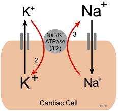
What is the function of the Na+/K+ ATPase pump?
It moves 3 Na+ ions out of the cell and 2 K+ ions into the cell, maintaining the concentration gradients.
The pump keeps Na+ concentration low in neuron
K+ diffuses back into neuron
→ Re-establishes resting membrane potential
Signal travels away from cell body towards axon terminals
No decay
AP propagation
What is the effect of K+ leak channels on resting potential?
They allow K+ to leave the neuron, contributing to the negative resting potential.
What is the net force acting on sodium ions (Na+) at rest? Why do these ions move into the cell?
Large net force
Na+ wants to move into the cell due to both concentration gradient and electrostatic pressure.
What is the role of chloride ions (Cl-) in neuronal resting potential?
Cl- is generally equally distributed but is influenced by both concentration gradient and electrostatic pressure.
What is the role of K+ ions in a neuron?
K+ ions diffuse back into the neuron, re-establishing the resting membrane potential.
What is meant by AP propagation?
AP propagation refers to the signal traveling away from the cell body towards the axon terminals without decay.
Na+ ions spread away from site of AP which changes the charge in nearby area of cell be be more + charged (depolarised)
This triggers another action potential
Next AP occurs as previous AP starts to die out
APs are triggered one after another all the way to axon terminals
→ ‘mexican wave effect’
How do Na+ ions contribute to action potential propagation?
Na+ ions spread away from the site of the action potential, depolarising nearby areas of the cell and triggering subsequent action potentials.
What prevents action potentials from traveling backwards?
The refractory period prevents action potentials from traveling backwards and determines the upper limit on action potential frequency.
What is the function of neurotransmitters?
Neurotransmitters are released from vesicles in the terminal ends of axons to excite, inhibit, or modulate postsynaptic cells.
2 (or more) neurotransmitters are released from each neuron
Name 7 neurotransmitters.
Acetylcholine
Serotonin
Dopamine
Nor/epinenphrine
Endorphins
GABA
Glutamate
What role does acetylcholine play in the nervous system?
Acetylcholine is an excitatory neurotransmitter that regulates heart rate, blood pressure, gut motility, muscle contractions, memory, and learning.
Imbalances linked with Alzheimer’s disease, seizures and muscle spasms
Name 4 monoamines
Serotonin
Dopamine
Epinephrine (adrenaline)
Norepinephrine
Name some factors involved within serotonin
Serotonin is an inhibitory neurotransmitter that regulates mood, sleep patterns, libido, anxiety, appetite, and pain.
Imbalances include SAD, anxiety, fibromyalgia and chronic pain
Medications which regulate serotonin include selective serotonin reuptake inhibitors (SSRIs) and serotonin-norepinephrine reuptake inhibitors (SNRIs)
What is dopamine's role in the brain?
Dopamine is involved in the reward system, facilitating pleasure, heightened arousal, focus, concentration, and learning.
Dysfunctions of the dopamine system include Parkinson’s disease, Sz, bipolar disease, restless legs syndrome and ADHD
What is the function of epinephrine and norepinephrine?
They are responsible for the 'fight-or-flight response' to fear and stress, increasing heart rate, breathing, blood pressure, and focus.
Excess epinephrine can lead to high blood pressure, diabetes, heart disease and other health problems.
As a drug, epinephrine is used to treat anaphylaxis, asthma attacks, cardiac arrest and severe infections
What are endorphins and their role in the body?
Endorphins are pain relievers that contribute to the perception of pain and create 'feel good' feelings.
Low levels may play a role un fibromyalgia and some types of headaches
Name 2 amino acids
Glutamate
Gamma-aminobutryic acid (GABA)
What is glutamate's role in the brain?
Glutamate is the most common excitatory neurotransmitter and plays a key role in cognitive functions like thinking, learning, and memory.
Imbalances associated with Alzheimer’s disease, dementia, Parkinson’s disease and seizures
What is GABA and its function?
GABA is the most common inhibitory neurotransmitter in the brain, regulating brain activity to prevent anxiety, irritability, and seizures.
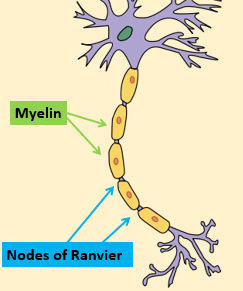
Outline structural features of glial cells (support cells)
Contain fatty tissue (myelin) which wraps around neuron axons
Forms insulating coating (myelin sheath)
Schwann cells - wrap individual axons
Oligodendrocyte - wrap several axons
Axons then become myelinated
→ Cells jump down the axon - nodes of Ranvier
Outline the anatomy of a myelinated axon
Yellow areas: Schwann cells, production of myelin sheaths
Purple areas: ‘naked’ axon - nodes of Ranvier
Ions can only cross the membrane at nodes of Ranvier
No ion leakage outside of nodes of Ranvier, so influence of AP spreads quicker in myelinated axons
Outline differences between unmyelinated and myelinated axons
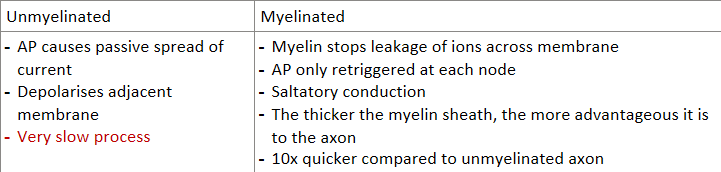
What are the advantages of myelinated axons compared to unmyelinated axons?
Myelinated axons conduct action potentials faster due to saltatory conduction.
Much more economical: less Na+ enters during APs, more efficient AP regeneration
The process where action potentials jump from node to node in myelinated axons between gaps (nodes of Ranvier)
Hereditary: Tay-sachs disease, Niemann-pick disease, Gaucher disease, and Hurler syndrome
Stroke
Infections: viruses, bacteria
Immune disorders
Metabolic disorders
Nutritional deficiencies (e.g. lack of vitamin B12)
Poisons, e.g. carbon monoxide
Drugs or medications, e.g. antibiotic ethambutol
Excessive use of alcohol