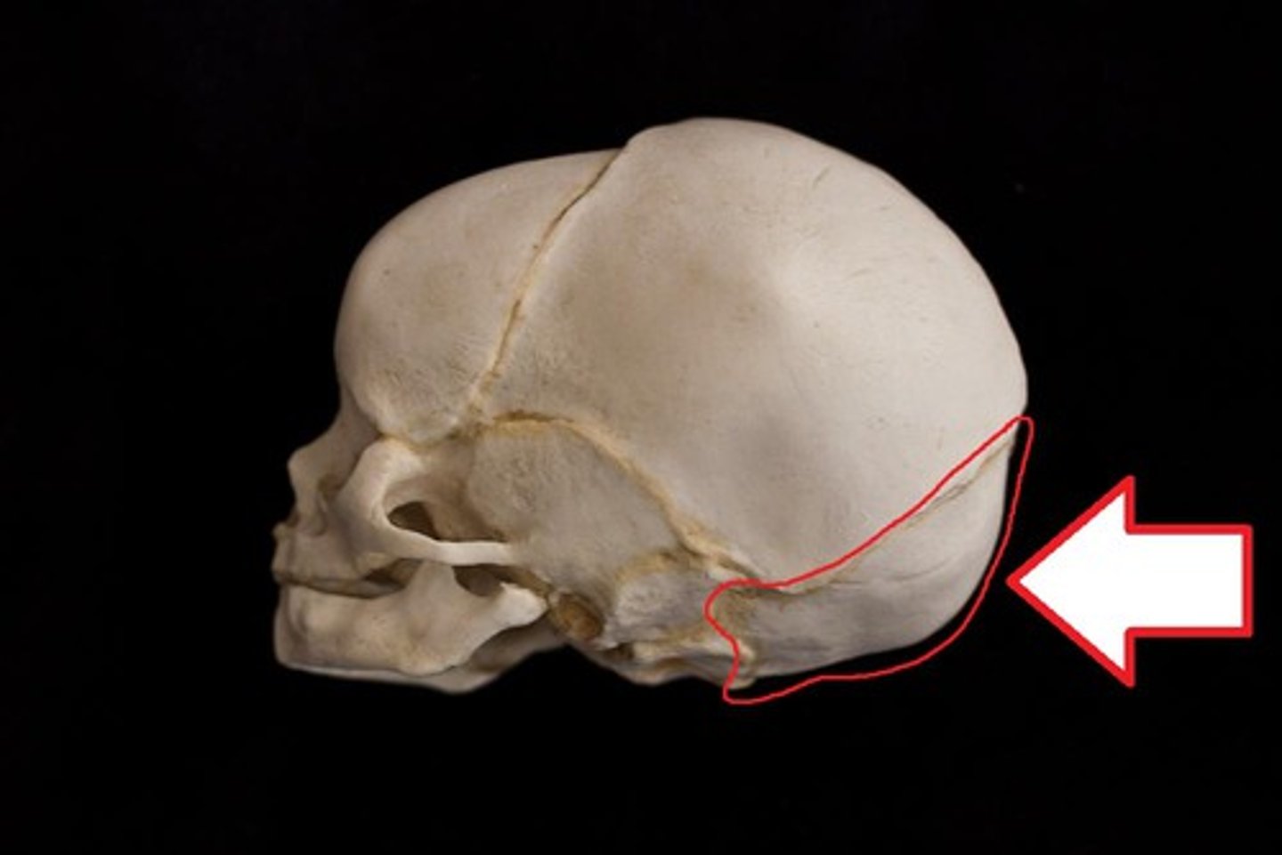
Occipital Bone
1/5
There's no tags or description
Looks like no tags are added yet.
Name | Mastery | Learn | Test | Matching | Spaced |
|---|
No study sessions yet.
6 Terms
Occipital Bone
The occipital bone is set at the rear of the cranium and articulates with the temporals, sphenoid, parietals, and the uppermost vertebra, the atlas.
(Better pictures:
https://www.bartleby.com/107/31.html)
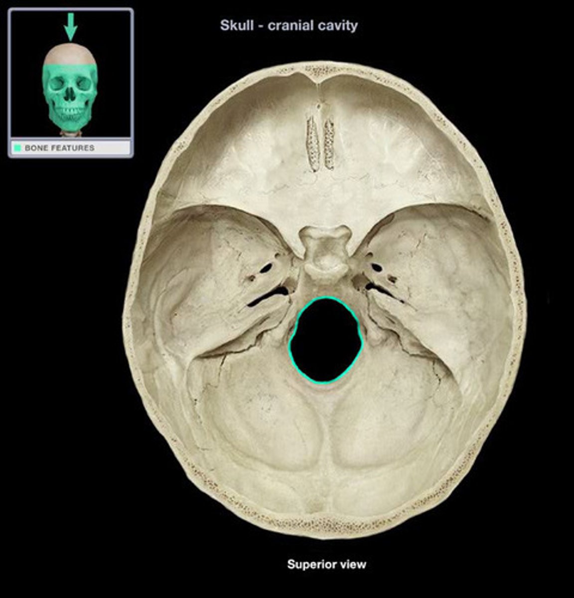
Foramen Magnum
The large hole in the occipital through which the brain stem passes inferiorly into the vertebral canal.
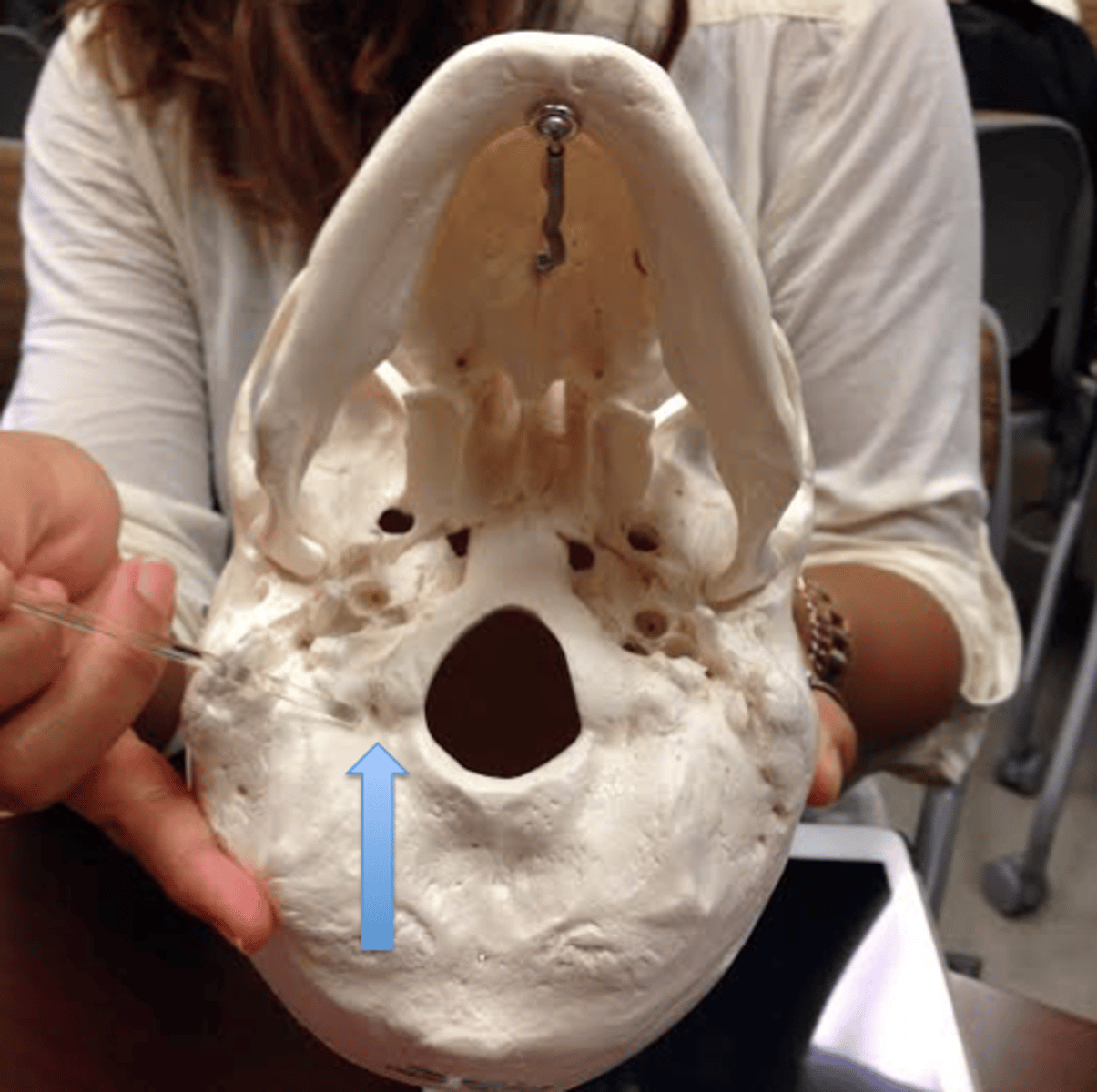
Occipital Condyles
The Condyles are raised oval structures on either side of the foramen magnum. Their inferior surfaces are convex. The articular surfaces of these condyles fit into the concave facets of the atlas vertebra.

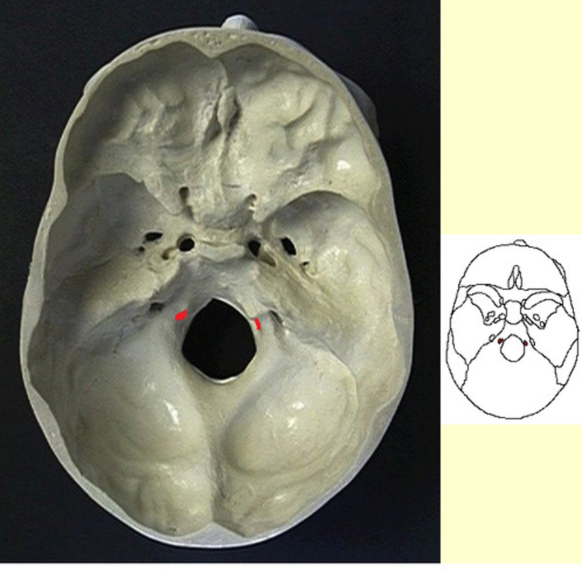
Condyloid Fossa
These are ectocranial depressions immediately posterior to the condyles. The fossae receive the posterior margin of the superior facet of the atlas vertebra when the head is extended backward.
(in the picture,it's the tiny hole)
Hypoglossal Canal
Tunnels through the anterior part of the base, therefore superior in placement, of each condyle. These canals give exit to hypoglossal nerves, cranial nerve twelve, and entrance to arteries.
Siding the Occipital
Isolated fragments of the occipital are easily sided by locating the lambdoid suture.
-For isolated condyles, the edge of the foramen magnum is medial and somewhat posterior to the condylar body centers.
-The condylar fossa is posterior, and the hypoglossal canals tunnel from anterolateral to posteromedial.