Week 1 - Cell to Cell communication
1/10
There's no tags or description
Looks like no tags are added yet.
Name | Mastery | Learn | Test | Matching | Spaced | Call with Kai |
|---|
No analytics yet
Send a link to your students to track their progress
11 Terms
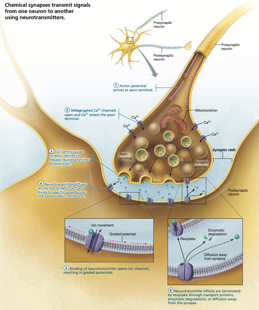
Location of cell-to-cell communication in neurons
Pre-synaptic axon terminal
Synapse
Post-synaptic dendrites
Pre-synaptic Axon terminal stage
Vesicles containing neurotransmitters lay stationary at the axon terminal
Axon potential arrives at the axon terminal
Electrical signals & voltage changes along the cell membrane cause voltage-gated Ca2+ & Na+ channels to open
Ca2+ and Na+ floods the inside of the axon terminal. (Ca2+ always in excess extracellularly)
Ca2+ sensing protein “synaptotagmin” found on the vesicles’ membranes cause the vesicles to fuse with the membranes of the axon terminal, releasing the inner neurotransmitters via exocytosis.
Excess Ca2+ inside the pre-synaptic terminal is removed by mitochondria or expelled via Ca2+ pumps
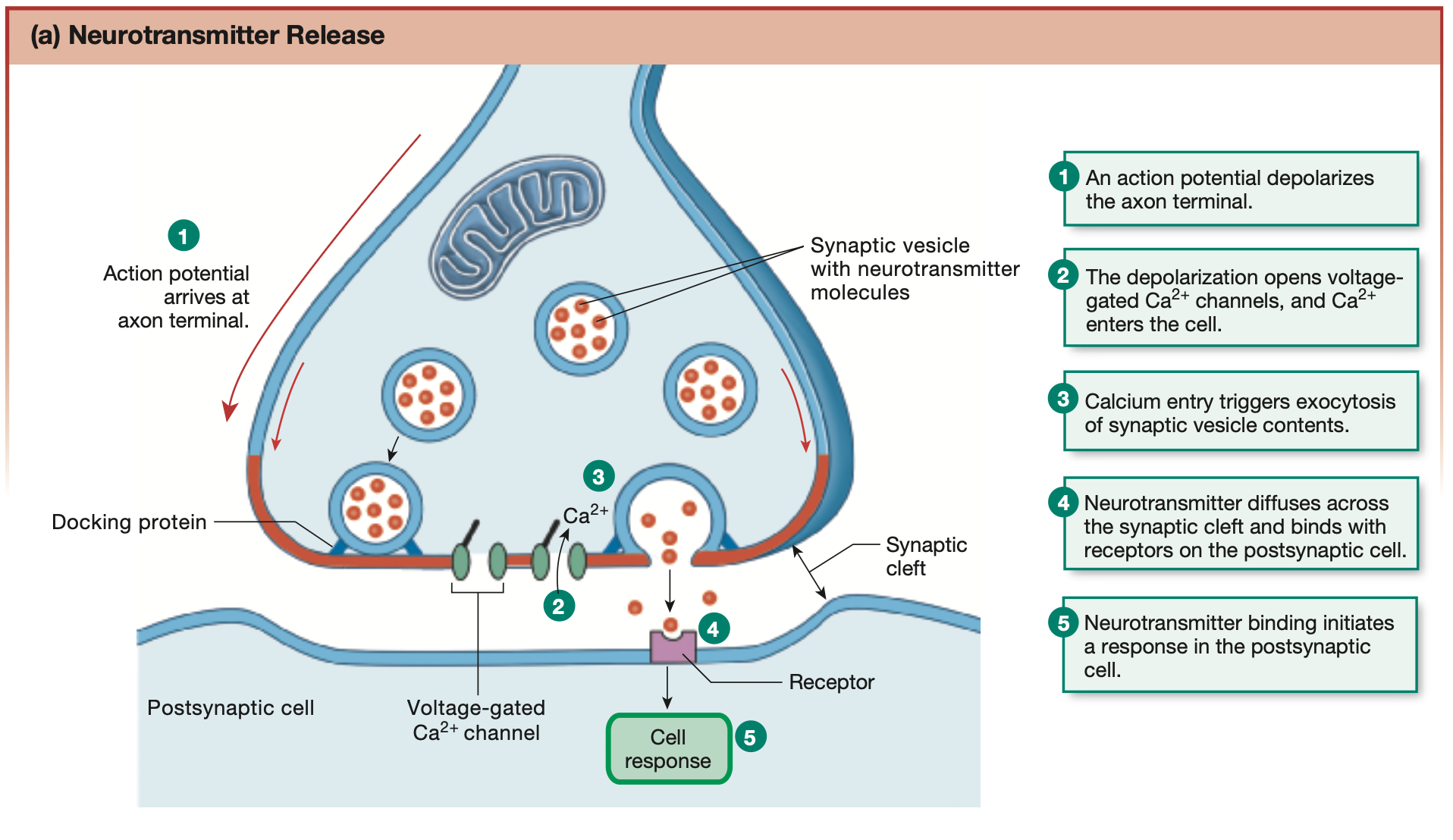
Post-synaptic dendrite stage
Neurotransmitters cross the synaptic cleft, binding to receptor sites found on the dendrites of post-synaptic neurons
Neurotransmitters can bind to chemically-gated/ligand-gated ion channels, permitting the passage of K+, Ca2+, Na+ or Cl- ions, creating graded potentials. Can be excitatory or inhibitory
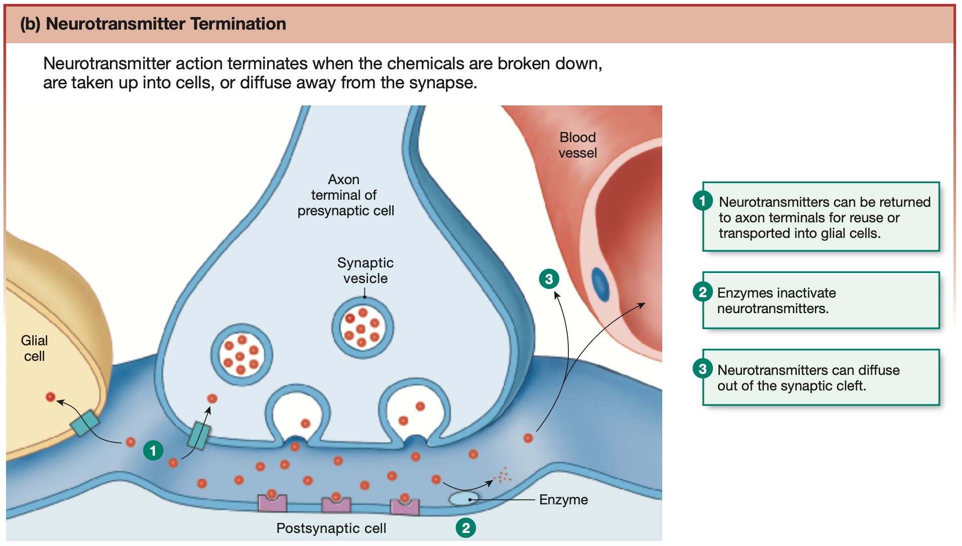
After effect is received, neurotransmitter is removed from the receptor sites of post-synaptic dendrites via one of 3 methods
3 methods of Neurotransmitter termination:
Reuptake
Degredation
Diffusion
Reuptake
Neurotransmitters can be reuptaken and stored in the astrocytes or pre-synaptic axon terminal again.
Degredation: Enzymes
Broken down by enzymes found on the membrane of post-synaptic dendrites or in the synaptic cleft.
Diffusion
Neurotransmitters can travel away from the synaptic cleft into areolar tissue or blood vessels.
Synaptic Transmission Diagram
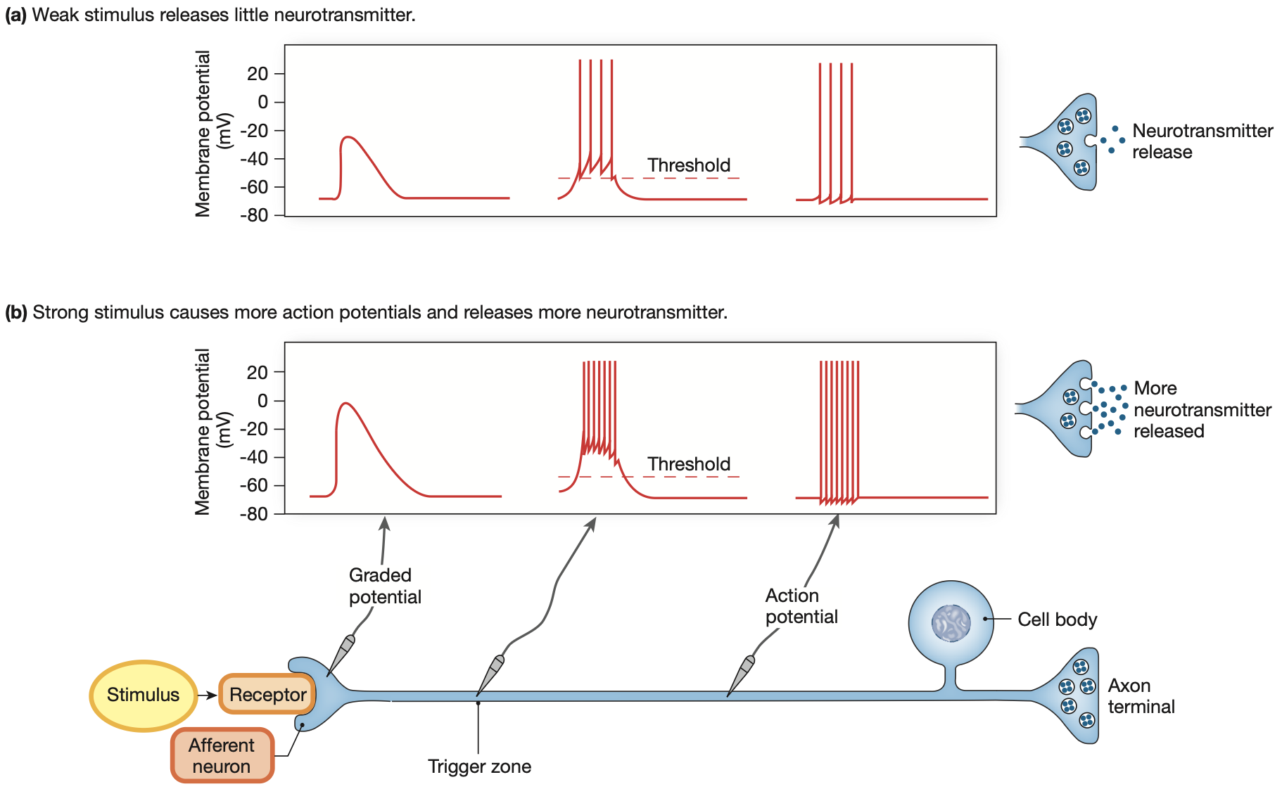
Coding action potentials
The stronger the stimulus, the more action potentials propagated through a neuron for a longer duration, hence the more neurotransmitters released over a period of time.
Coding action potentials: transforming mechanical stimulus of the cell membrane into an electrical stimulus.
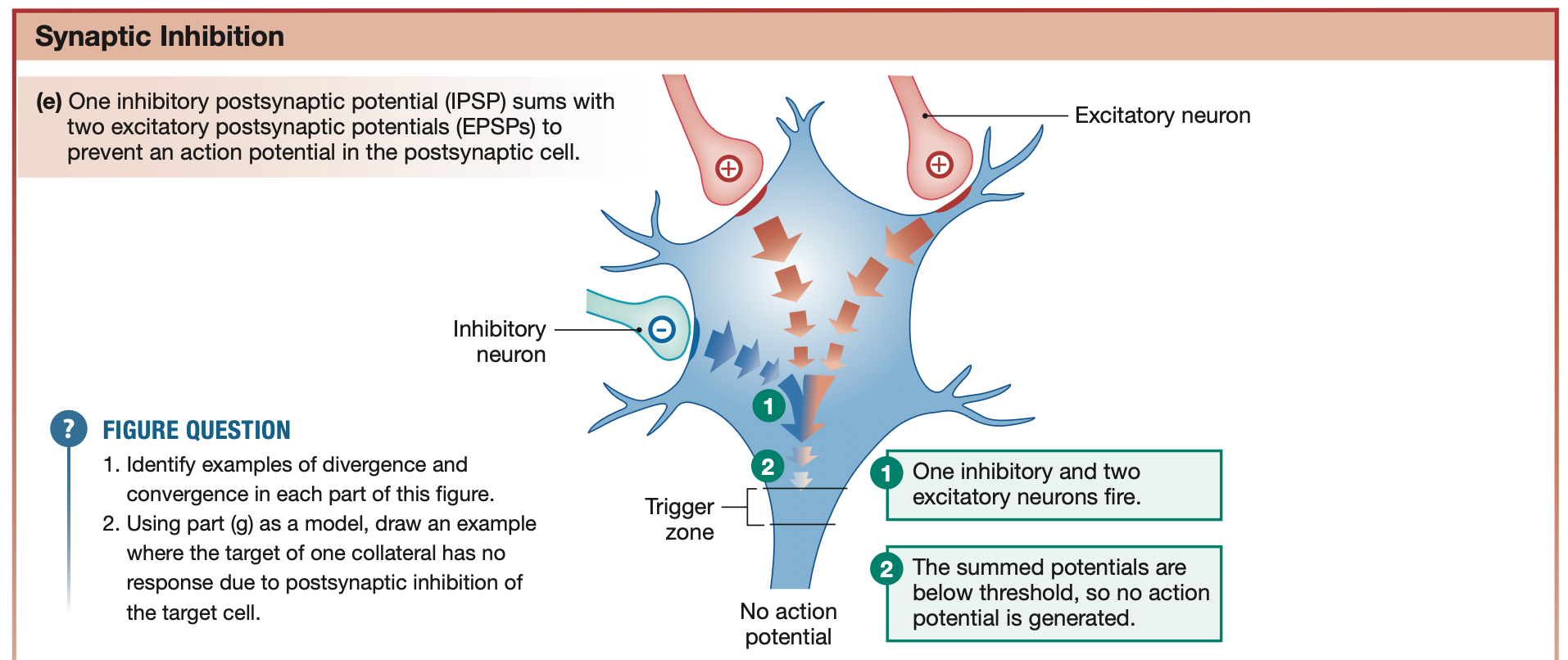
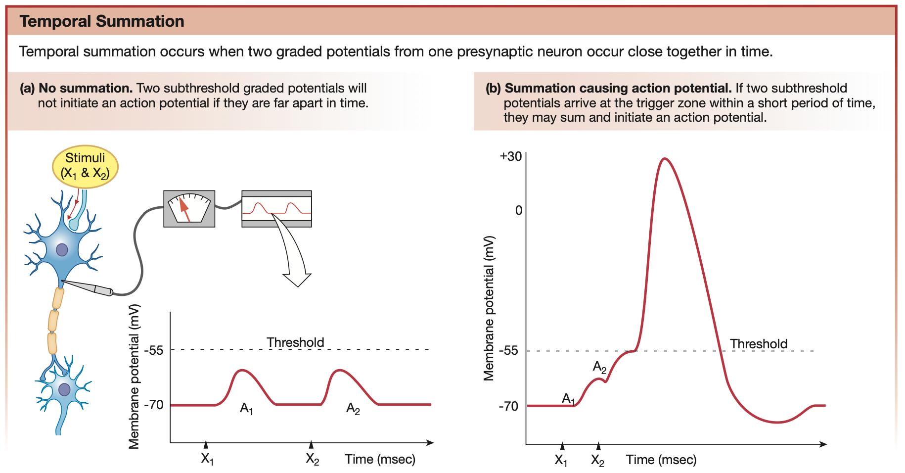
2 forms of Integration for Synaptic Signalling Summation
Temporal summation
Spatial summation
Temporal Summation
Graded potentials that happen close enough in time together can summate to reach the threshold potential and trigger an action potential
Spatial Summation
One post-synaptic neuron receives graded potentials from multiple pre-synaptic neurons at the same time, in the same space. Simultaneous graded potentials can combine (or summate) to reach the threshold potential.
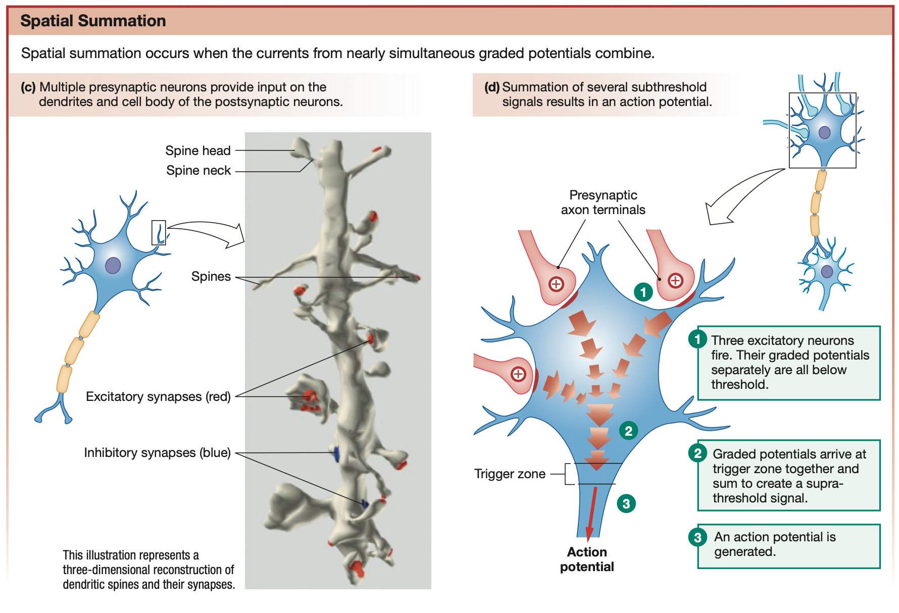
Excitatory Graded potentials (EPSPs)
EPSP - Excitatory post-synaptic potentials
Graded potentials that depolarise the cell membrane (make inside more positive)
Brings it closer to threshold potential
Can summate with other graded potentials to create an action potential
Multiple EPSPs are needed to propagate into an action potential
Red in diagram

Inhibitory Graded potentials (IPSPs)
IPSP - Inhibitory post-synaptic potentials
Graded potentials that hyperpolarise the cell membrane (make inside more negative)
Brings it further away from threshold potential
Can prevent graded potentials from summating and becoming action potentials
Blue in diagram