Muscles of the Head, Neck, and Torso (Part 1)
1/13
There's no tags or description
Looks like no tags are added yet.
Name | Mastery | Learn | Test | Matching | Spaced | Call with Kai |
|---|
No analytics yet
Send a link to your students to track their progress
14 Terms
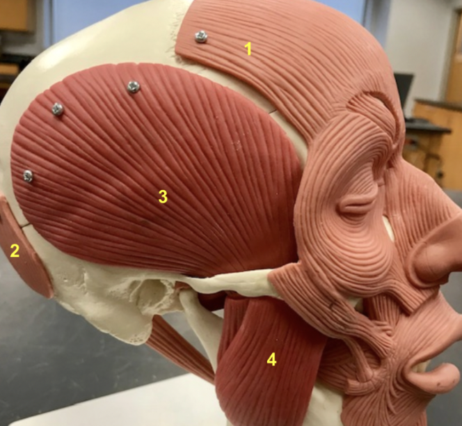
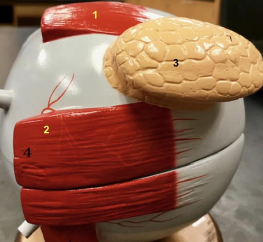
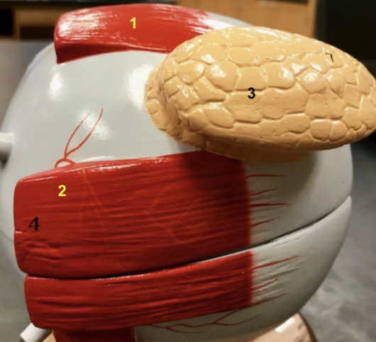
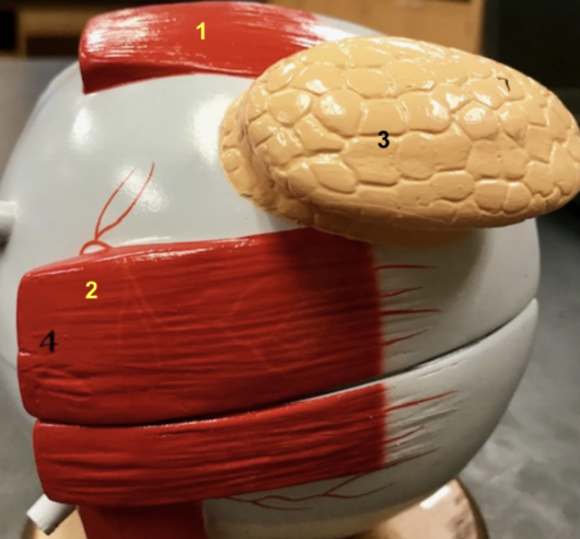
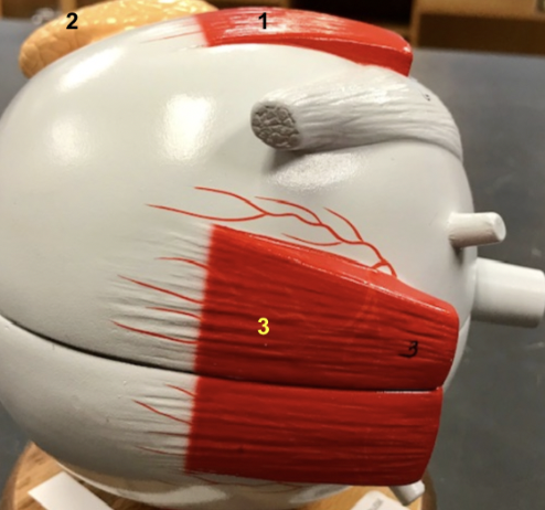
This model shows key head muscles, which two muscles are represented by 1 and 2?
The epicranius or occipitofrontalis [frontal (1) and occipital bellies (2)] raises the eyebrows and wrinkles the forehead.
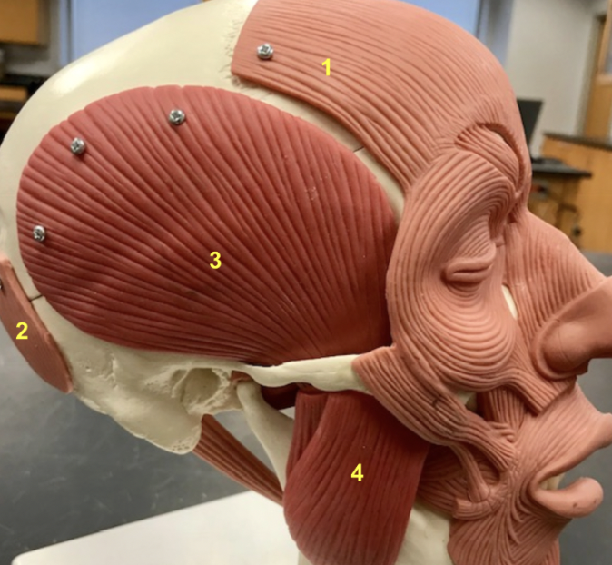
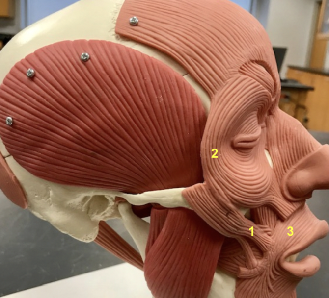
This model shows key head muscles, which muscle is represented by 3?
The temporalis (3) elevates the mandible, originating from the parietal bone and inserting on the mandible's coronoid process.
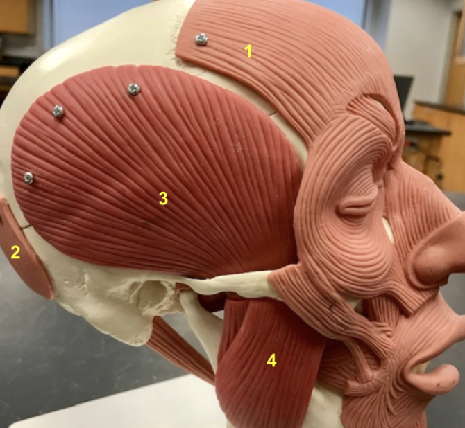
This model shows key head muscles, which muscle is represented by 4?
The masseter (4), also elevating the mandible, originates from the zygomatic arch and inserts on the ramus and angle of the mandible .
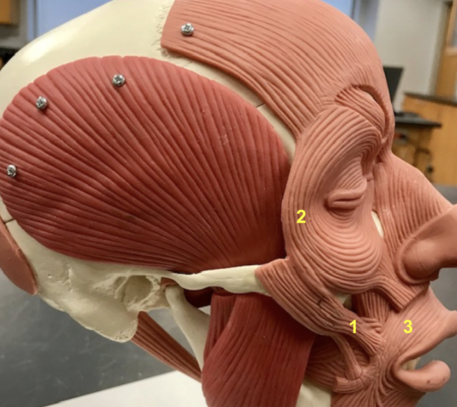
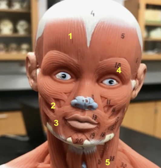
Which muscle is represented by 1?
The zygomaticus major (1) originates from the zygomatic bone and inserts at the corner of the mouth, pulling it superiorly (upward) and laterally (outward) in a smile.
Which muscle is represented by 2?
The orbicularis oculi (2) encircles the eye and closes it.

Which muscle is represented by 3?
The orbicularis oris (3) surrounds the mouth. It closes and puckers the lips.
This model shows the epicranius (frontalis)(1), zygomaticus major (2), orbicularis oris (3), and orbicularis oculi (4). Which muscle is represented by 5?
the sternocleidomastoid (5). It originates from the manubrium of the sternum and the medial end of the clavicle and inserts on the mastoid process of temporal bone. It flexes the neck when both sides contract, and when one side contracts, it causes contralateral rotation and ipsilateral flexion.
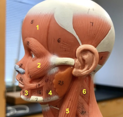
Which muscle is represented by 6?
Splenius capitis (6), When working together, the left and right splenii capites act to extend the neck, moving the head up. When a single splenius capitis contracts, it causes ipsilateral rotation.
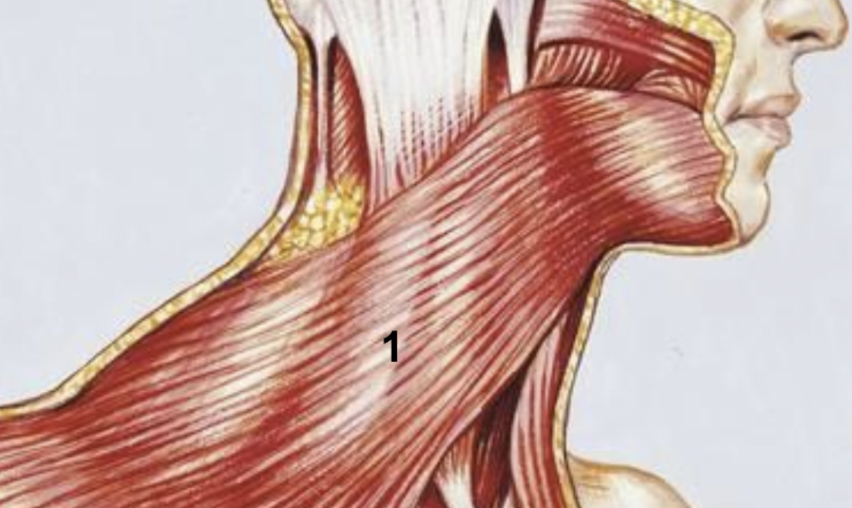
Which muscle is represented by 1?
The platysma (1), a superficial muscle that acts to depress the mandible (open the mouth) and tighten the skin in the neck.
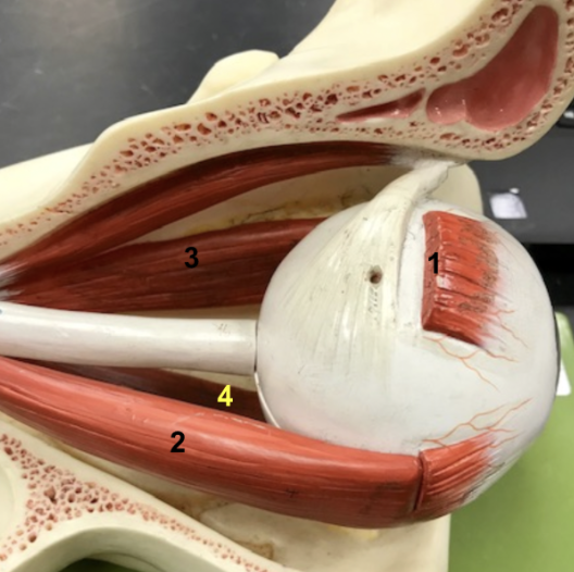
This is the lateral view of the eyeball. Which muscle is represented by 1?
Superior rectus (1), it functions to elevate the eye (look up).
This is the lateral view of the eyeball. Which muscle is represented by 2?
Lateral rectus (2), it functions to abduct the eyeball
What gland is depicted by 3?
lacrimal gland (3), it is only found on the lateral side of the eyeball.
This is the medial view of the eyeball, which muscle is being depicted by 3?
Medial rectus (3), it functions to adduct the eye.
In this model, we can see several muscles met previously including the Superior rectus (1), Lateral rectus (2), and Medial rectus (3). Which muscle is depicted by 4?
Inferior rectus (4), it functions to depress the eye.