Neuroscience - Ventricles, Cerebrospinal Fluid, & Meninges
1/115
Earn XP
Description and Tags
Name | Mastery | Learn | Test | Matching | Spaced | Call with Kai |
|---|
No analytics yet
Send a link to your students to track their progress
116 Terms
What are cranial meninges?
membranous coverings of brain
What are the functions of cranial meninges?
protect the brain
form supporting framework for blood vessels and venous sinuses
provide a fluid-filed cavity for the brain and spinal cord
What are the 3 layers of the cranial meninges?
dura mater
arachnoid mater
pia mater
What are the 2 layers of the dura mater?
periosteal (outer) layer
meningeal (inner) layer
What is the dural septa?
meningeal layer of dura gives rise to infoldings (septa) to divide cranial cavity into compartments and separate brain regions
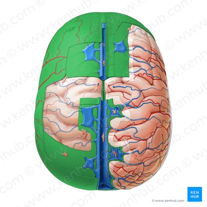
What are dural venous sinuses?
endothelium-lined spaces between periosteal and meningeal layers
drain venous blood from nervous system
What makes up the dura mater septa? (4)
falx cerebri
tentorium cerebelli
falx cerebelli
diaphragma sellae
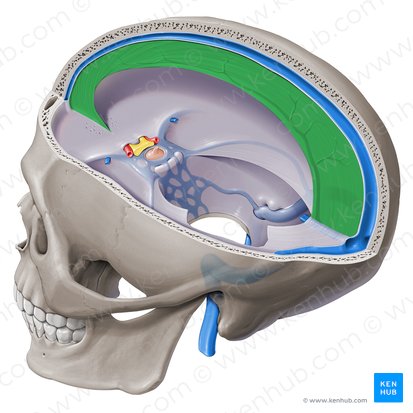
What is the falx cerebri?
lies in longitudinal fissure and separates right and left hemispheres

What is the falx cerebri continuous with?
tentorium cerebelli

What is the tentorium cerebelli?
separates occipital lobe from cerebellum
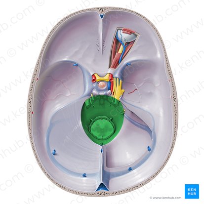
What is the tentorial notch of the tentorium cerebelli?
gap through which brainstem extends from posterior to middle cranial fossa
What is the falx cerebelli?
vertical dural infolding inferior to tentorium cerebelli that partially separates cerebellar hemispheres
What is the diaphragma sellae?
circular sheet of dura suspended to form a roof over hypophyseal fossa (sella turcica)
covers pituitary gland
Where do dural venous sinuses ultimately drain?
internal jugular vein
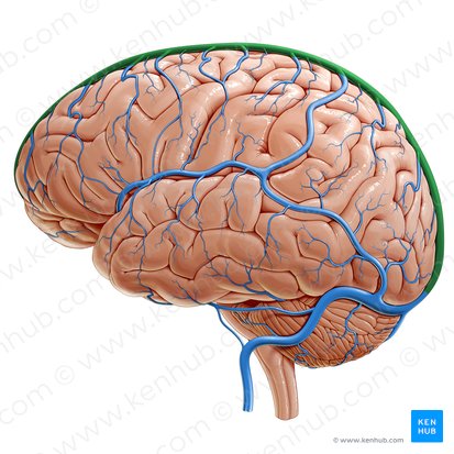
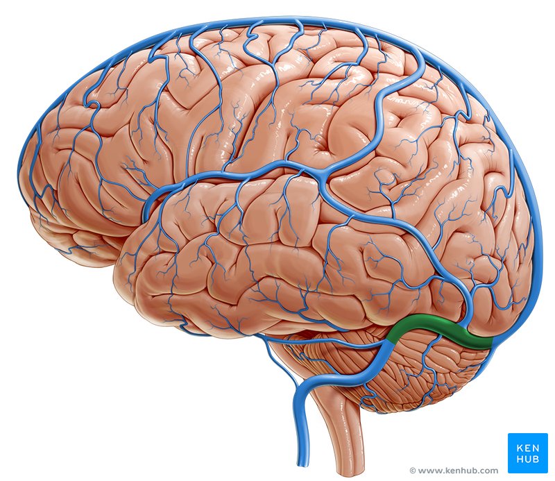
Where does venous outflow from the brain travel?
through superficial and deep cerebral veins and drains into dural venous sinuses which ultimately drain into internal jugular vein

What is the confluence of sinuses?
meeting place of superior sagittal, straight, and transverse sinuses
Where does the superior sagittal sinus usually drain into?
right transverse sinus
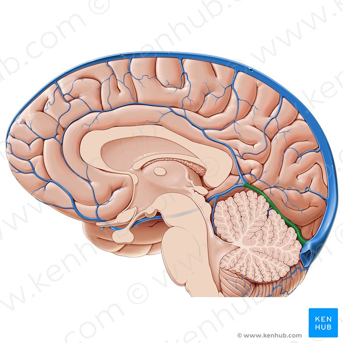
Where does the straight sinus usually drain into?
left transverse sinus
What are notable paired dural sinuses? (3)
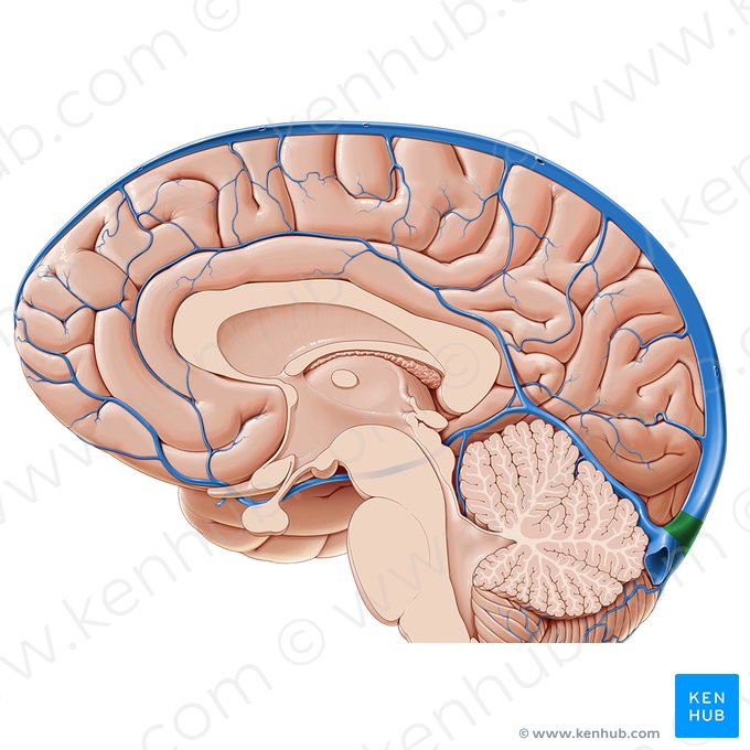
transverse
cavernous
sigmoid
What are notable unpaired dural sinuses? (3)
superior sagittal
inferior sagittal
straight
What do emissary veins connect?
dural venous sinuses with veins outside cranium
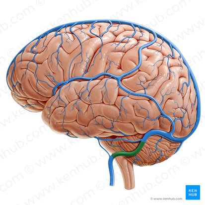
Where do the sigmoid sinuses drain?
internal jugular vein
Where is the confluence of sinuses located?
cruciform eminence of occipital bone
Where do the transverse sinuses usually drain?
sigmoid sinus
What is the blood supply to the dura of the anterior cranial fossa? (3)
cavernous portion of internal carotid artery
ethmoidal arteries
ascending pharyngeal artery
What is the blood supply to the dura of the middle cranial fossa?
middle meningeal artery
What is the blood supply to the dura of the posterior cranial fossa? (2)
branches of ascending pharyngeal artery
branches of occipital arteries
What is the blood supply to the spinal dura? (3)
vertebral, intercostal, and lumbosacral arteries
What part of the dura do tentorial and ethmoidal nerves ("meningeal branches" of V1) innervate?
part of dura mater of anterior and middle cranial fossa
anterior and posterior part of dura mater of tentorium cerebelli
Generally, what innervates the dura of the anterior and middle cranial fossae?
branches of trigeminal (CN V)
What innervates the dura of the posterior cranial fossa?
dorsal roots of C1-C3 (some fibers distributed via hypoglossal and vagus)

What are the two parts of the arachnoid mater?
arachnoid barrier cell layer
arachnoid trabeculae
What does the arachnoid barrier cell layer attach to?
dura mater

What does the arachnoid trabeculae layer attach to?
pia mater
What forms within the arachnoid mater layer?
CSF filled spaces

What holds the arachnoid mater against the dura mater?
pressure of CSF
What is another name for subarachnoid space?
leptomeningeal space
What makes up the leptominenges?
inner 2 meningeal layers (arachnoid and pia):
parietal part = arachnoid mater
visceral part = pia mater

What is the subarachnoid space?
space between arachnoid and pia that contains CSF, blood vessels, roots of cranial and spinal nerves as they enter/exit
What suspends the brain?
subarachnoid space

What are components of the subarachnoid space? (3)
arachnoid trabeculae
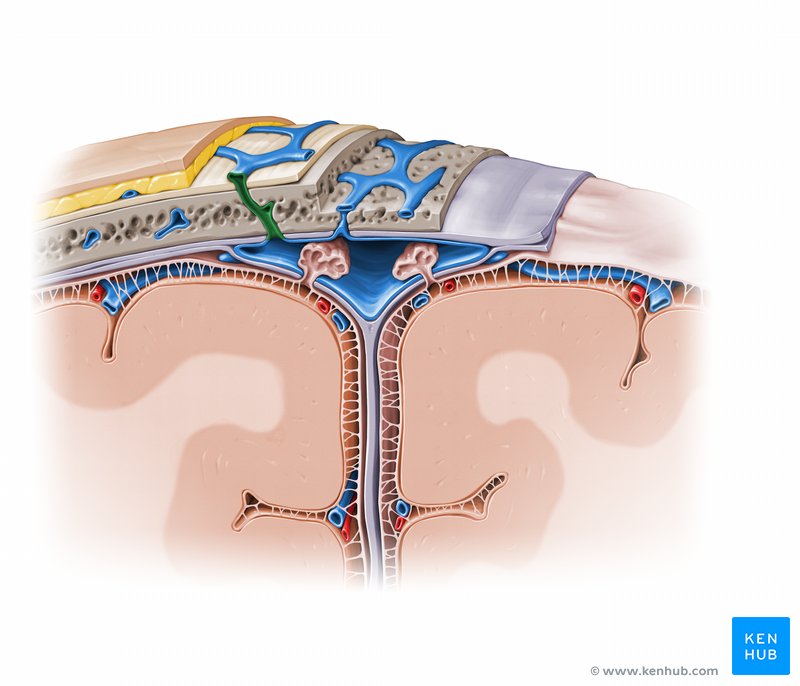
arachnoid villi
subarachnoid cisterns
What are arachnoid trabeculae?
fibroblasts that bridge subarachnoid space
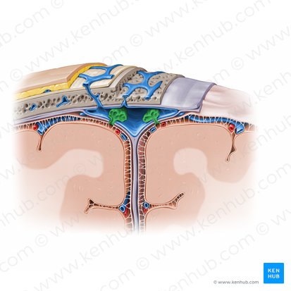
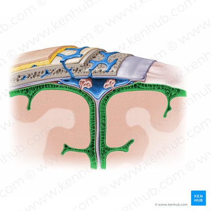
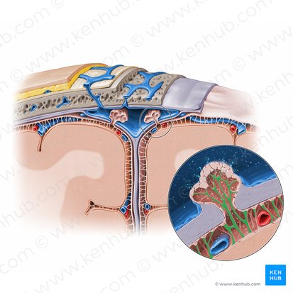
What are arachnoid villi?
small specialized portions of arachnoid that protrude through meningeal layer into venous sinuses

What is the purpose of arachnoid villi?
assist with transfer of CSF from subarachnoid space into venous system
How does the CSF travel through the arachnoid villi?
down a pressure gradient
What are subarachnoid cisterns?
enlarged regions of subarachnoid spaces (pools of CSF)
What are granular foveolae?
impressions of arachnoid villi on skull

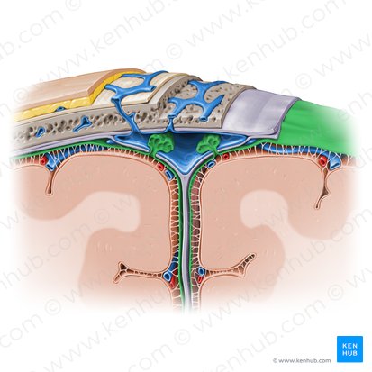
What makes up the pia mater?
layer of flat cells that closely follow the surface features of the brain and spinal cord
continuous with the surface of the brain and cannot be separated
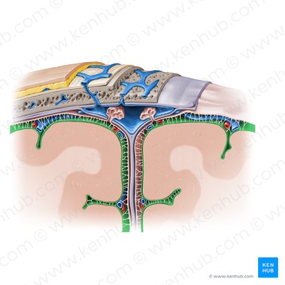
What separates the pia mater from the brain?
glial basement membrane
What are perivascular spaces of pia mater?
regions where small vessels penetrate the surface of the brain and spinal cord
What is another name form perivascular spaces of pia mater?
Virchow-Robin spaces
What structures of pia mater are analagous to arachnoid trabeculae in the spinal cord? (3)
denticulate ligaments
filum terminale internum (inside dural sac, still a pial structure)
filum terminale externum (actually a dural structure)
What is tethered spinal cord syndrome?
if a child has an abnormally thick filum terminale, it becomes stretched and taut as the spinal cord is tugged between the lower back and brain stem
What is meningitis?
infection of cranial meninges from viruses, bacteria, etc.
What is leptomeningitis?
infection of arachnoid and pia
What is pachymeningitis?
infection of dura
What is bacterial meningitis and what are symptoms of it?
fever, chills, neck stiffness, headache, decreased consciousness
vaccines are available
What is viral meningitis and what are symptoms of it?
fever, headaches, confusion, altered consciousness
no antiviral medications are available
most common in younger patients
How do you confirm a meningitis diagnosis?
with a lumbar puncture to assess CSF composition
Can meningitis be fatal?
yes
What during development gives rise to ventricles and the central canal of the spinal cord?
neural tube
What are the ventricles that the neural tube gives rise to during development? (4)
lateral ventricles
third ventricle
cerebral aqueduct
fourth ventricle
What is the choroid plexus?
secretes CSF to fill ventricles and subarachnoid space
What secondary region of the brain are the lateral ventricles associated with?
telencephalon

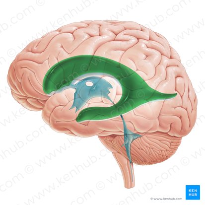
How many lateral ventricles are there?
2 (one in each cerebral hemisphere)

What are the features of the lateral ventricles? (5)
anterior horn
body
atrium
posterior horn
inferior horn
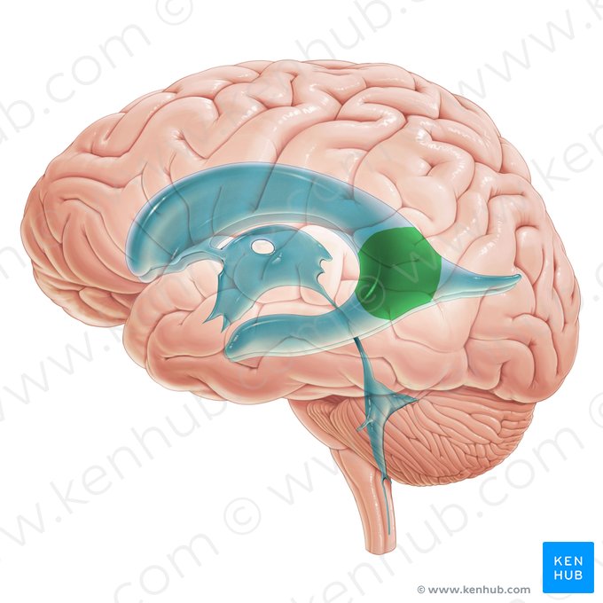
What is in the atrium of the lateral ventricles?
glomus choroideum (glomus)- large clump of choroid plexus

What is the medial wall of the lateral ventricle?
septum pellucidum and fornix

What is the roof of the lateral ventricle?
corpus callosum

What is the floor of the lateral ventricle?
thalamus
What is the lateral wall of the lateral ventricle?
caudate nucleus
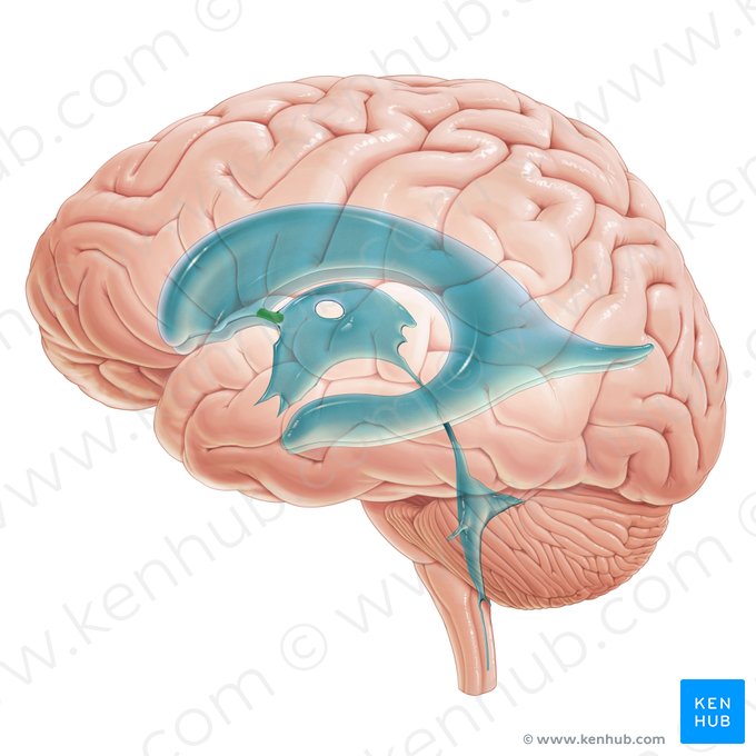
What is the interventricular foramina (of Monro)?
connects lateral ventricles with third ventricle (bilateral)
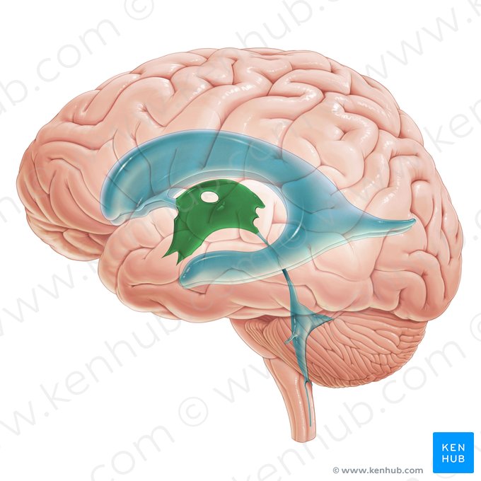
What secondary region of the brain is associated with third ventricle?
diencephalon

What is the anterior wall of the third ventricle?
lamina terminalis and anterior commissure

What is the posterior wall of the third ventricle?
pineal gland and posterior commissure

What are the lateral walls of the third ventricle?
thalamus and hypothalamus

What is the roof of the third ventricle?
tela choroidea

What is the floor of the third ventricle?
optic chiasm, pituitary gland, and mammillary bodies

What are the 5 recesses of the third ventricle?
infundibular
supraoptic
anterior
suprapineal
pineal

What secondary brain region is associated with the cerebral aqueduct?
mesencephalon
What ventricle does not have a choroid plexus?
cerebral aqueduct

What is the cerebral aqueduct?
very narrow midline channel between third and fourth channels
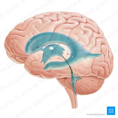
What surrounds the cerebral aqueduct?
periaqueductal gray matter
Where is the periaqueductal gray matter (PAG) located?
midbrain
What is the function of periaqueductal gray matter (PAG)?
receive somatosensory input
high levels of opiate receptor binding activity
suppression and modulation of pain
What is periaqueductal gray matter interconnected with?
hypothalamus and thalamus
has projections to brainstem nuclei

What secondary brain regions is the fourth ventricle associated with?
metencephalon and myelencephalon

Where is the fourth ventricle found?
forms cavity of hindbrain
extends into cerebellum
What are the 3 sites that CSF can leave the fourth ventricle and enter the subarachnoid space?
Foramen of Magendie (median aperture)
Foramina of Luschka (paired lateral apertures)

What are the lateral walls of the fourth ventricle?
superior and inferior cerebellar peduncles and cuneate and gracile tubercles

What is the roof of the fourth ventricle?
cerebellum

What is the floor of the fourth ventricle?
rhomboid fossa (pons and medulla)
What is the ependyma?
lining of specialized epithelial cells that helps to form the choroid plexus
What specialized epithelial cells make up the ependyma?
simple cuboidal epithelium
What does the ependyma line?
ventricles and central canal of spinal cord
What helps with movement of CSF?
cilia on ependymal cells
How does the ependyma layer help form components of the blood-CSF barrier?
form an interface between CSF-filled ventricles and the blood
What is the development of the choroid plexus?
roof of fourth ventricle is lined with an internal layer of the ependyma and the outer layer of the pia
blood vessels invaginate this membrane to form the choroid plexus
Where is the choroid plexus found?
floors of the bodies and inferior horns of lateral ventricles
roofs of third and fourth ventricles
What is the structure of the choroid plexus?
series of folds (villi)