Laboratory 10: The Digestive System and Laboratory 11: Respiratory System
1/212
There's no tags or description
Looks like no tags are added yet.
Name | Mastery | Learn | Test | Matching | Spaced | Call with Kai |
|---|
No analytics yet
Send a link to your students to track their progress
213 Terms
alimentary canal
a tube that extends from the mouth to the anus (AKA gastrointestinal tract)
oral (buccal) cavity
non-keratinized Stratified Squamous Epithelia
vestibule
space between lips and teeth
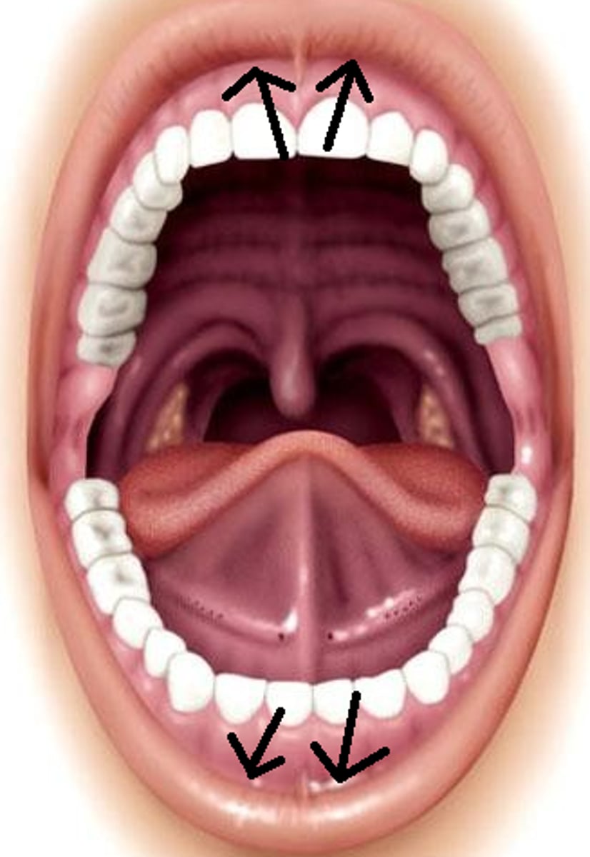
labial frenulum
fold that connects lips to gums
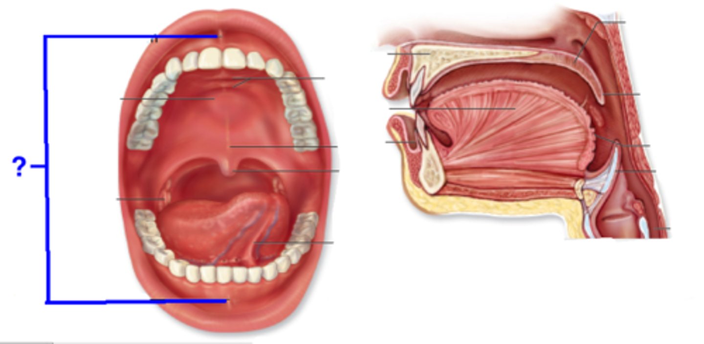
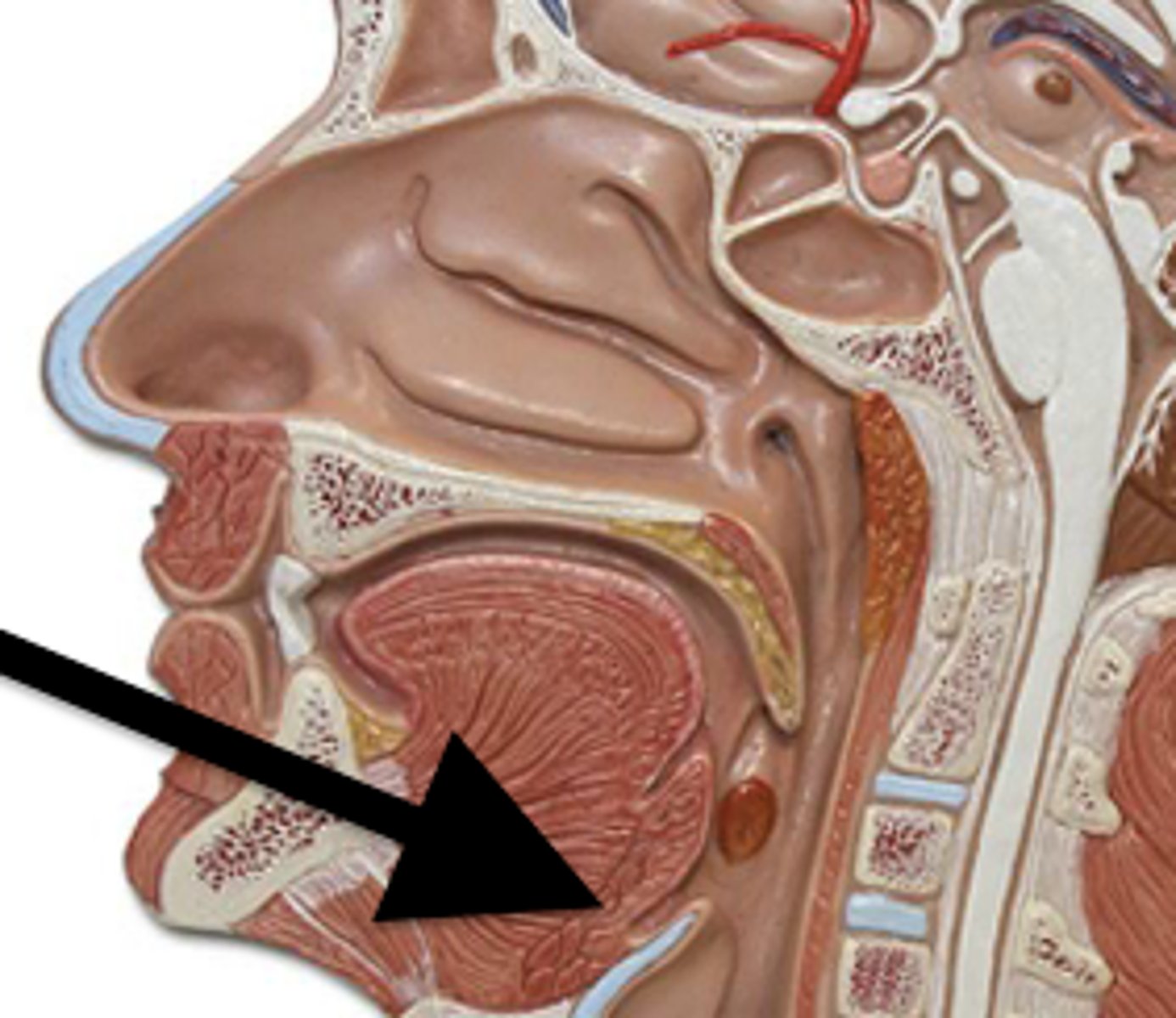
tongue
manipulates food for chewing and swallowing
lingual frenulum
attaches tongue to base of mouth
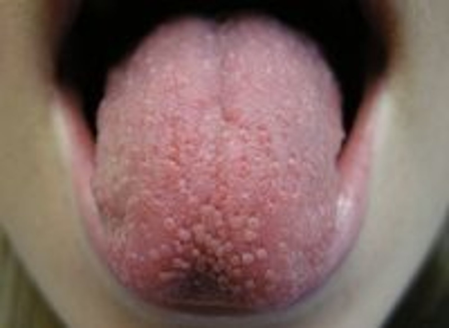
Papillae
bumps on tongue
filiform papillae
no taste buds, roughen tongue, smaller
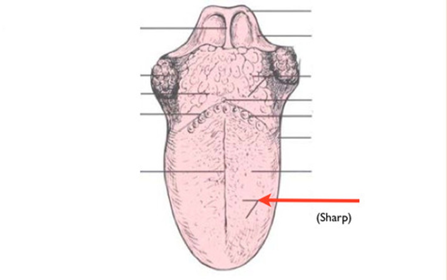
fungiform papillae
taste buds, slightly larger (look like mushrooms)
foliate papillae
slit like, lateral on tongue, taste buds in childhood but not adulthood
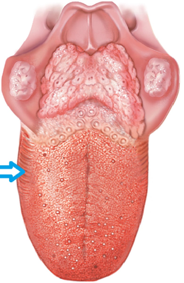
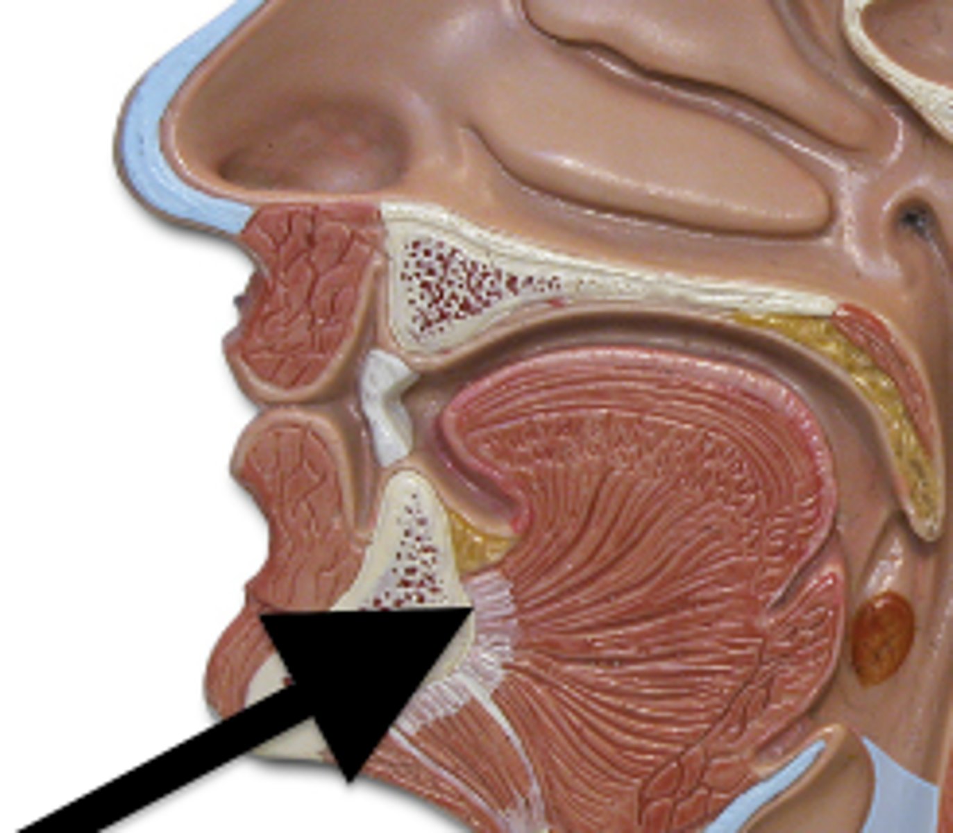
circumvallate papillae
largest, v shape on posterior tongue with taste buds
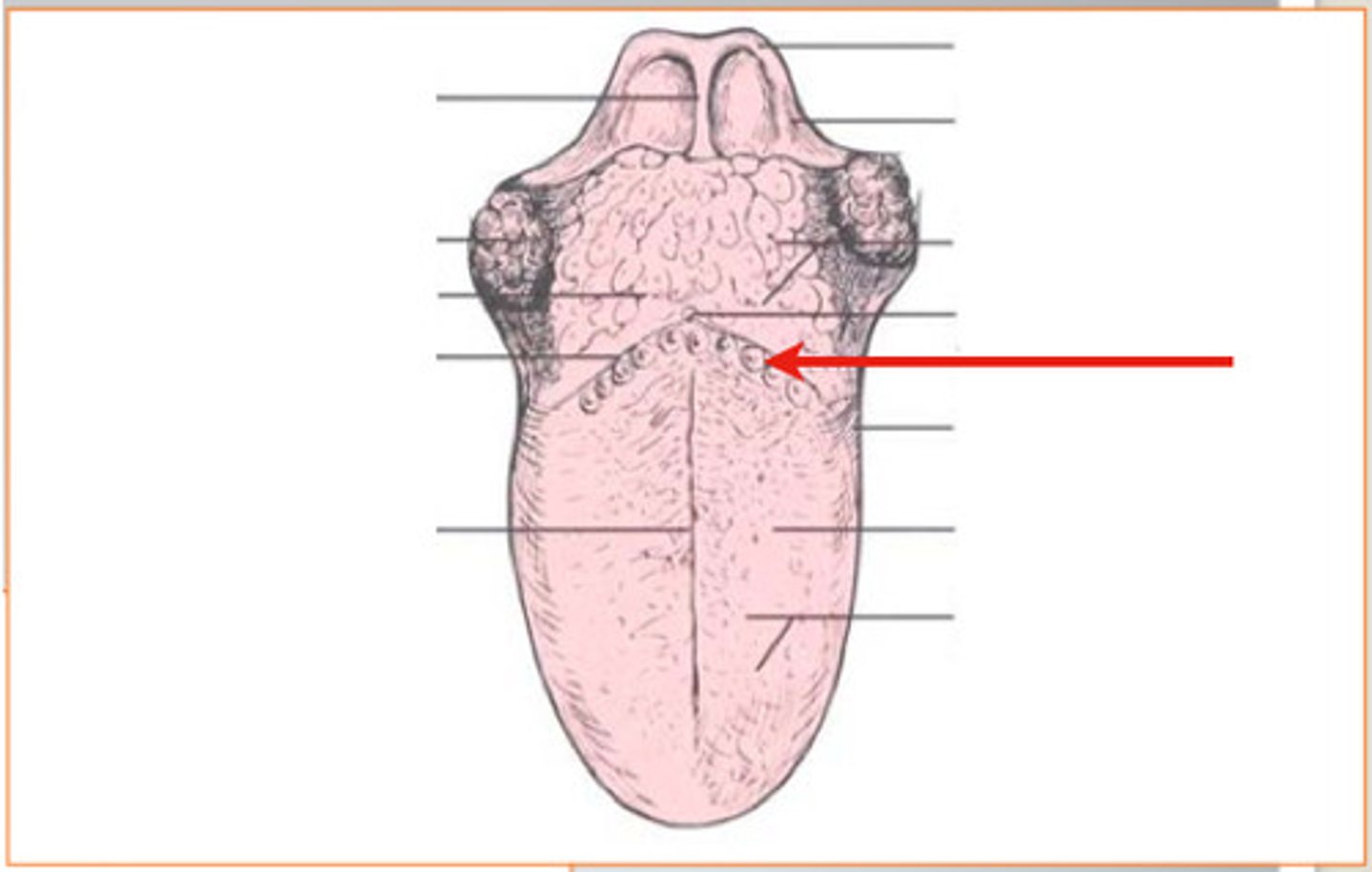
sulcus terminalis
marks border between mouth and pharynx
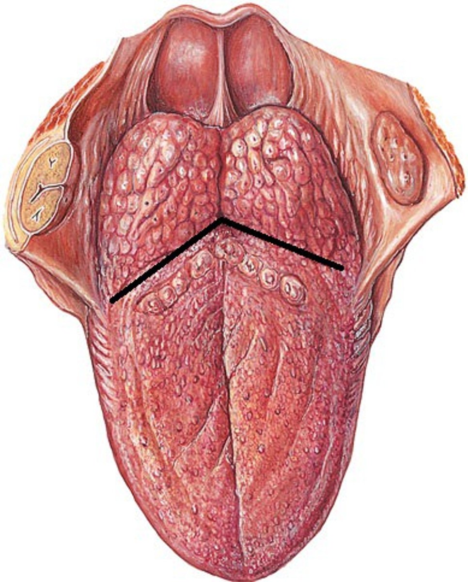
lingual tonsils
located at the base of the tongue
palate
roof of the mouth
hard palate
anterior 4/5: palatine process of maxilarry bones
posterior 1/5: horizontal plaate of palatine bone
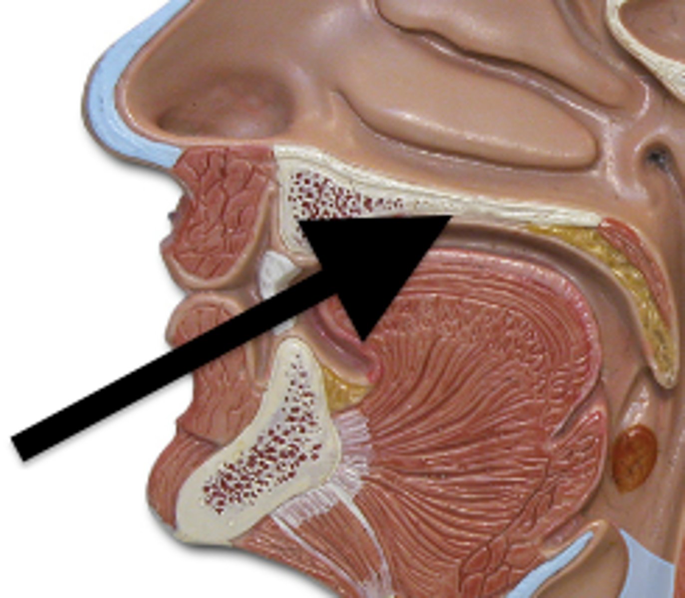
soft palate
muscular posterior (back) portion of the palate
uvula
closes off nasopharynx during swallowing
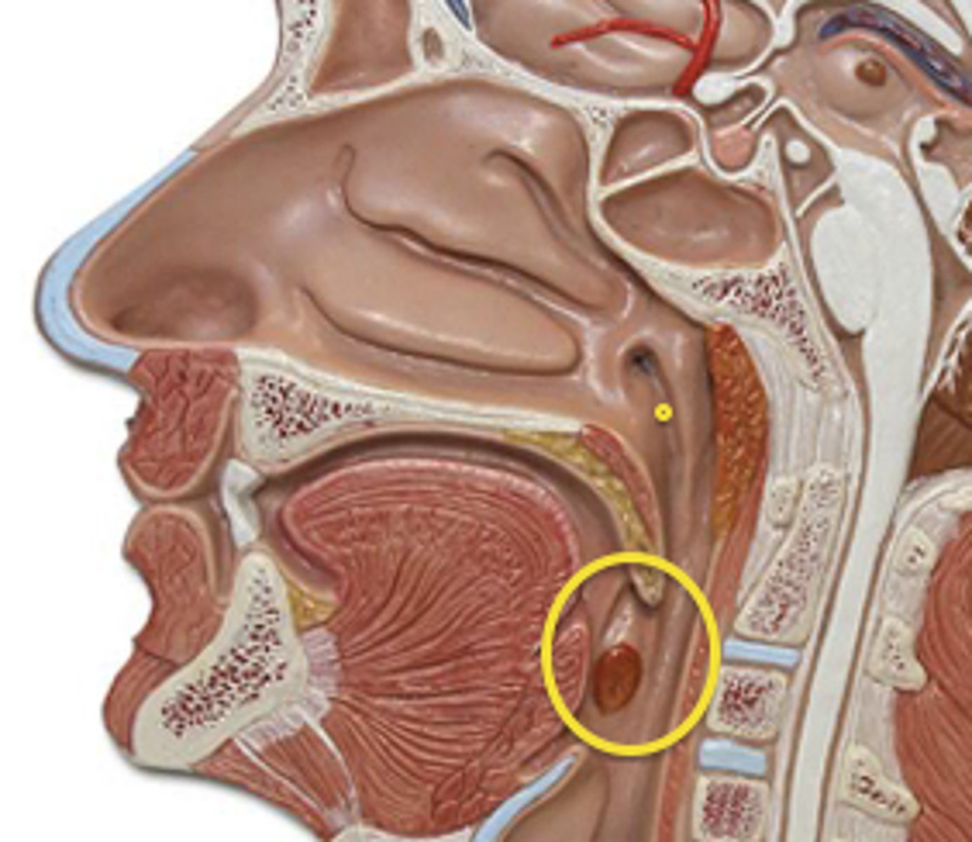
palatine tonsils
inferior to uvula, lateral sides of throat
gingivae
gums
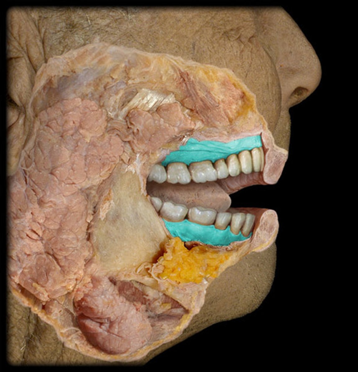
teeth
hard bony projections in the jaws for masticating (chewing) food
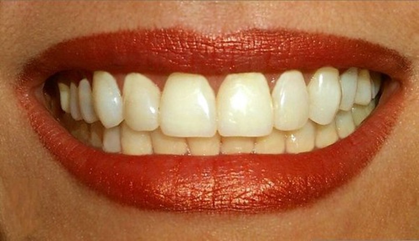
Incisors
front teeth
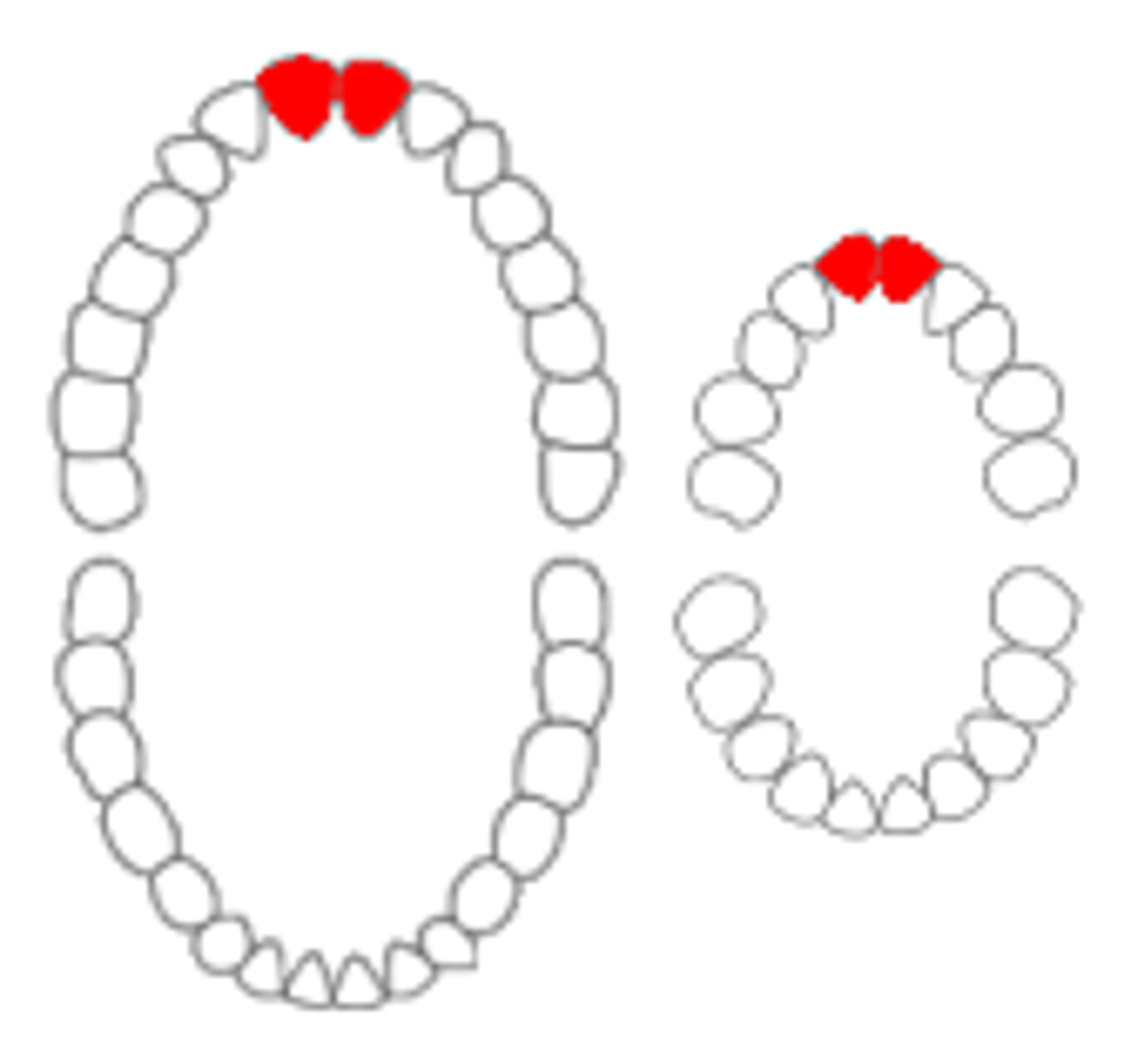
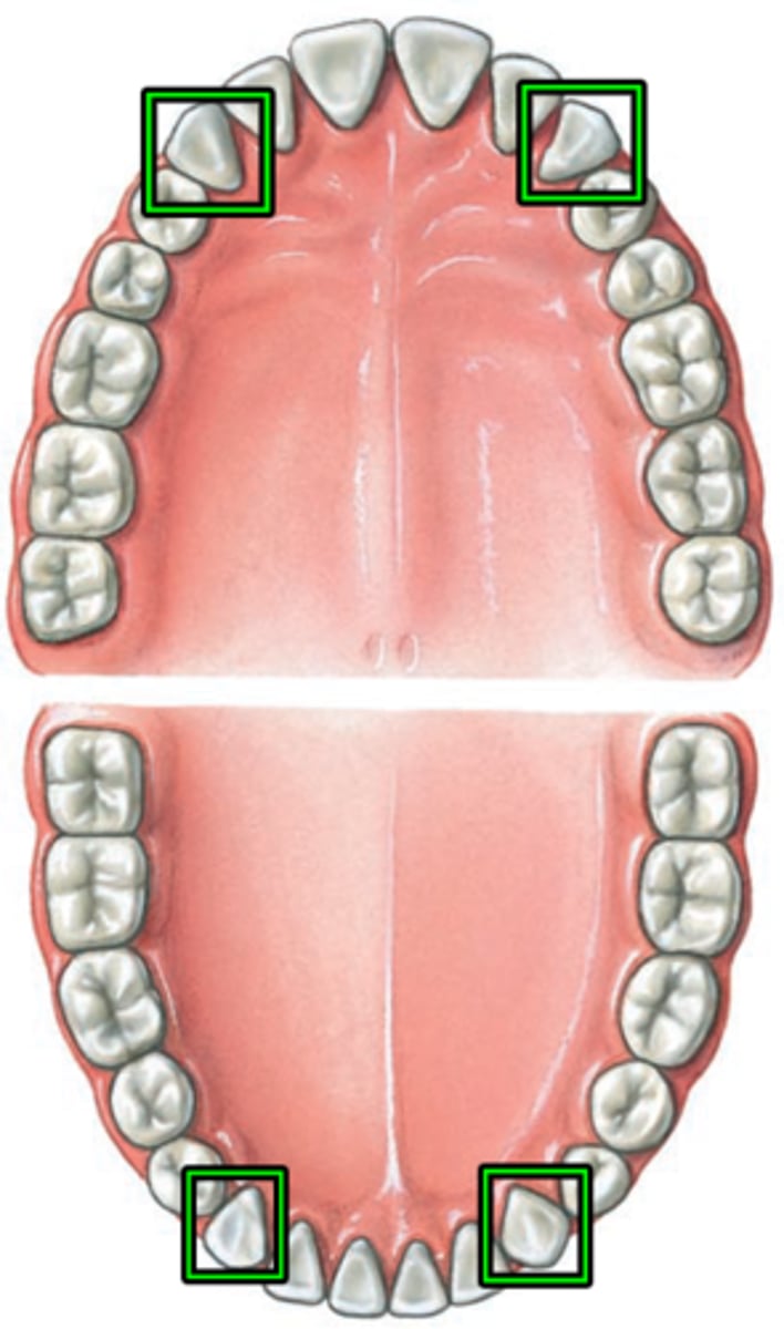
canines (cuspids)
2 pairs in anterior corners of mouth, responsible for holding, tearing, and piercing
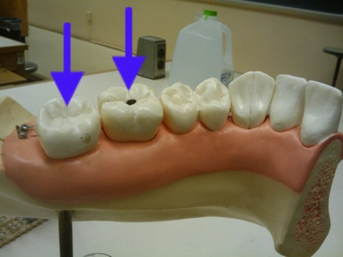
bicuspids (premolars)
-Flattened crowns
-Prominent ridges
-Used to crush, mash, and grind
-Have one or two roots

Molars
Back teeth that grind food
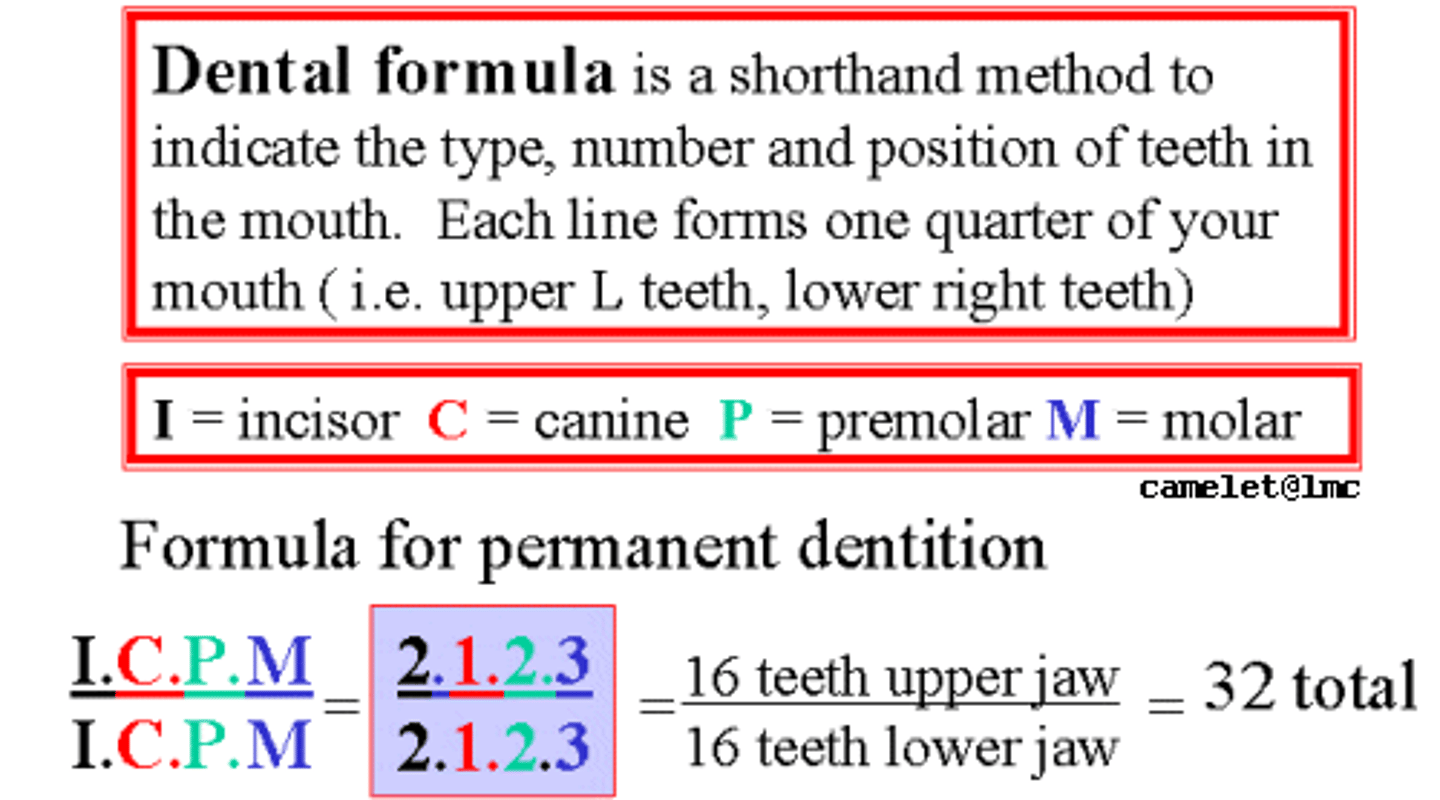
Dental formula
The numerical description of a species' teeth, listing the number, in one quadrant of the jaws, of incisors, canines, premolars, and molars.
Adult: 2I, 1C, 2P, 3M (32 total)
Deciduous: 2I, 1C, 2M (20 total)
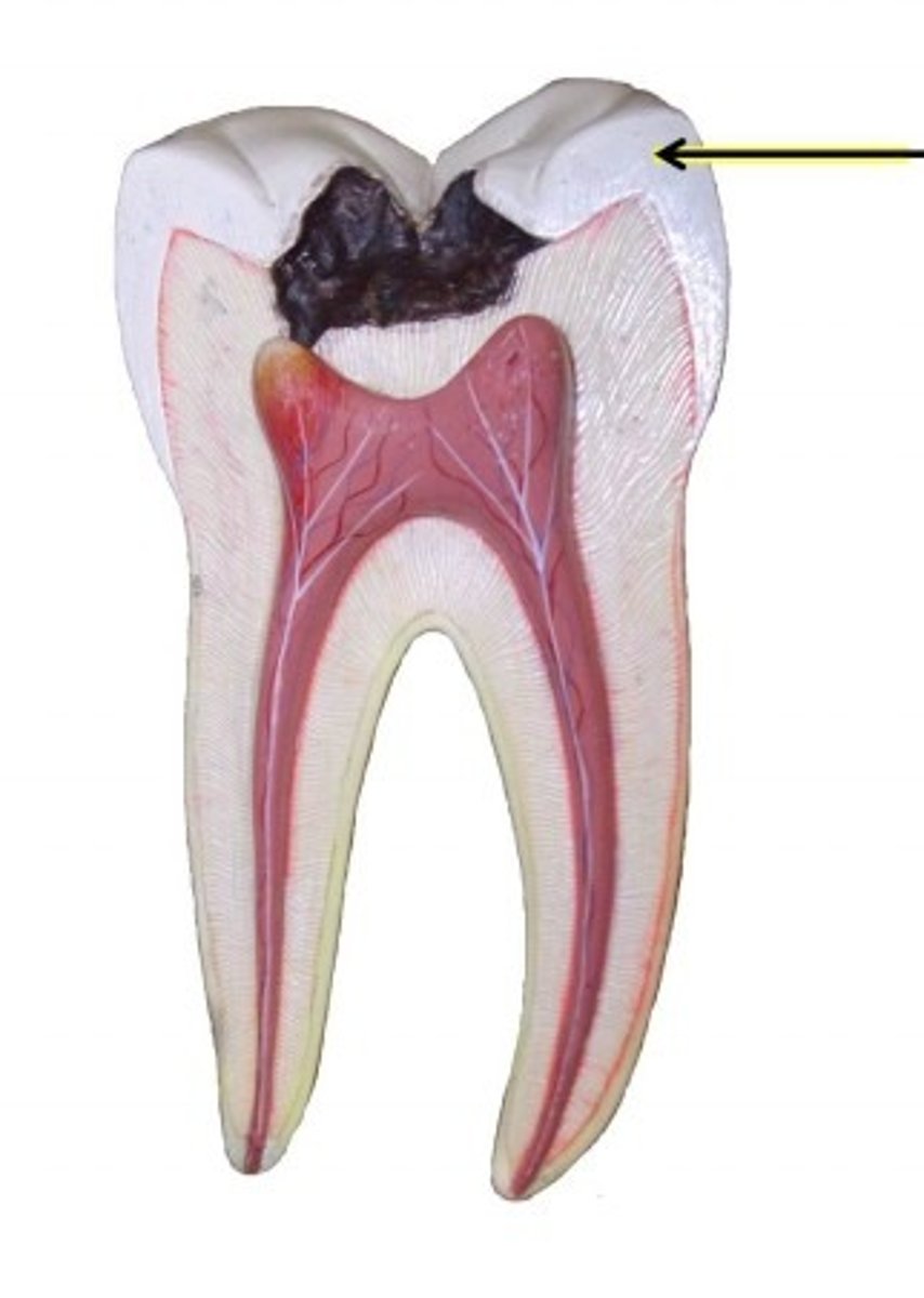
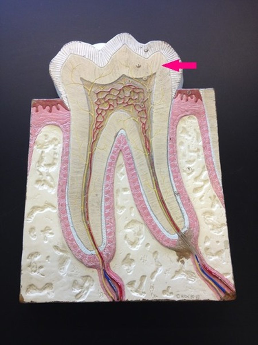
crown
Visible part of the tooth. pulp cavity, enamel, dentin
pulp cavity
contains blood vessels and nerves
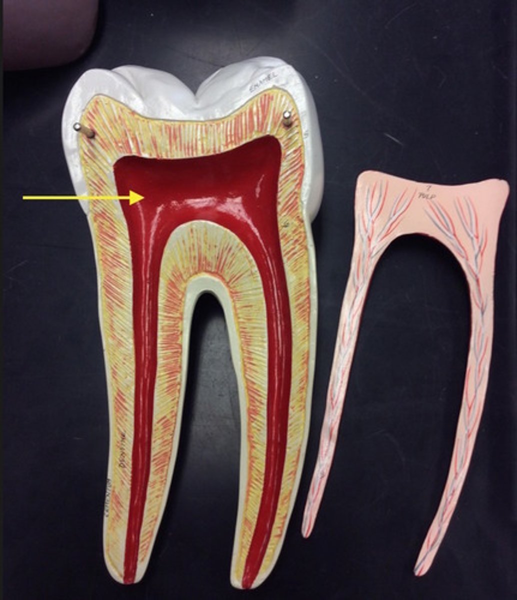
enamel
hard, outermost layer of a tooth
Dentin
Dense tissue forming the bulk of a tooth.
neck of tooth
where the crown and root meet
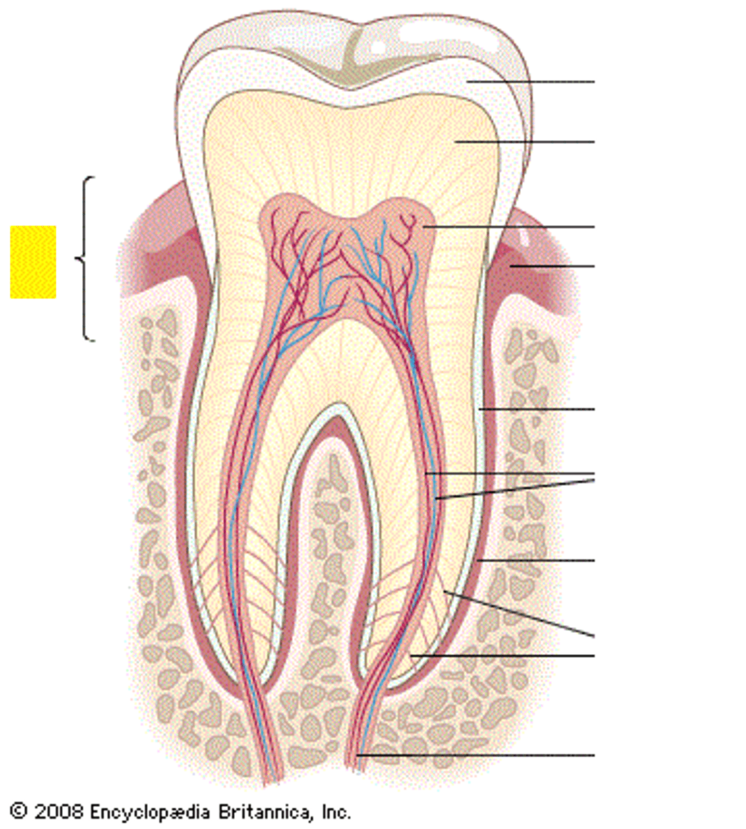
root of tooth
below the gum line. cementum, periodontal ligament, root canal
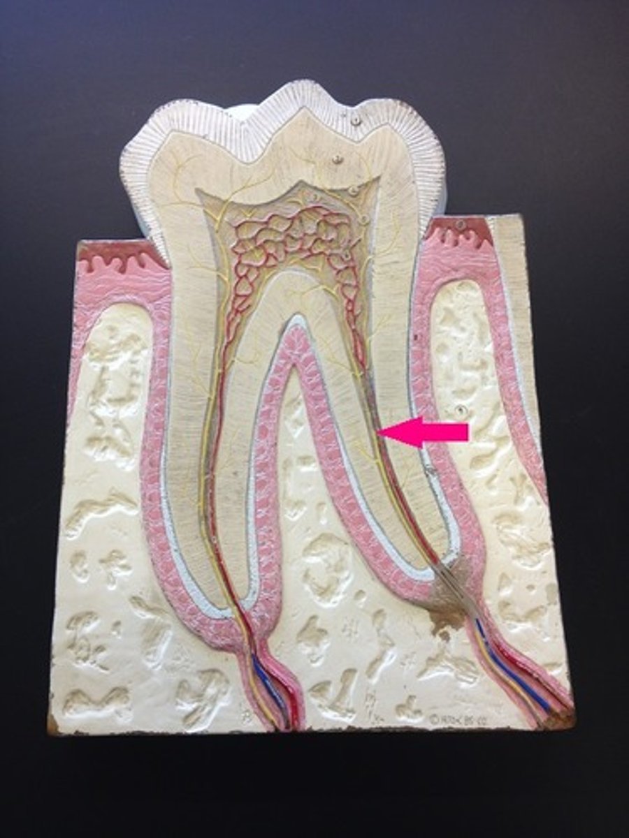
Cementum
Specialized, calcified connective tissue that covers the anatomic root of a tooth.
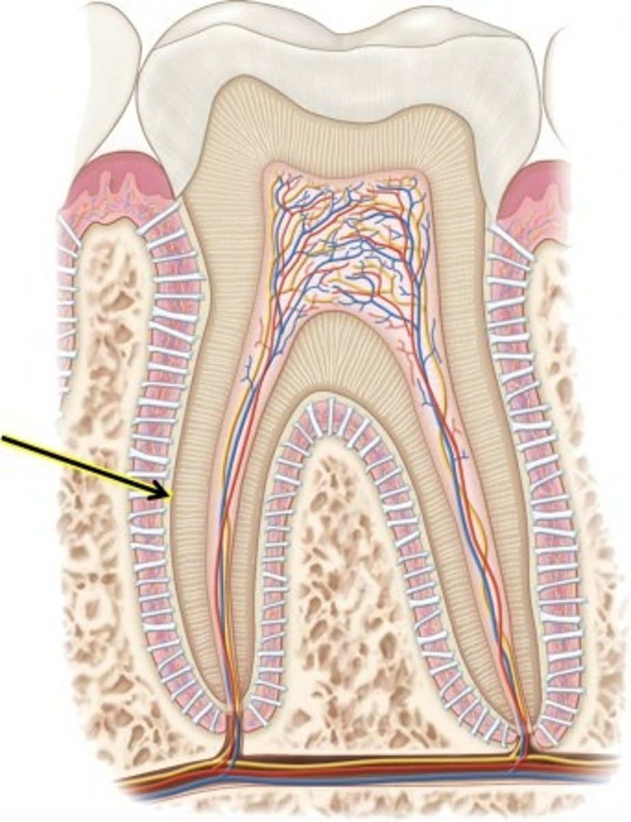
periodontal ligament
attaches tooth to jaw
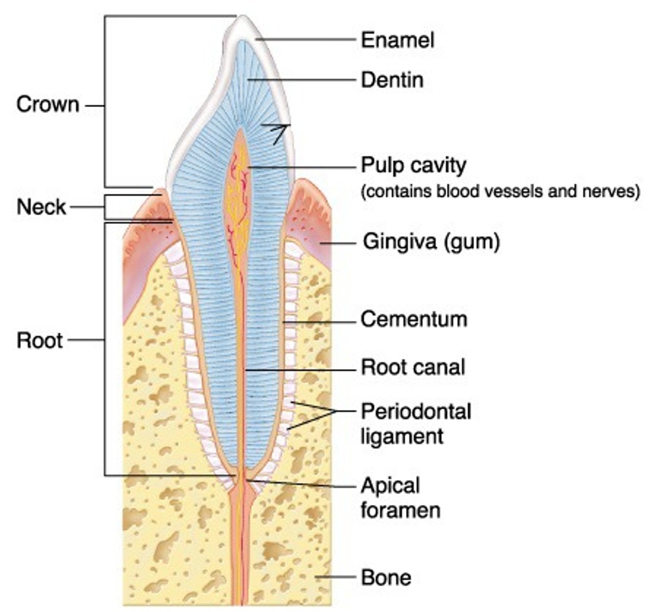
root canal
where the pulp cavity extends into the root

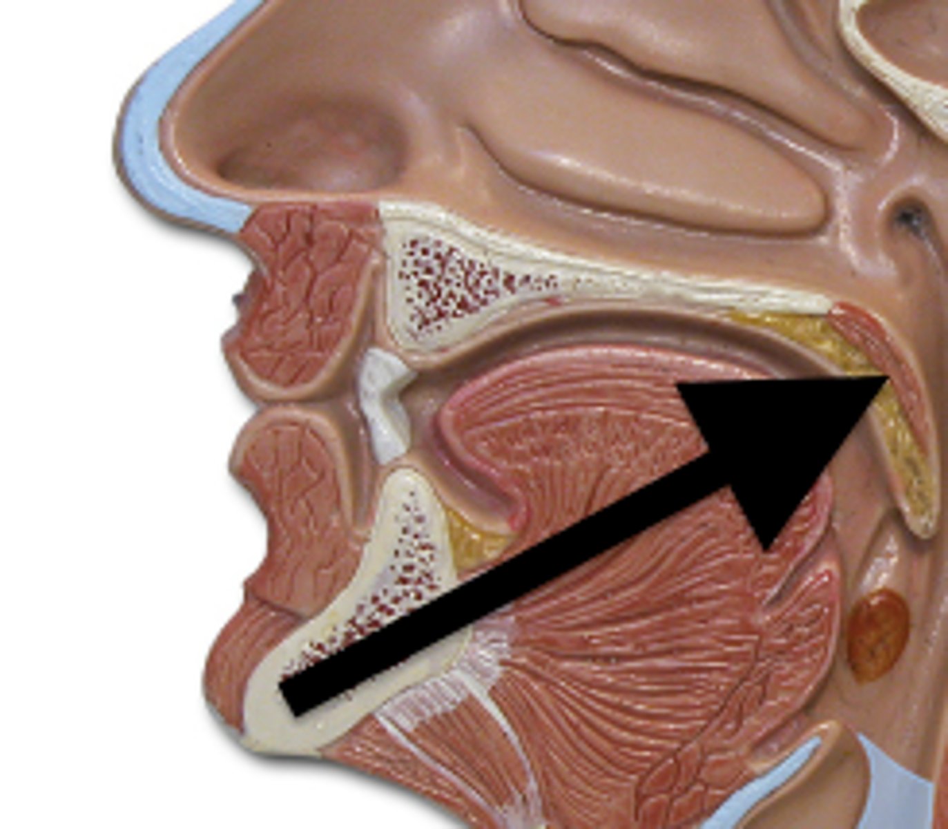
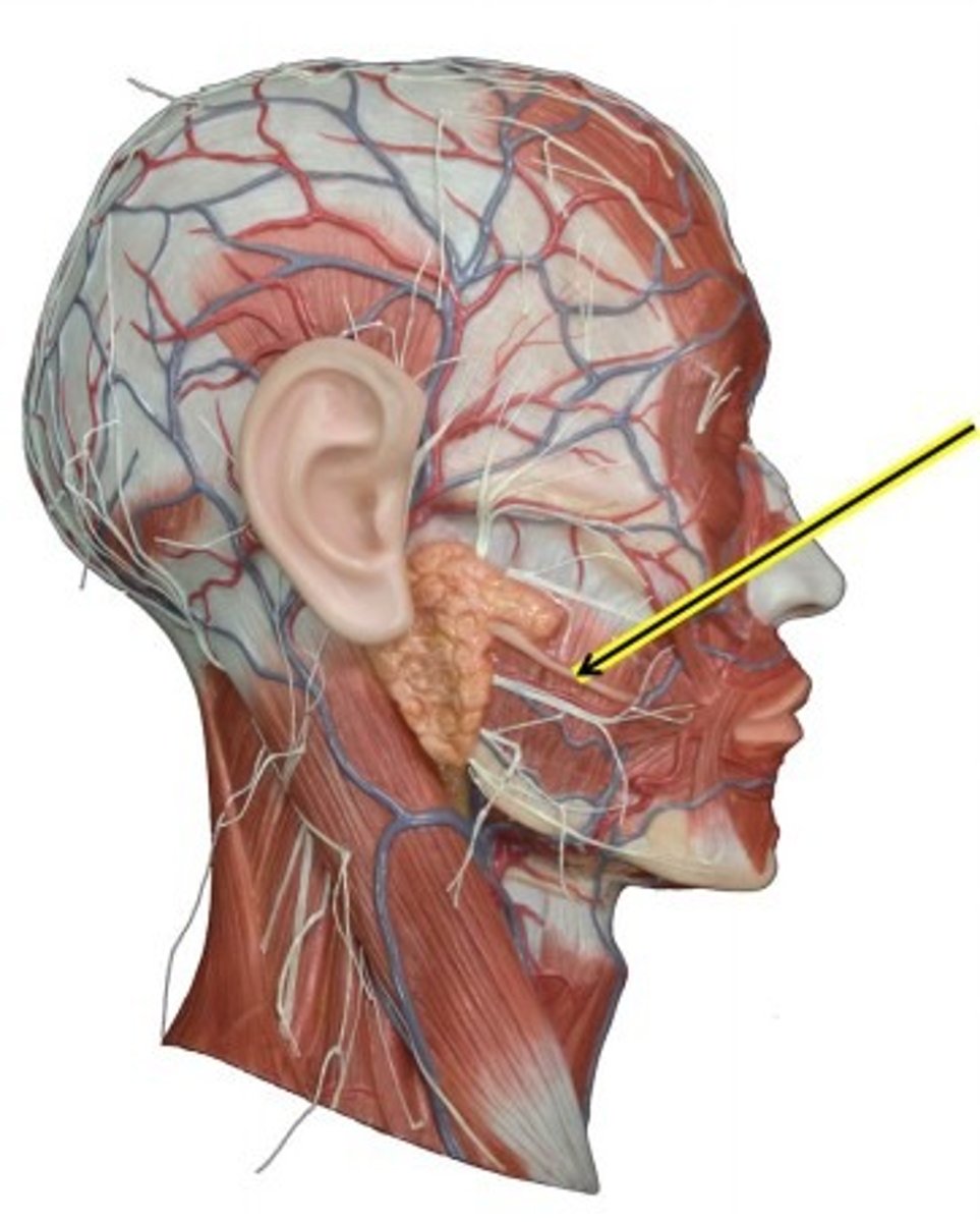
salivary glands
stratified cuboidal epithelium
parotid salivary gland
glossopharyngeal nerve CN IX
secretes serous fluid
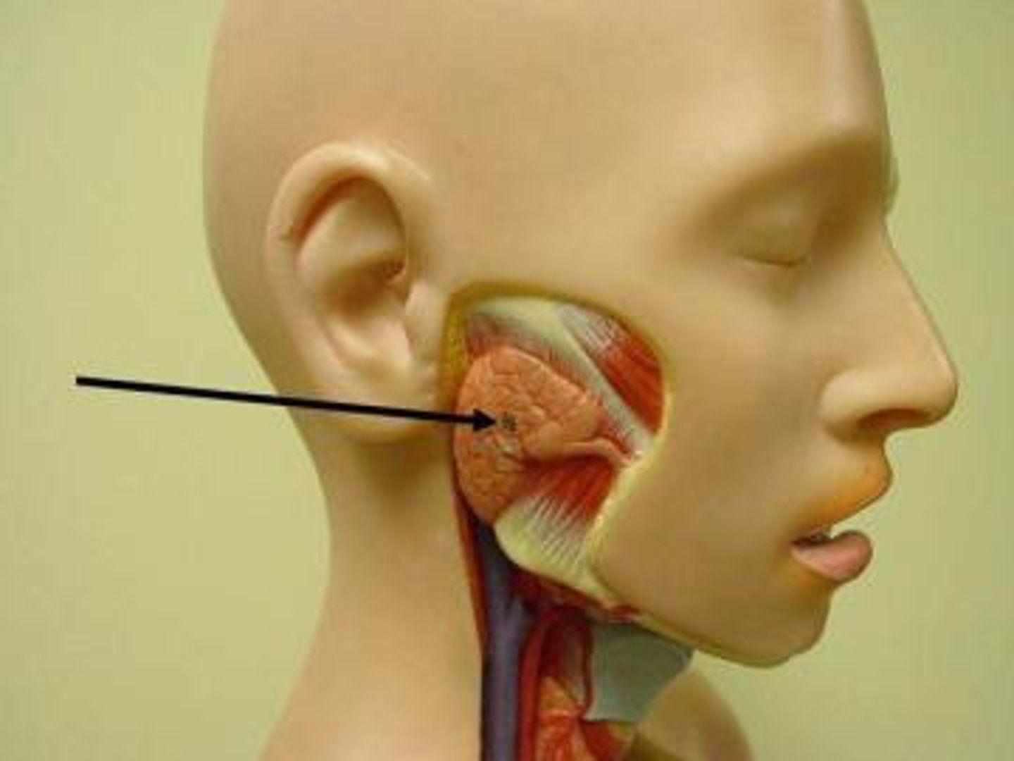
parotid salivary duct
brings saliva to mouth from parotid gland (under ear)
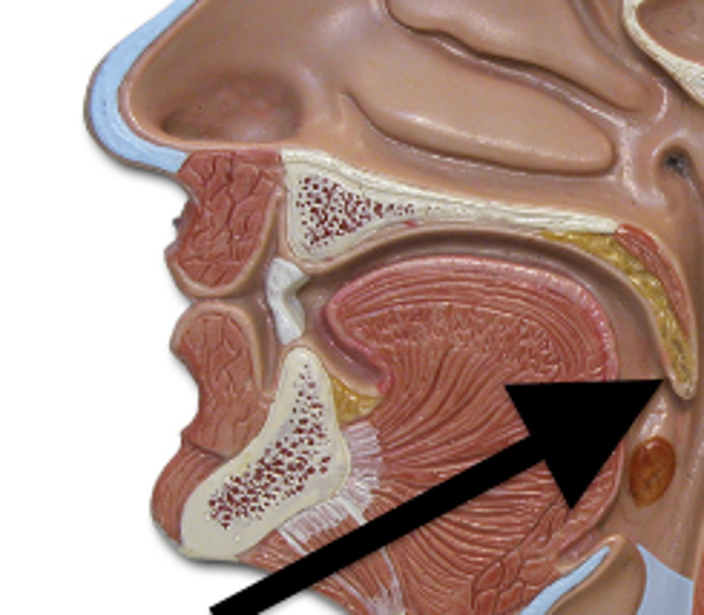
submandibular salivary gland
Facial nerve CN VII
secretes mucous and serous fluid
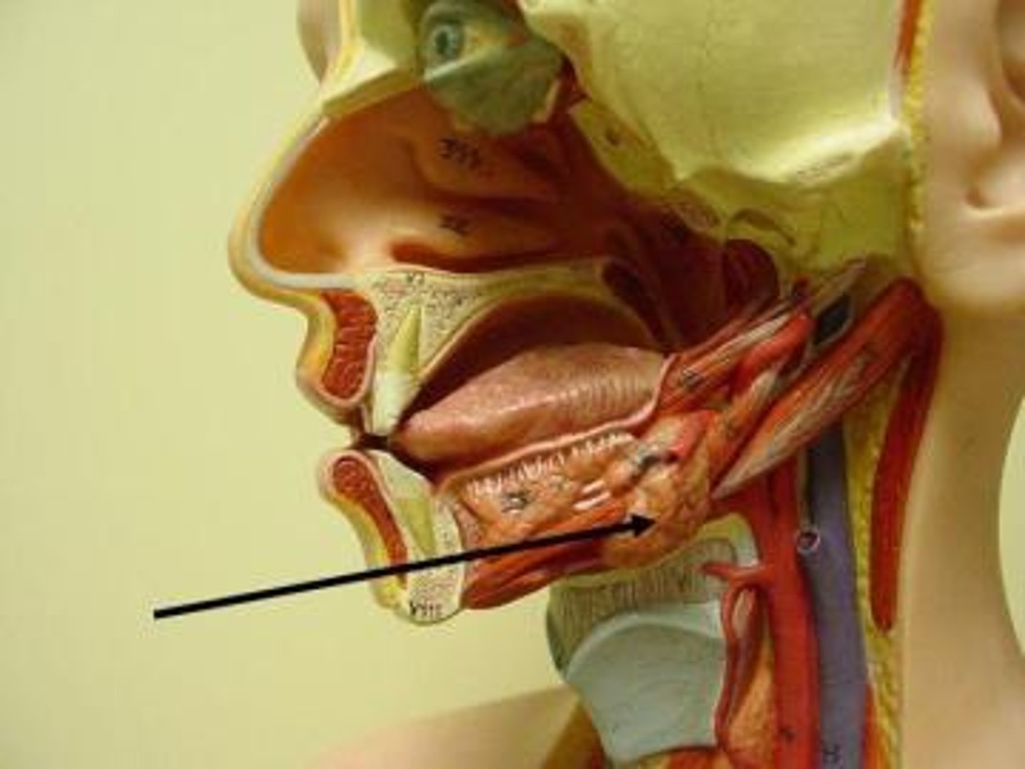
submandibular salivary duct
Straight, flat duct that arises from deep part of submandibular gland and heads toward the anterior aspect of the floor of the mouth
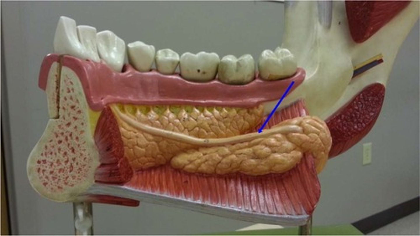
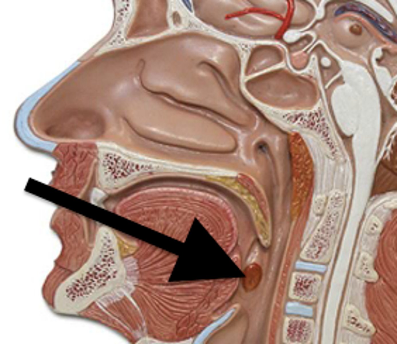
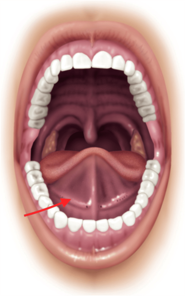
sublingual salivary gland
Facial nerve CN VII. Secretes mucous
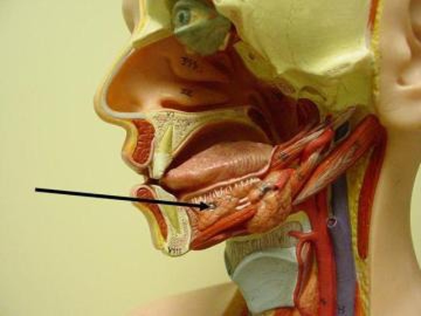
sublingual salivary duct
Are located beneath the tongue, superior to the sublingual glands in the anterior portion of the floor of the mouth. 10-12 ducts
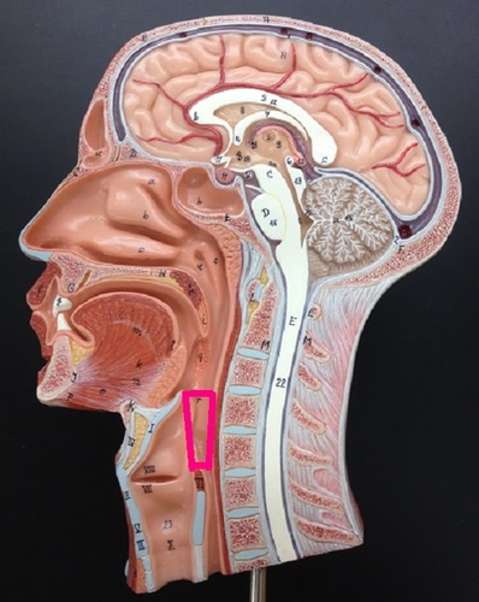
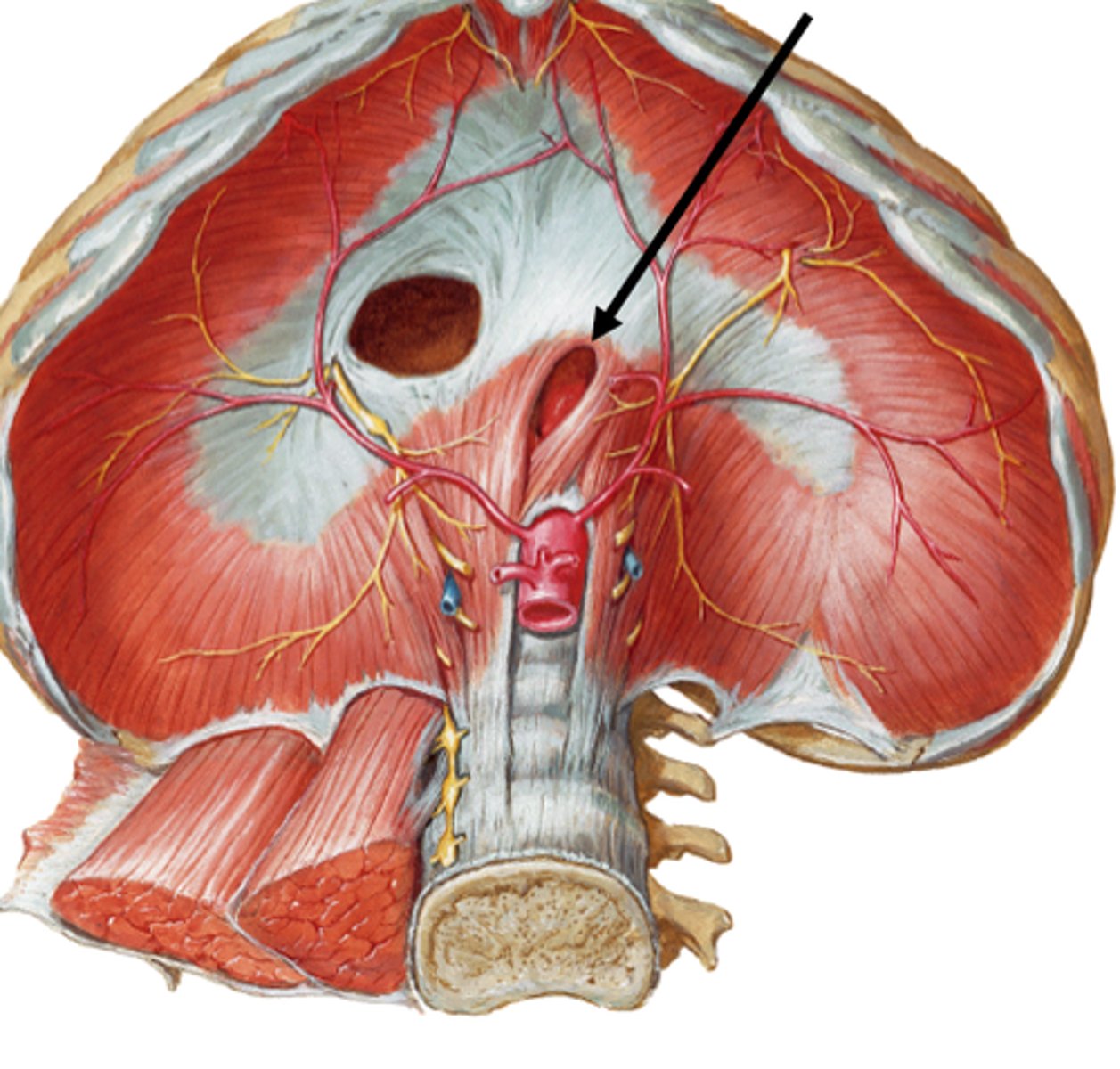
Pharynx
throat
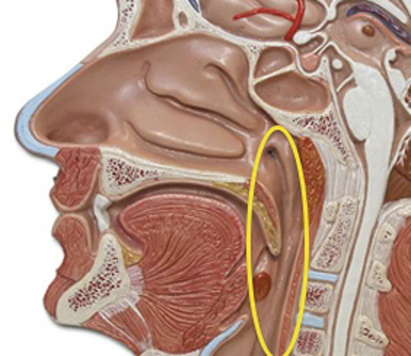
nasopharynx
inferior sphenoid bone to soft palate
Tissue: ciliated pseudostratified columnar epithelia
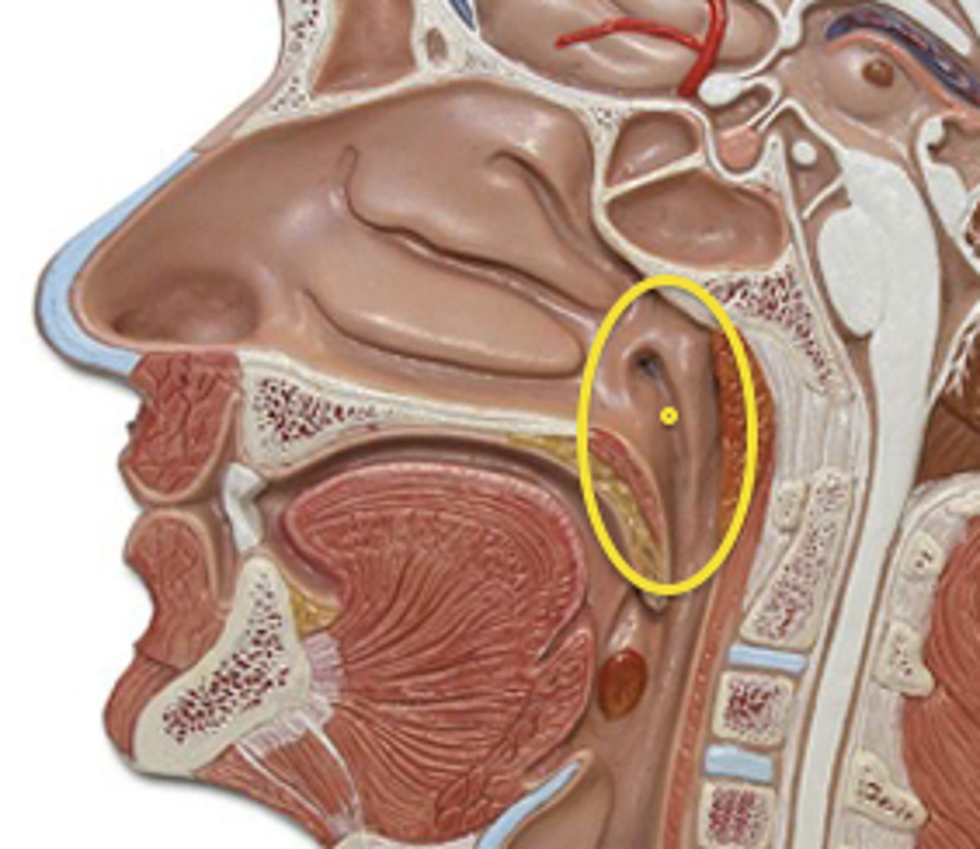
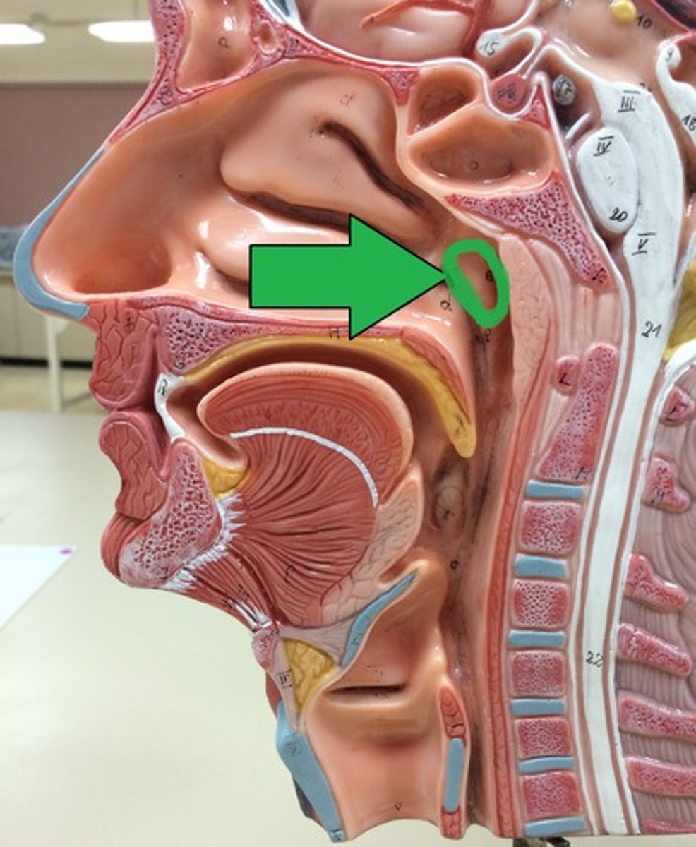
pharyngeal tonsil (adenoids)
single tonsil on wall of nasopharynx
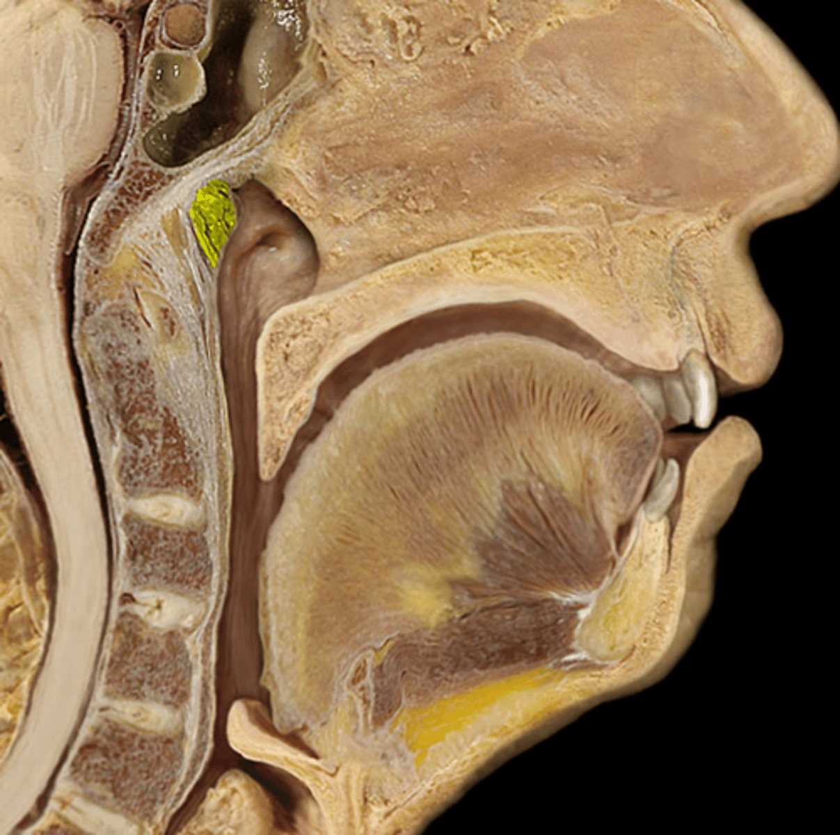
tubal tonsils
surround the openings of the auditory tubes into the pharynx
oropharynx
soft palate to epiglottis
non-keratinized Stratified Squamous Epithelia
laryngopharynx
epiglottis to inferior side of cricoid cartilage
non-keratinized Stratified Squamous Epithelia
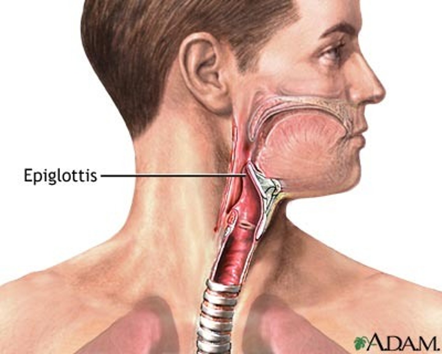
Epiglottis
flap of elastic cartilage that directs food
Elastic Cartilage
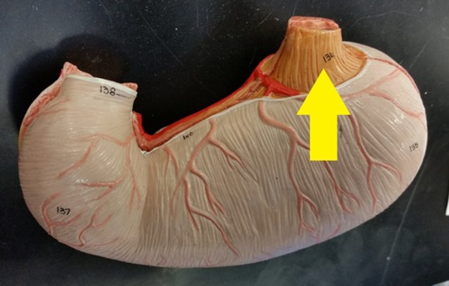
Esophagus
A muscular tube that connects the mouth to the stomach.
non-keratinized Stratified Squamous Epithelia
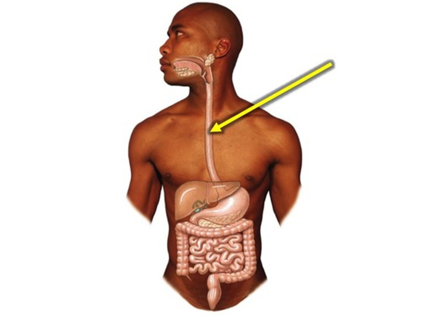
esophageal hiatus
opening in the diaphragm allowing the esophagus to pass through and enter abdomen
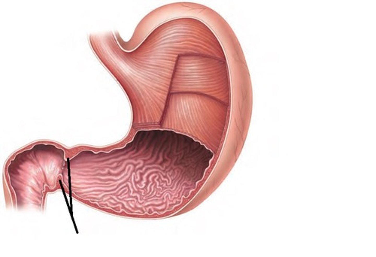
cardiac sphincter
helps prevent regurgitation of stomach contents. Closes off the stomach.
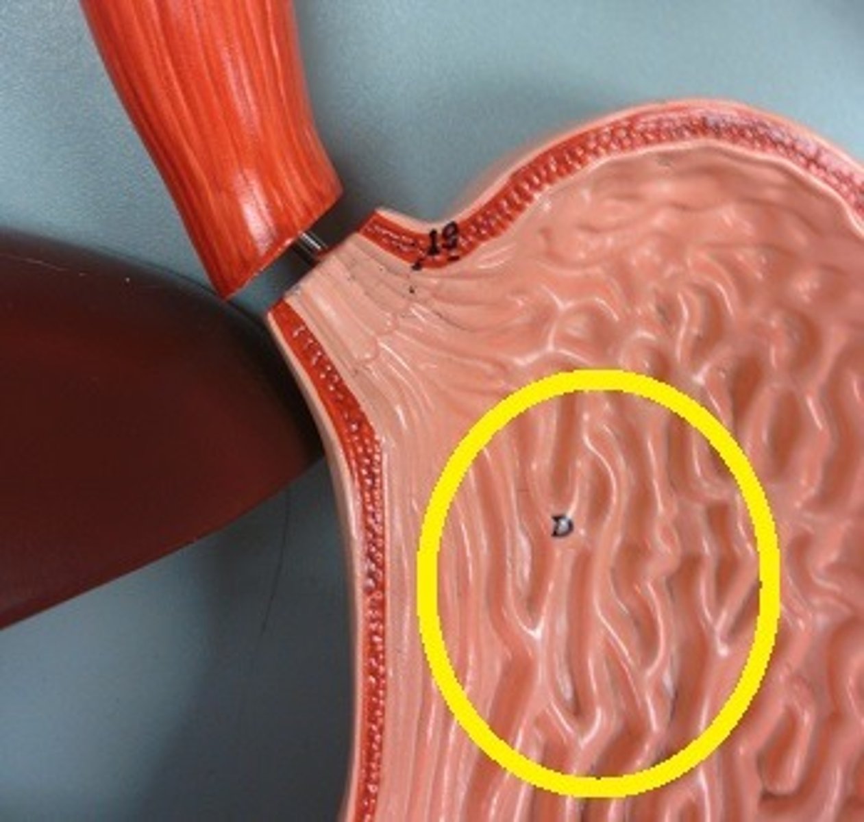
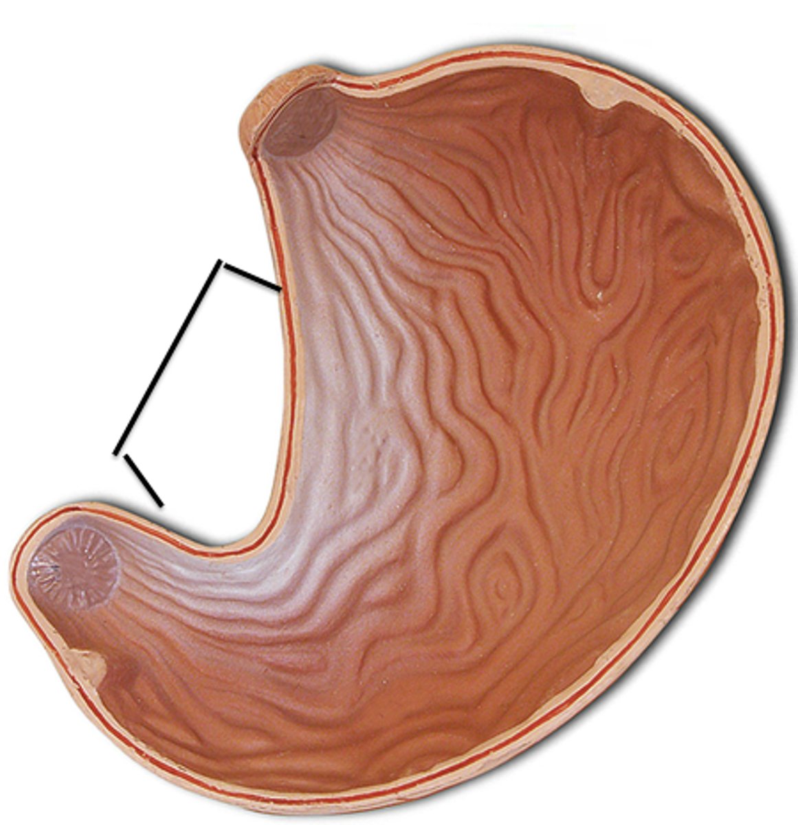
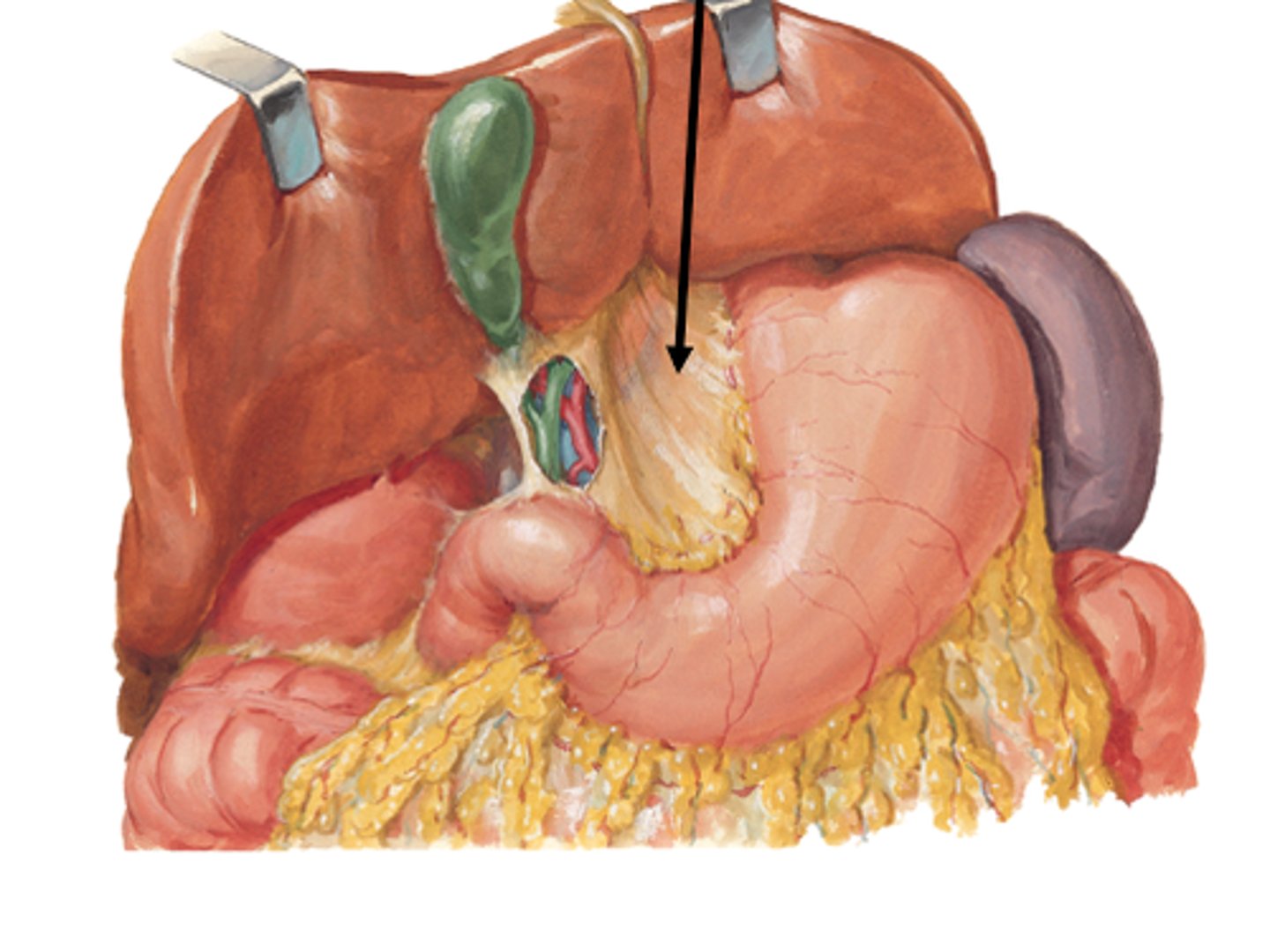
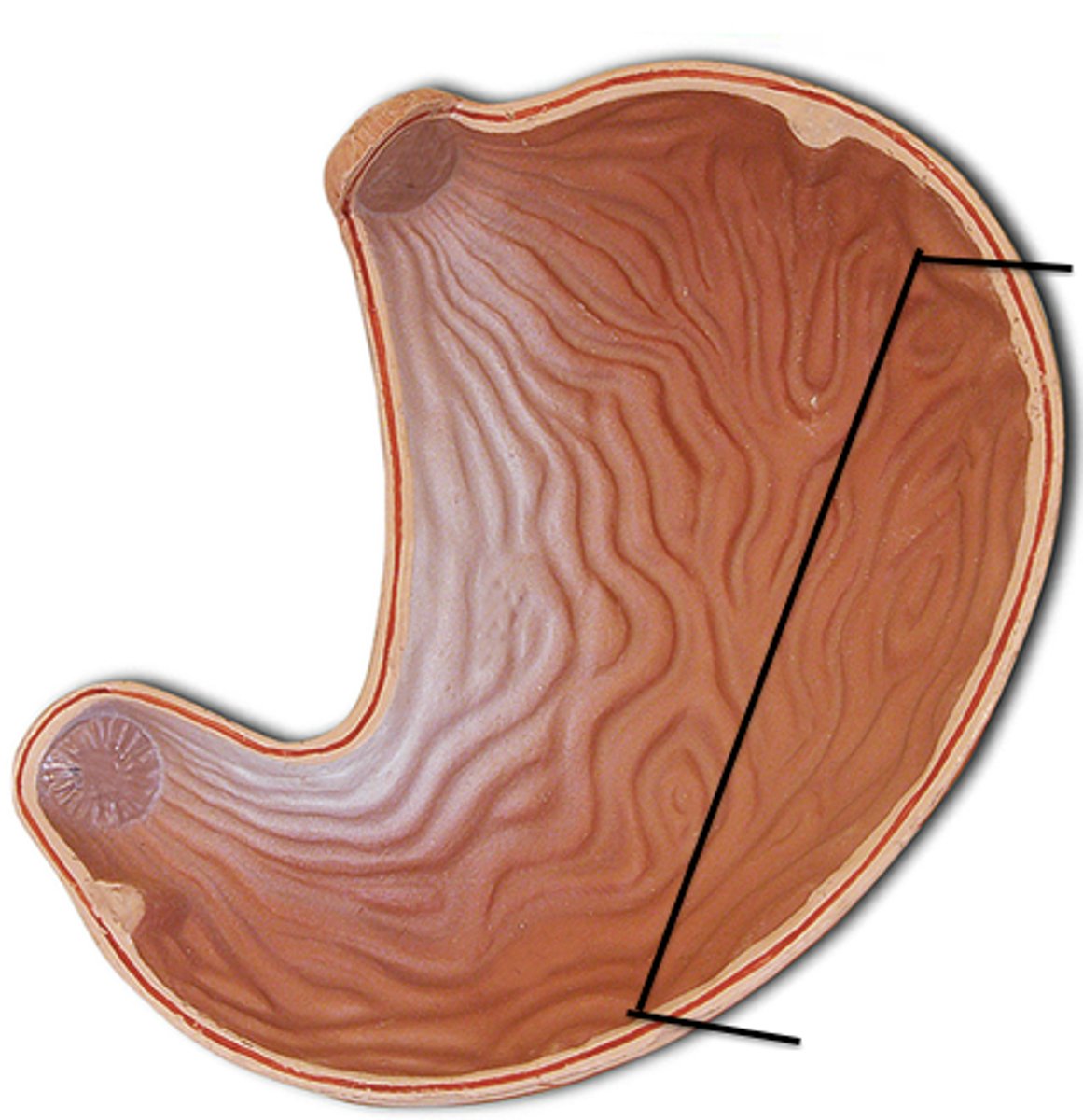
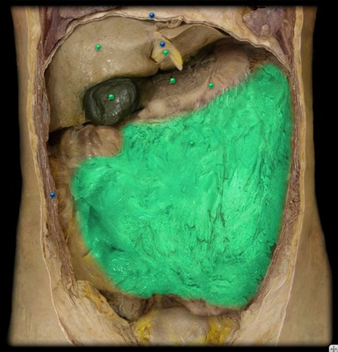
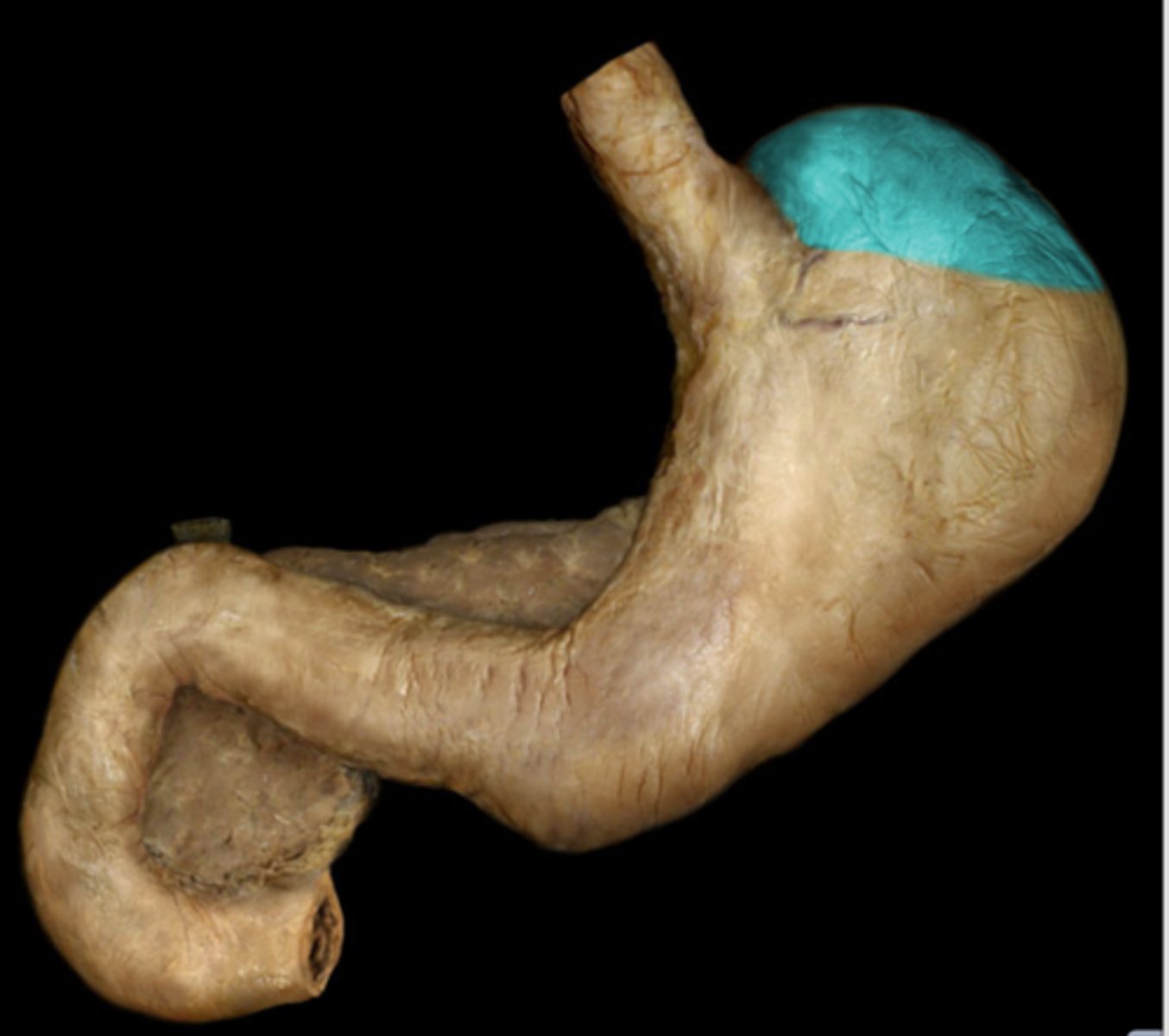
stomach
large muscular sac that continues the mechanical and chemical digestion of food
Lined by Non-Ciliated Simple Columnar Epithelia with Microvilli
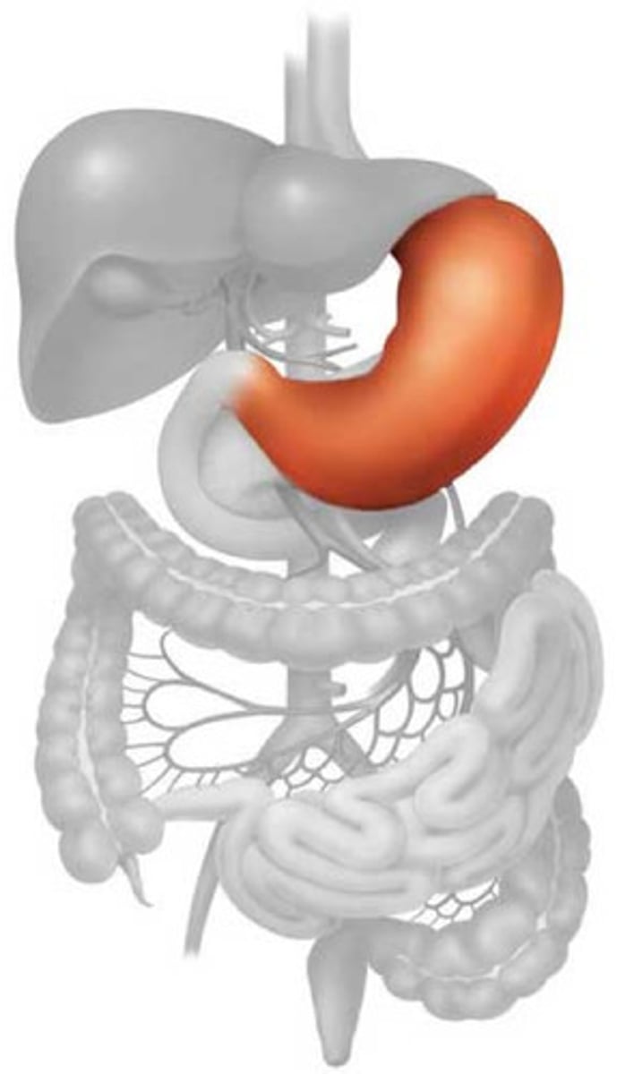
rugae
help stomach expand to allow more food to come in
lesser curvature
curve on superior side of stomach
lesser omentum
attaches stomach to liver
greater curvature
curve on inferior side of stomach
greater omentum
part of the peritoneum attached to the stomach and to the colon and covering the intestines
stomach regions
cardia, fundus, body, pyloric
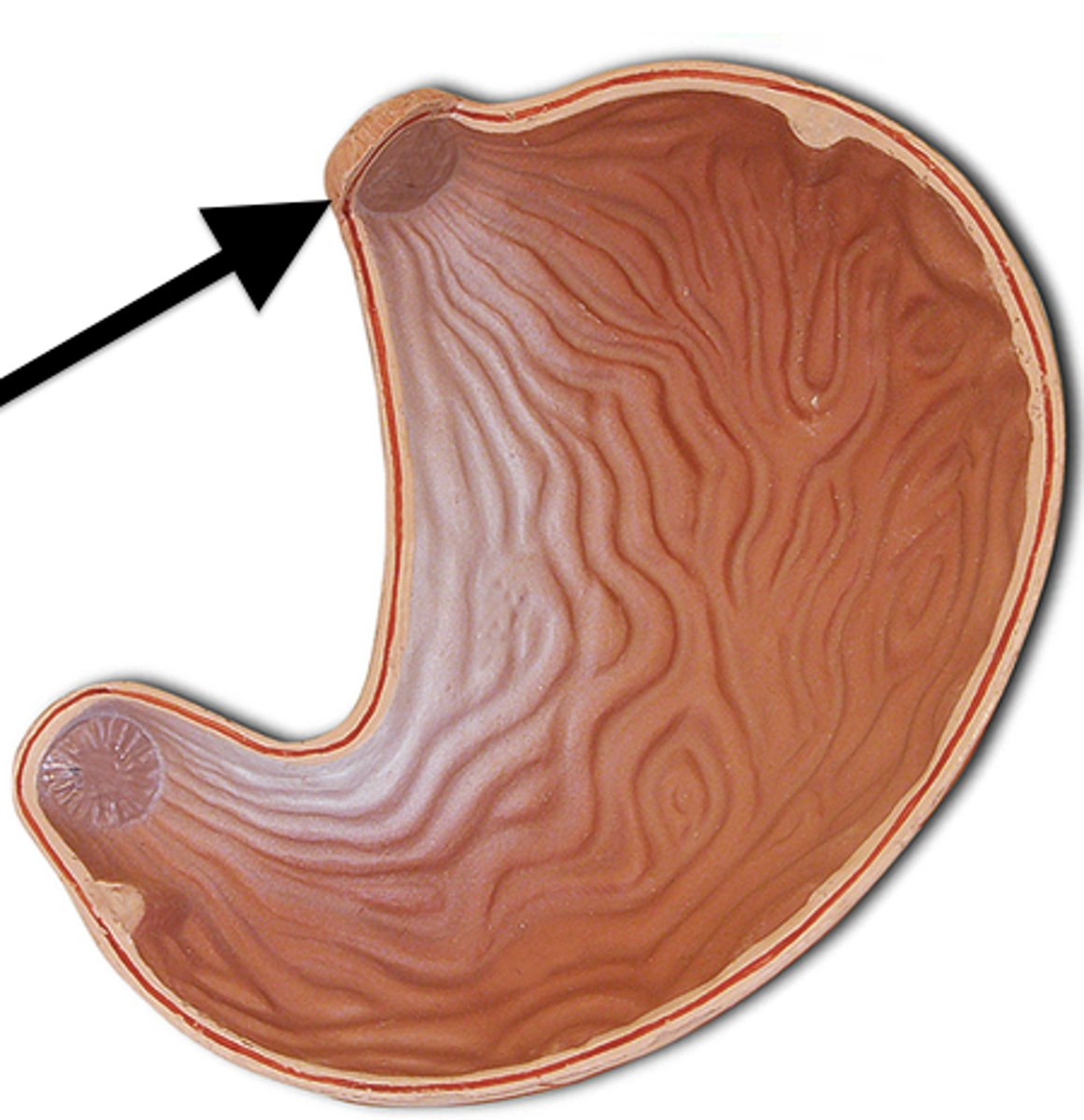
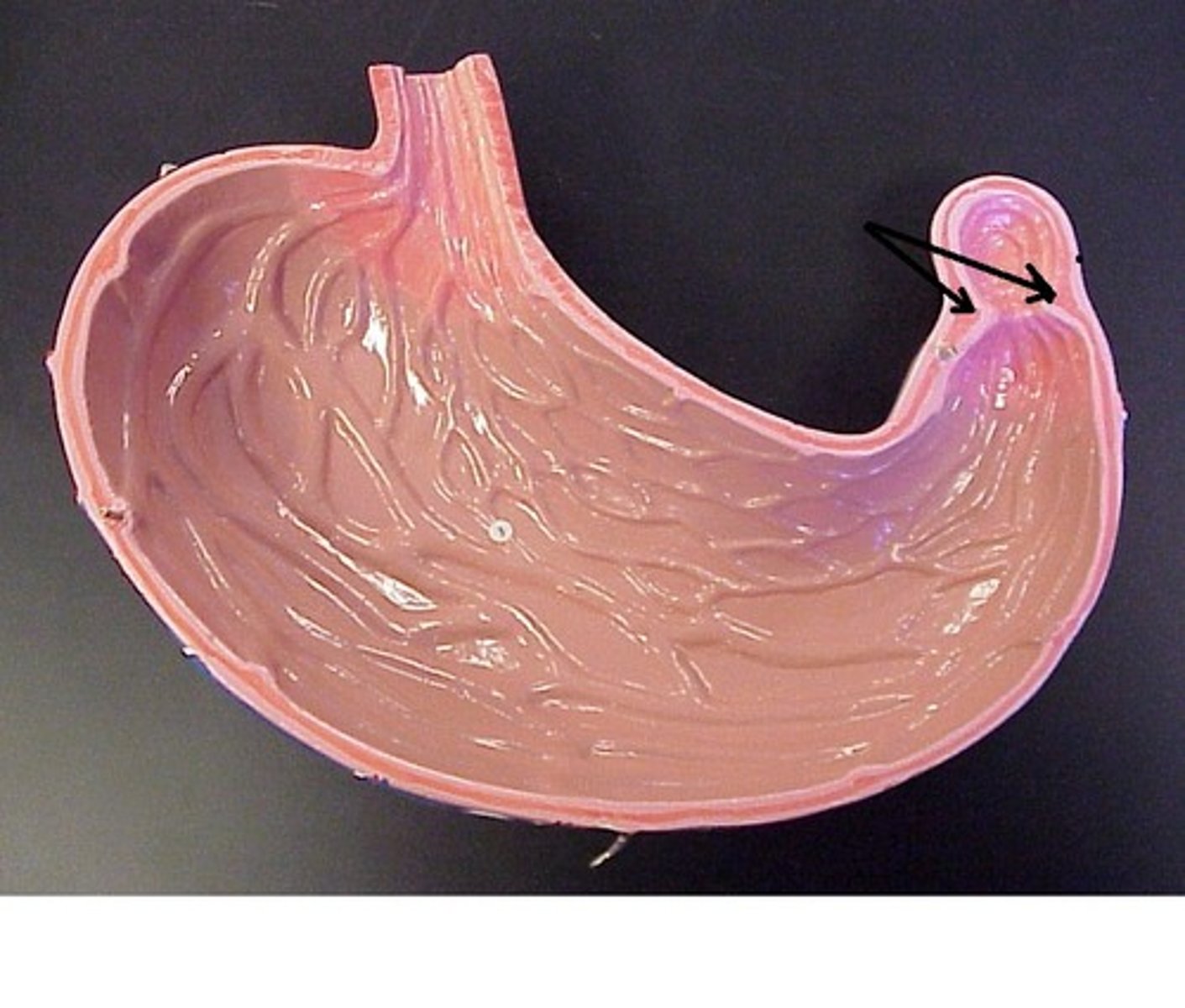
cardia region
where the esophagus enters the stomach
fundus region
balloons out toward left side
body of stomach
middle section
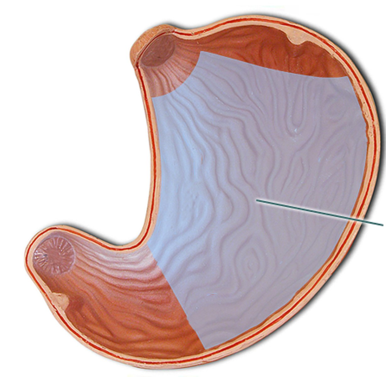
pyloric region
narrower pouch at the inferior end
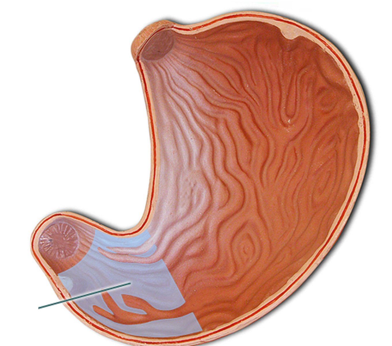
pyloric canal
space where it starts to narrow
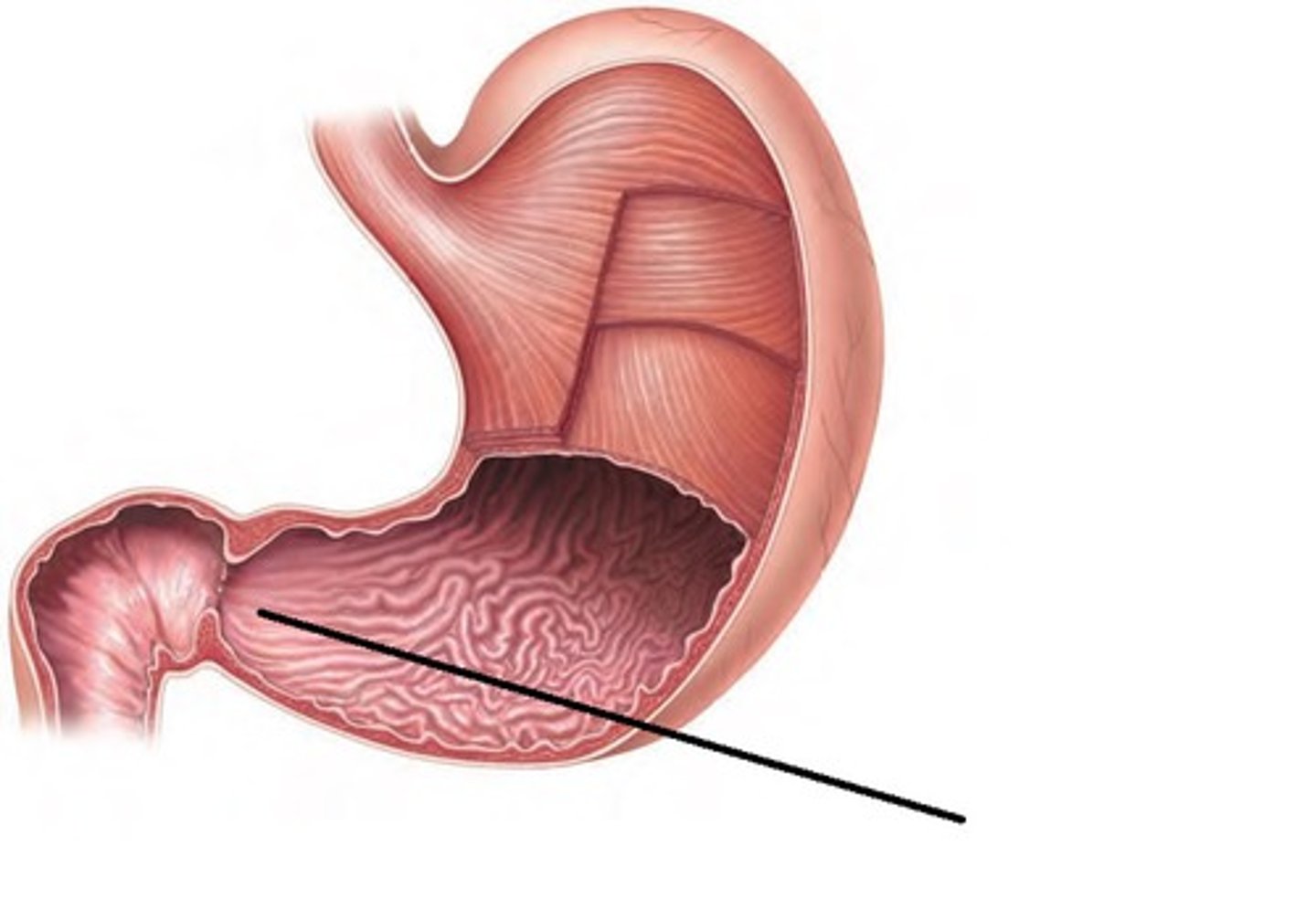
pylorus
space that the sphincter surrounds
pyloric sphincter
closes opening between stomach and small intestine
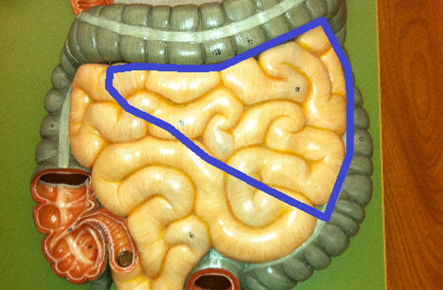
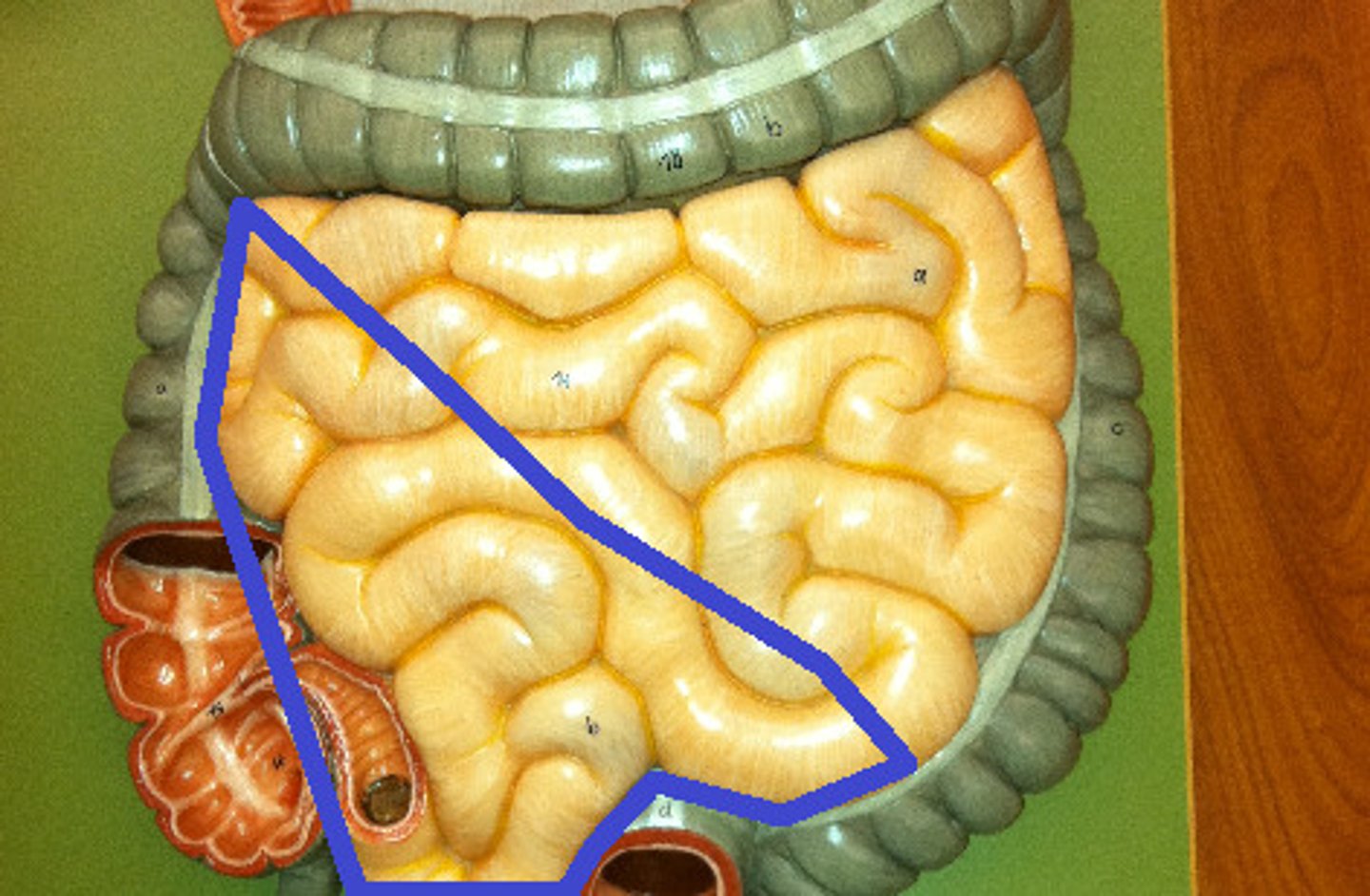
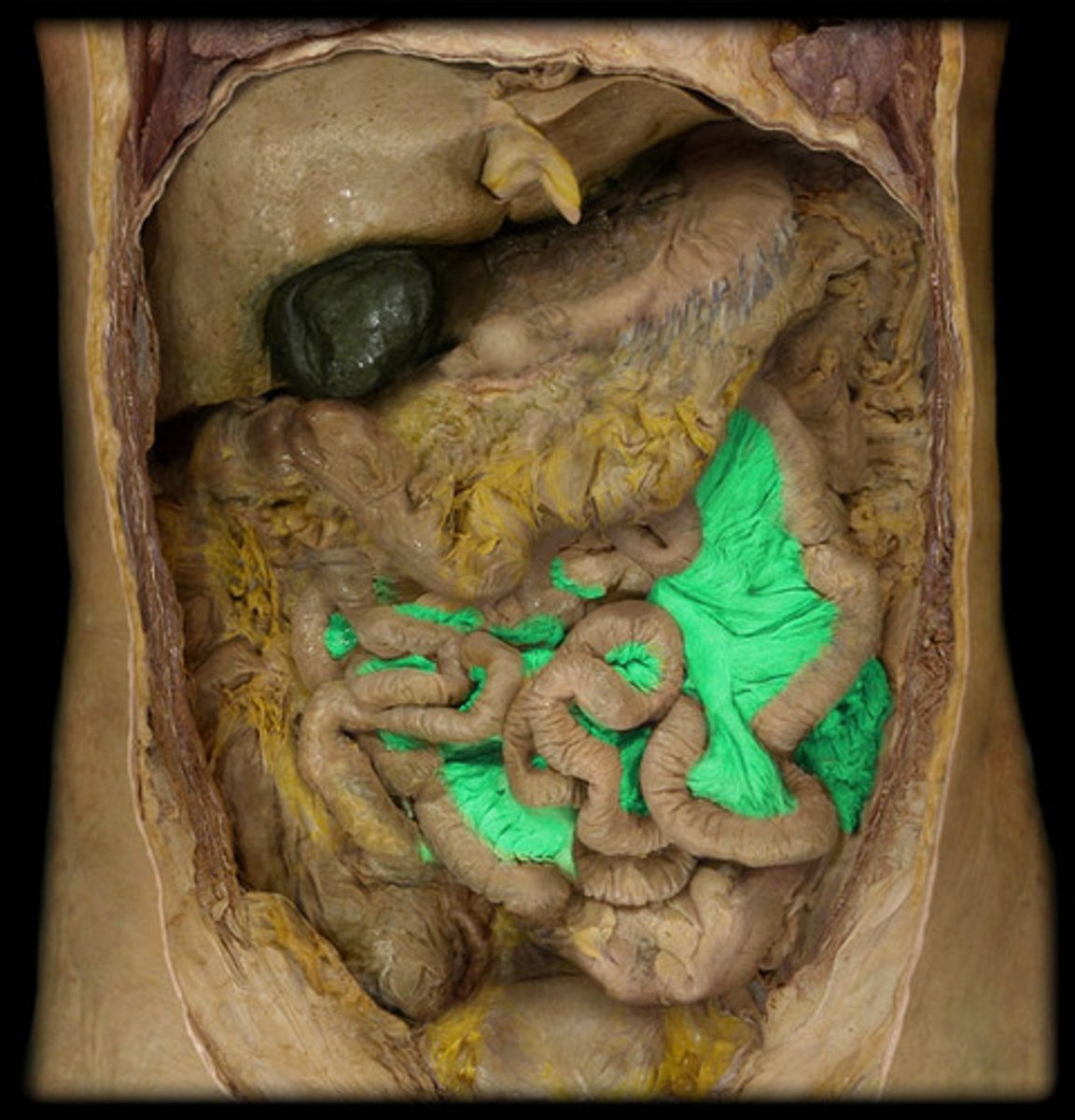
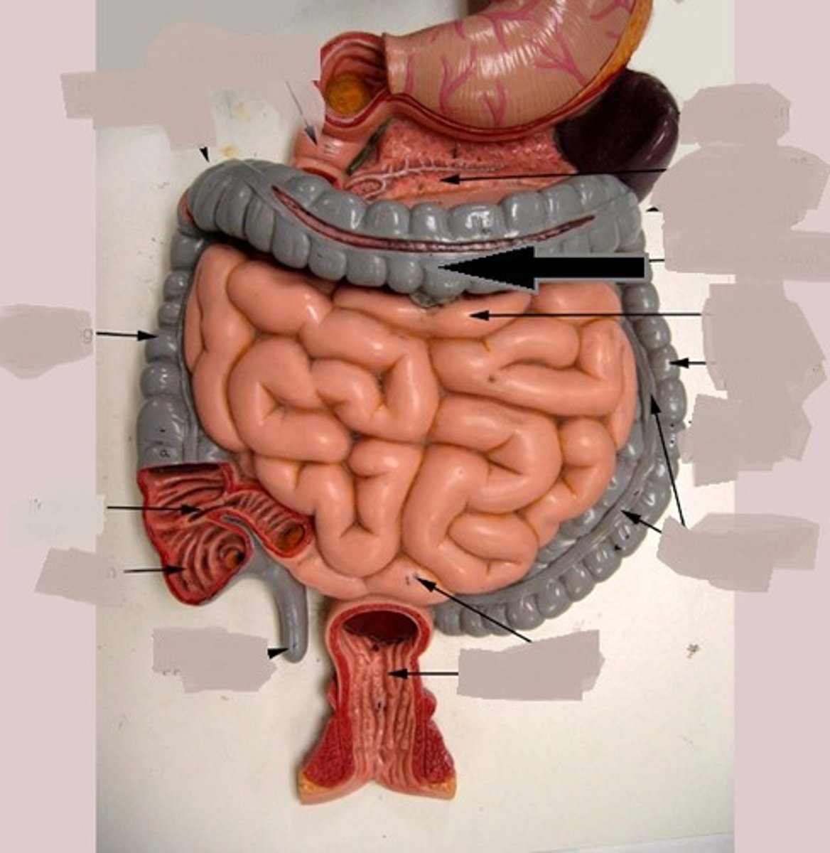
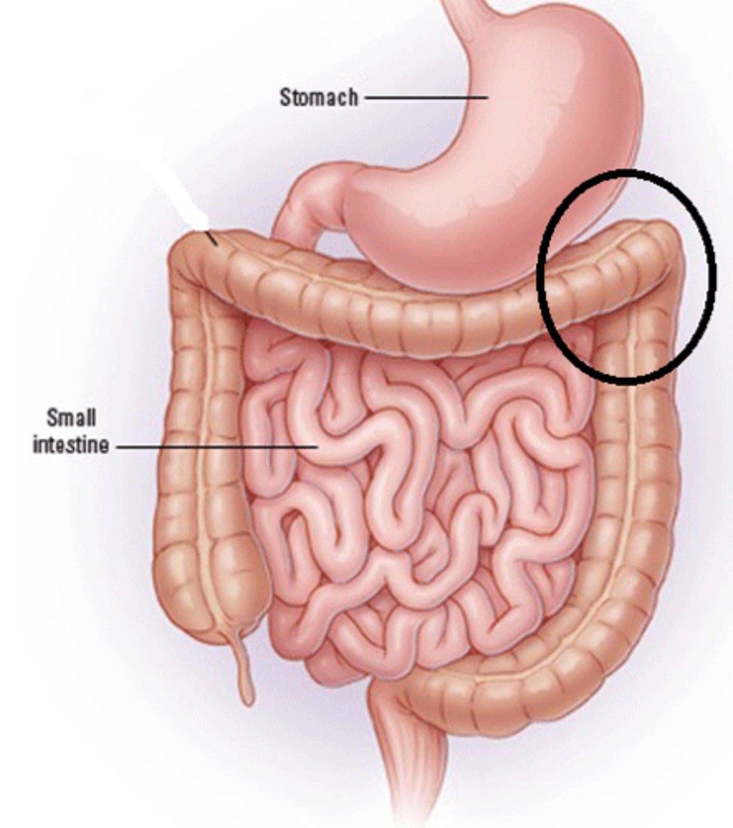
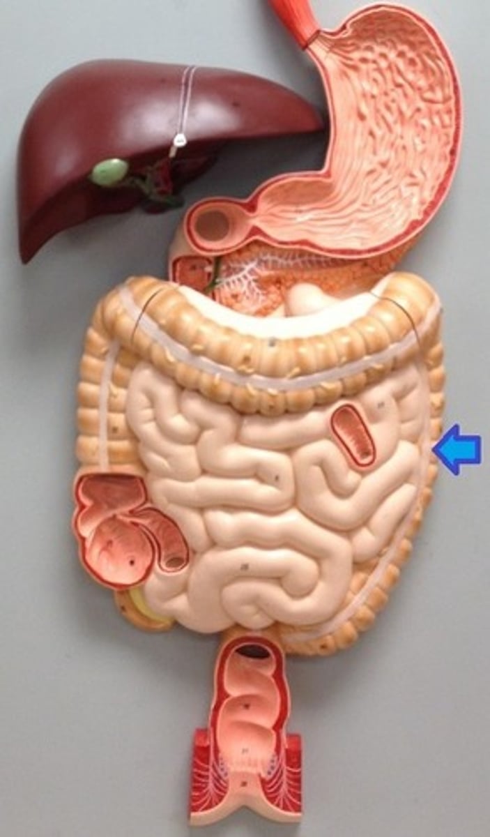
small intestine
Digestive organ where most chemical digestion and absorption of food takes place
Tissue: non-ciliated simple columnar epithelia with microvilli
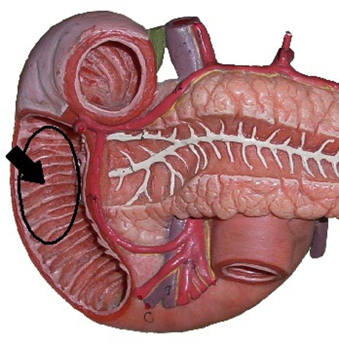
Duodenum
first portion of the small intestine. right side
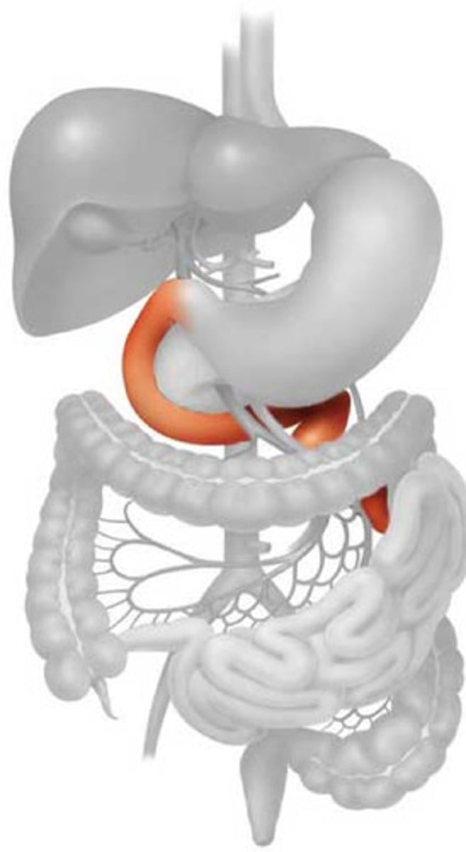
Jejunum
upper left
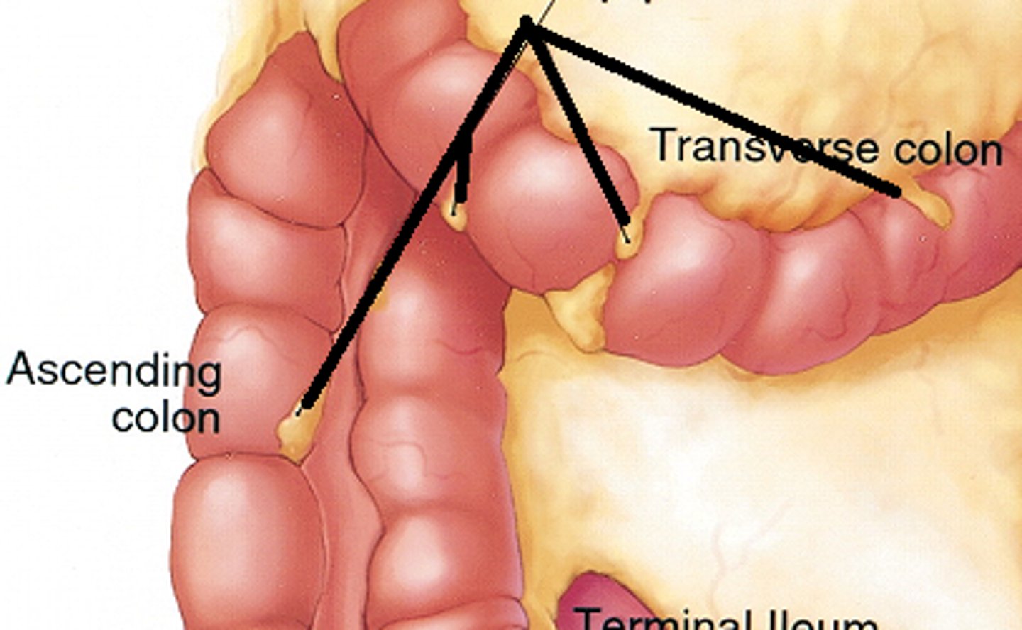
Ileum
third part of the small intestine
plicae circulares
folds that increases surface area for nutrient absorption
mesentary
fatty tissue attaching stomach and duodenal wall and to self
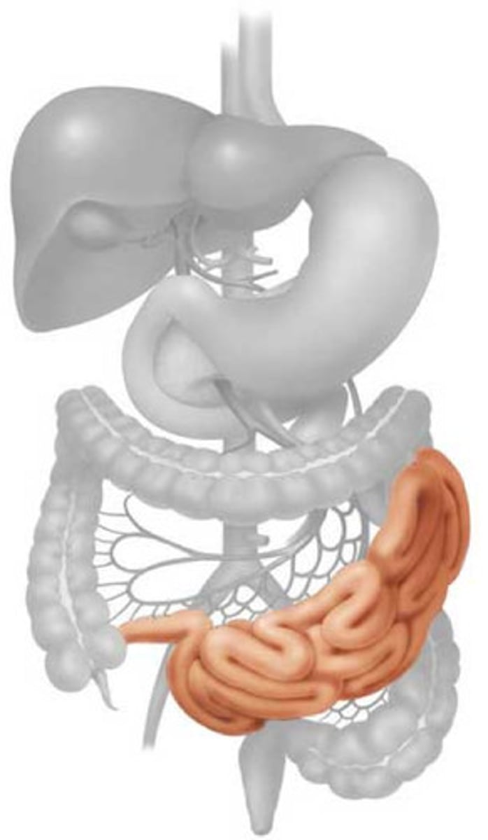
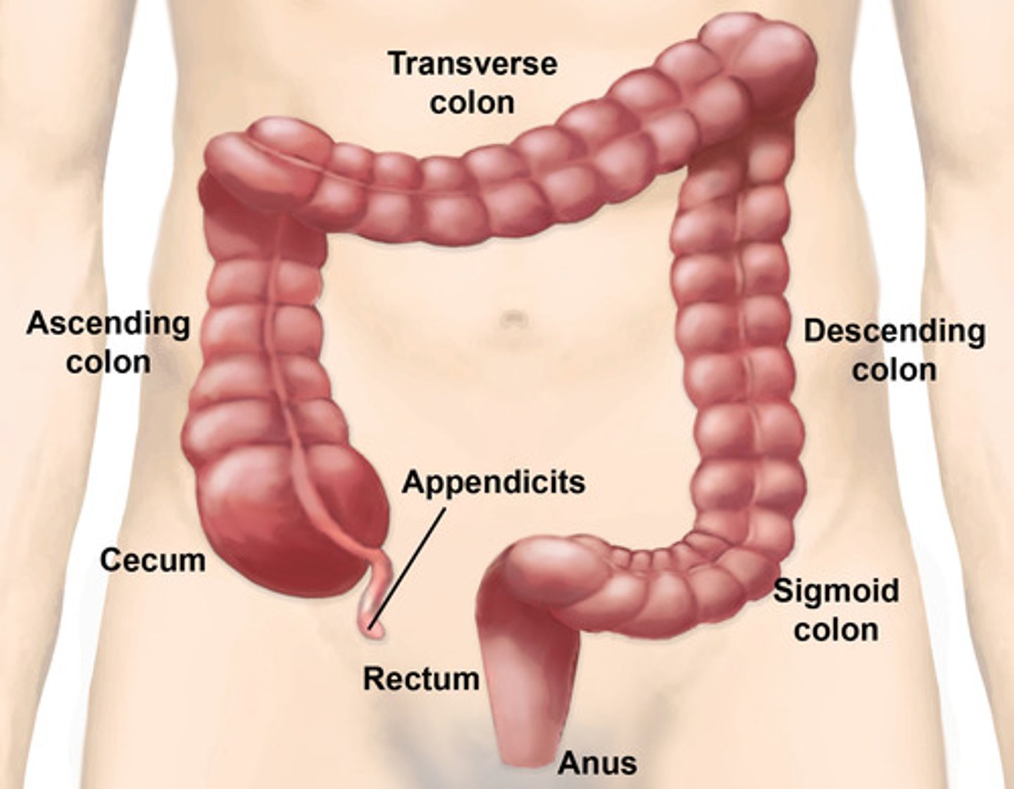
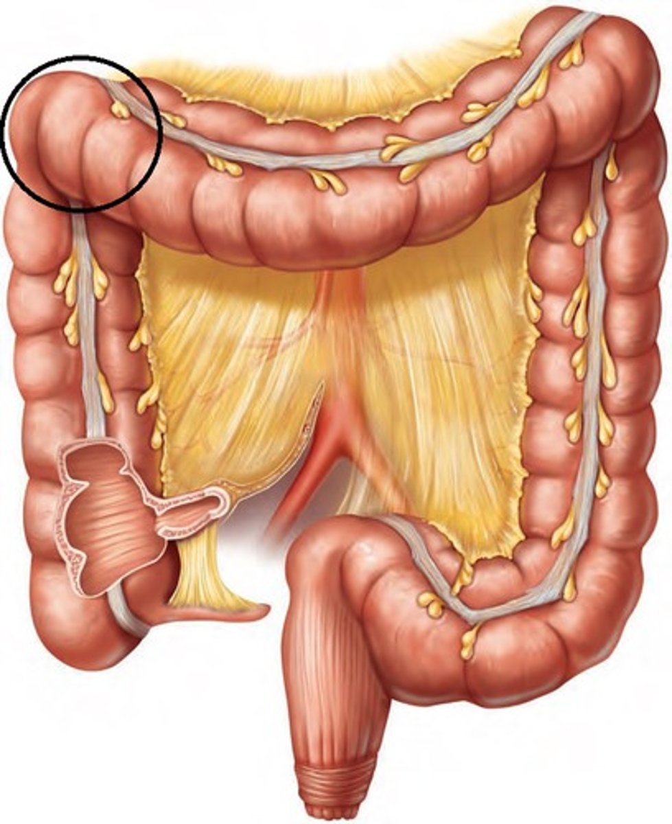
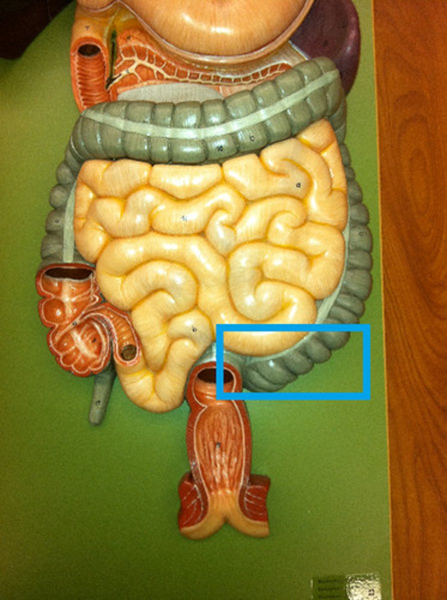
large intestine
Absorbs water and forms feces
Tissue: non-ciliated simple columnar epithelia with microvilli
taeniae coli
helps large intestines contract into haustra
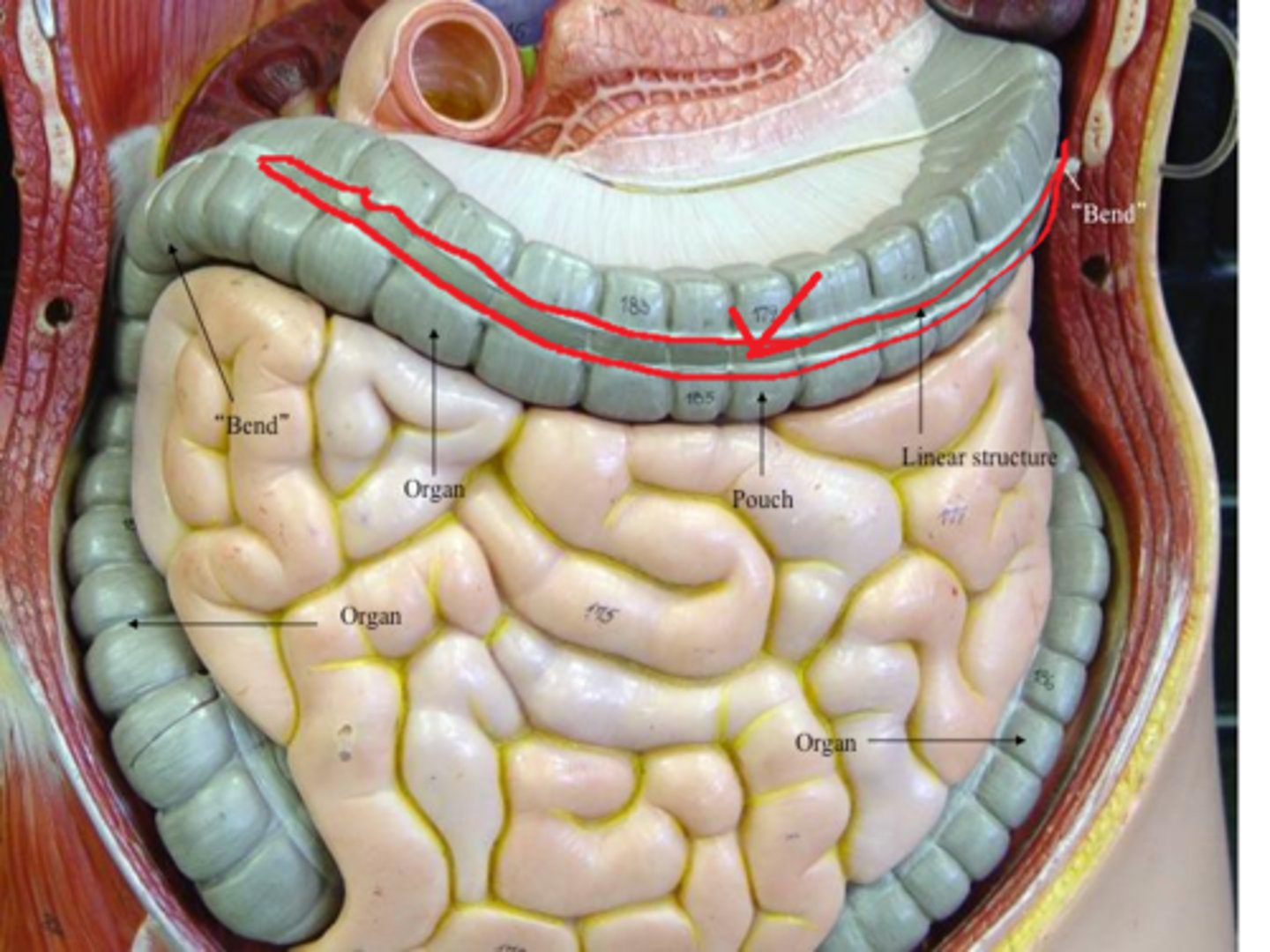
Haustra
pouches that form in the large intestine
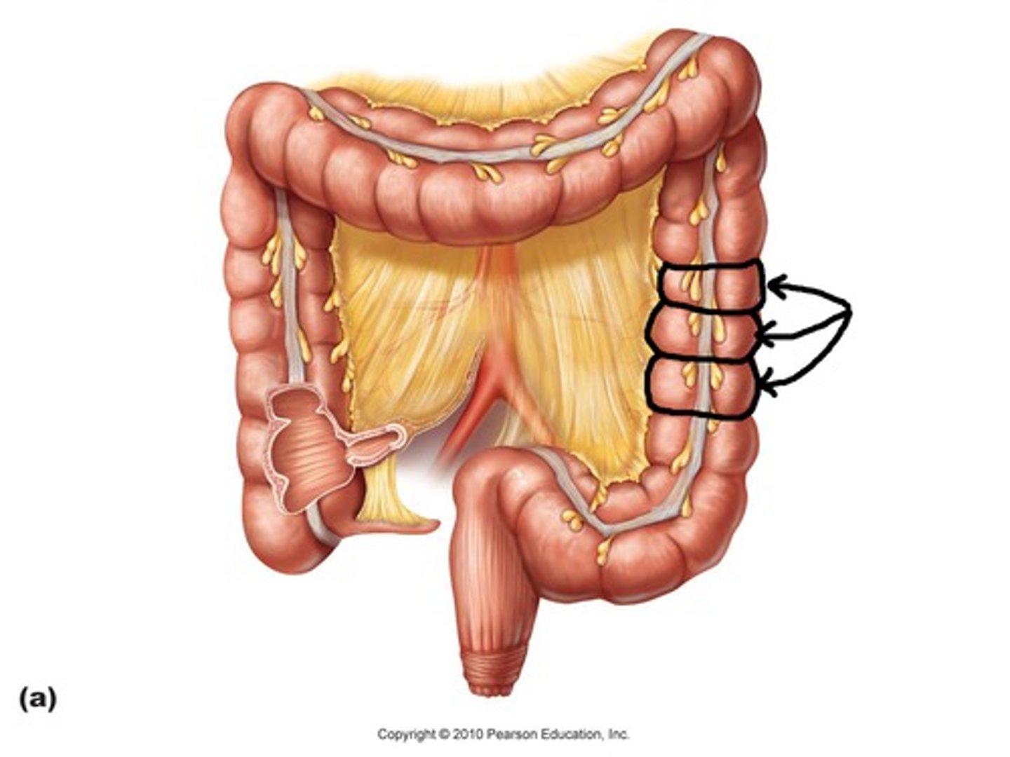
epiploic appendages
fat-filled pouches of visceral peritoneum
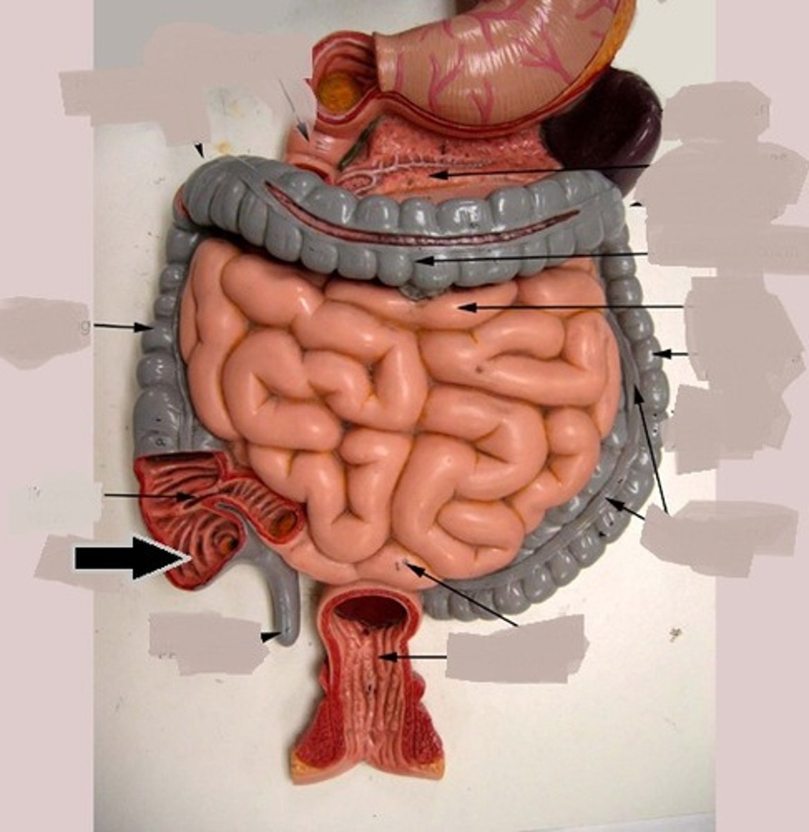
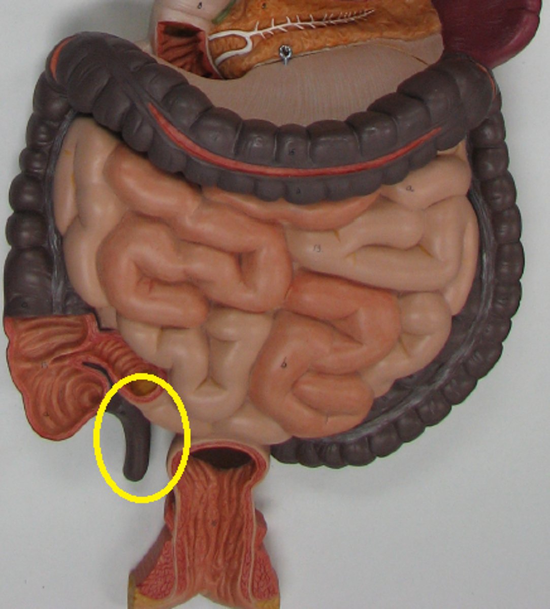
Cecum
first part of the large intestine. Big bag
ileocecal valve
helps control flow of ileum of small intestine and cecum of large intestine
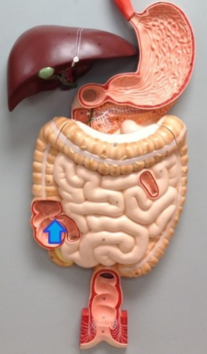
vermiform appendix
hangs from the lower portion of the cecum
ascending colon
the part of the large intestine that ascends from the cecum to the transverse colon
hepatic flexure
Bend between the ascending colon and the transverse colon.
transverse colon
passes horizontally from right to left toward the spleen
splenic flexure
area of the colon that bends downward near the spleen
descending colon
portion of the colon that extends downward from the transverse colon
sigmoid colon
an S-shaped structure that continues from the descending colon above and joins with the rectum below
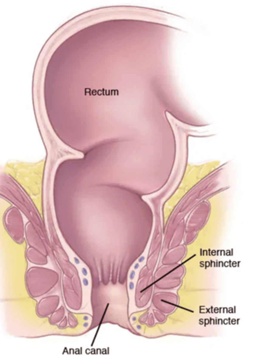
Rectum
A short tube at the end of the large intestine where waste material is compressed into a solid form before being eliminated

anal canal
region, containing two sphincters, through which feces are expelled from the body
non keratinized stratified squamous epithelium
anal columns
small longitudinal folds in the anal canal

anus
A muscular opening at the end of the rectum through which waste material is eliminated from the body
keratinized stratified squamous epithelium

internal anal sphincter
smooth muscle, involuntary

external anal sphincter
skeletal muscle, voluntary
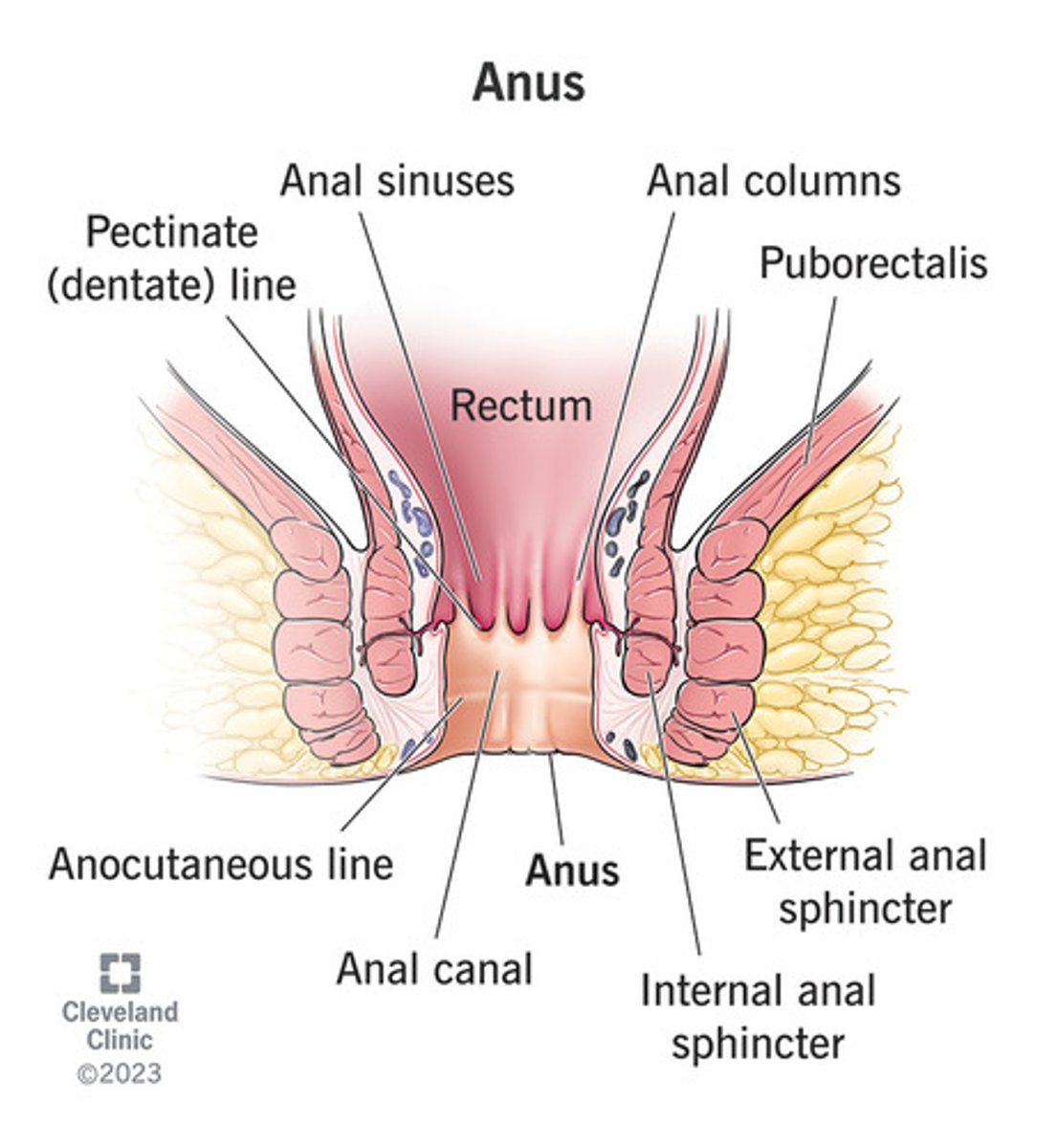
anal orifice
opening of the anus

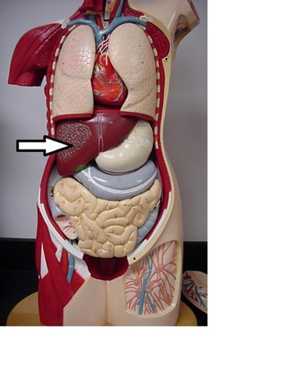
Accessory Organs
help aide in digestion, but no food passes through
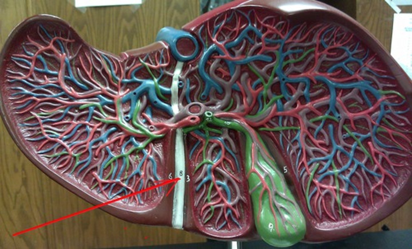
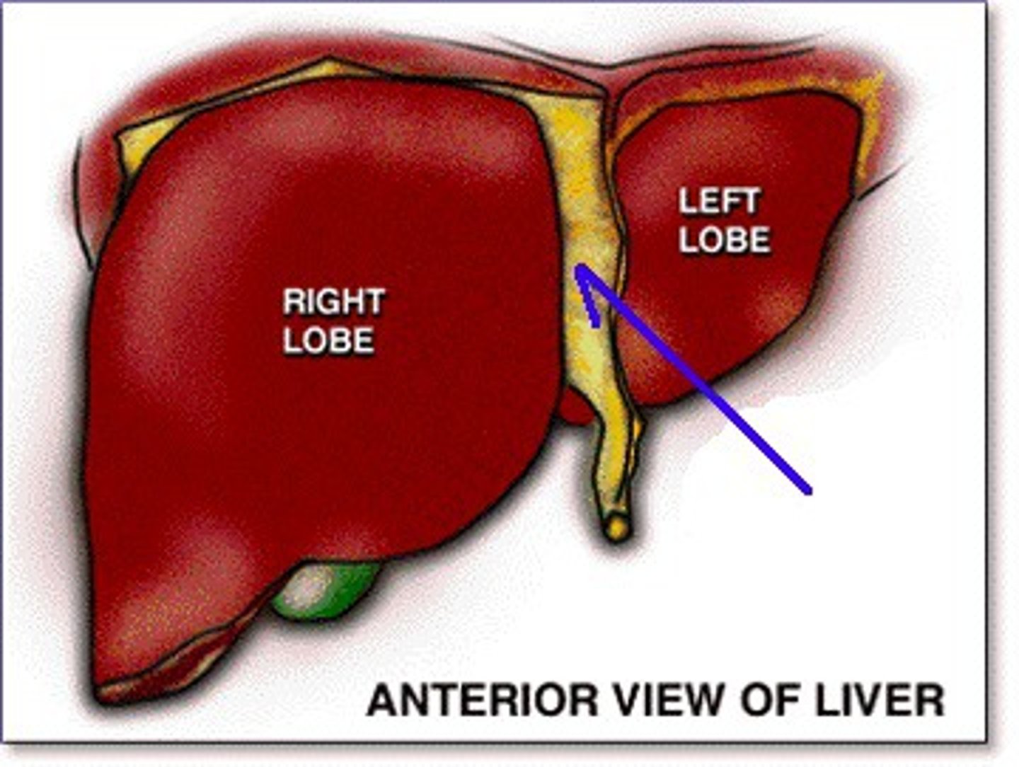
liver
4 lobes
Reticular Connective Tissue
right lobe
largest lobe of the liver
left lobe
smaller lobe of liver
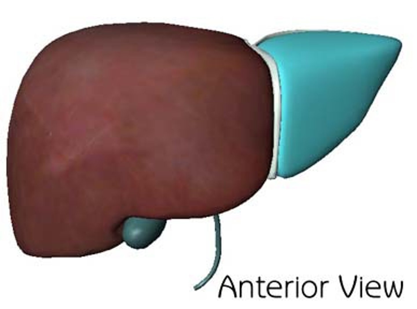
quadrate lobe
the medial segment of the left lobe. anterior and cube shaped
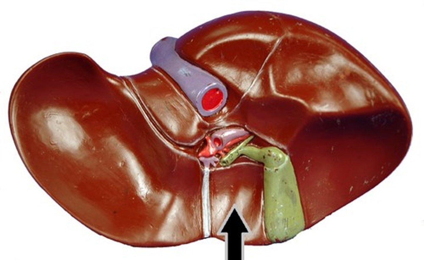
caudate lobe
smallest lobe of the liver situated on the posterior side
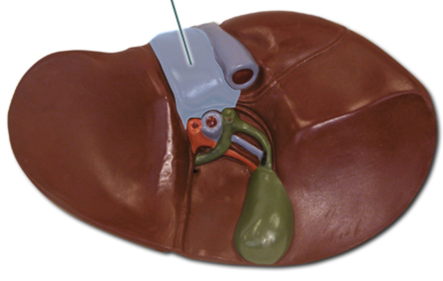
falciform ligament
divides right and left
coronary ligament
attaches liver to diaphragm. closer to heart.
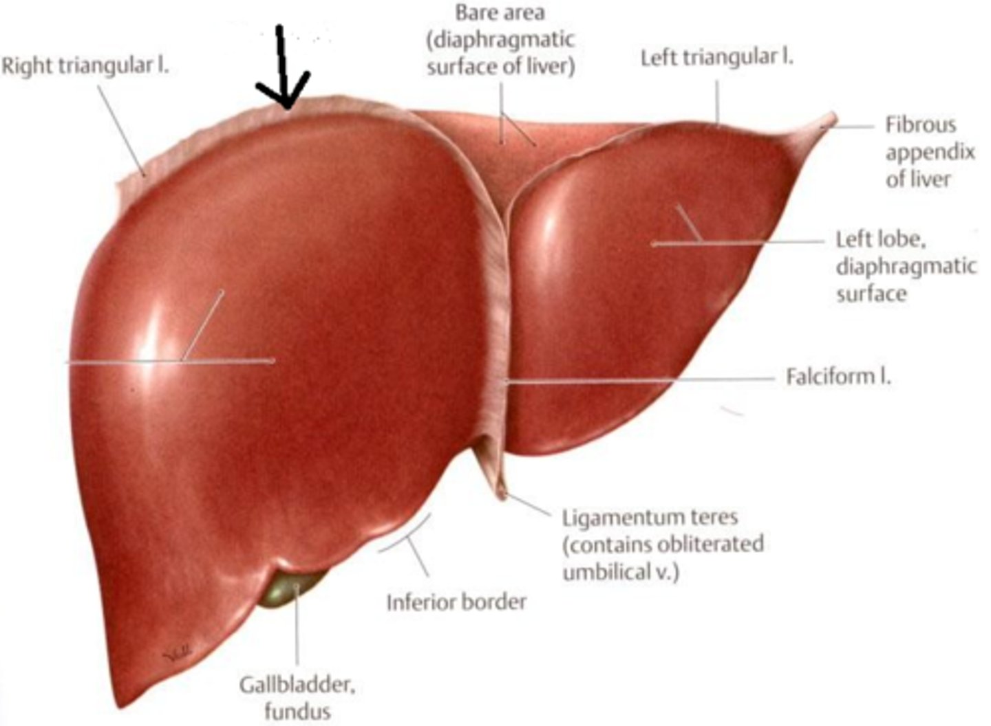
ligamentum teres (round ligament)
fetal reminent of umbilical vein