Articulation, Resonance, and Ingestion
1/133
There's no tags or description
Looks like no tags are added yet.
Name | Mastery | Learn | Test | Matching | Spaced | Call with Kai |
|---|
No analytics yet
Send a link to your students to track their progress
134 Terms
Articulation
process of bringing mobile and immobile articulators into contact to shape the sounds of speech
Resonation
the sound quality given to voiced sounds by the vocal tract
Deglutition
swallowing
Mastication
chewing
Vocal Tract
Larynx creates voicing —> pharynx —> oral/nasal cavity
Mobile articulators
lips, tongue, mandible, soft palate (velum), pharynx, cheeks, and fauces
immobile articulators
alveolar ridge, hard palate, teeth
velum (soft palate)
changes direction of air flow to oral or nasal cavities
pharynx
moves to change resonance and shape of sound
fauces
alveolar ridge of maxillae
ridge behind teeth
hard palate (palatine processes of the maxilla and horizontal plates of the palatine bones)
roof of mouth
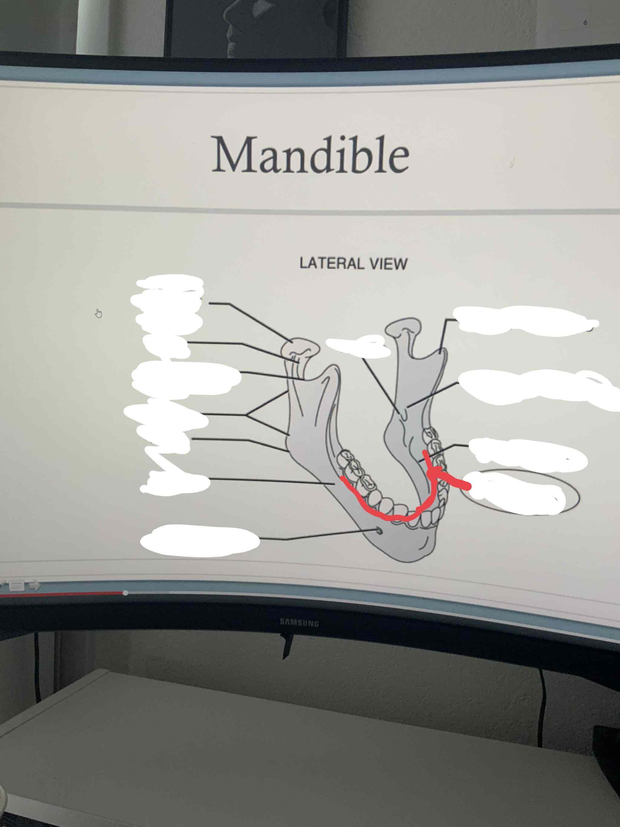
Mandible
lower jaw. uses large movements for chewing, small movements for speech
Condylar Process
articulates with the temporal bone forming the temp oro mandibular joint. top connector of the mandible
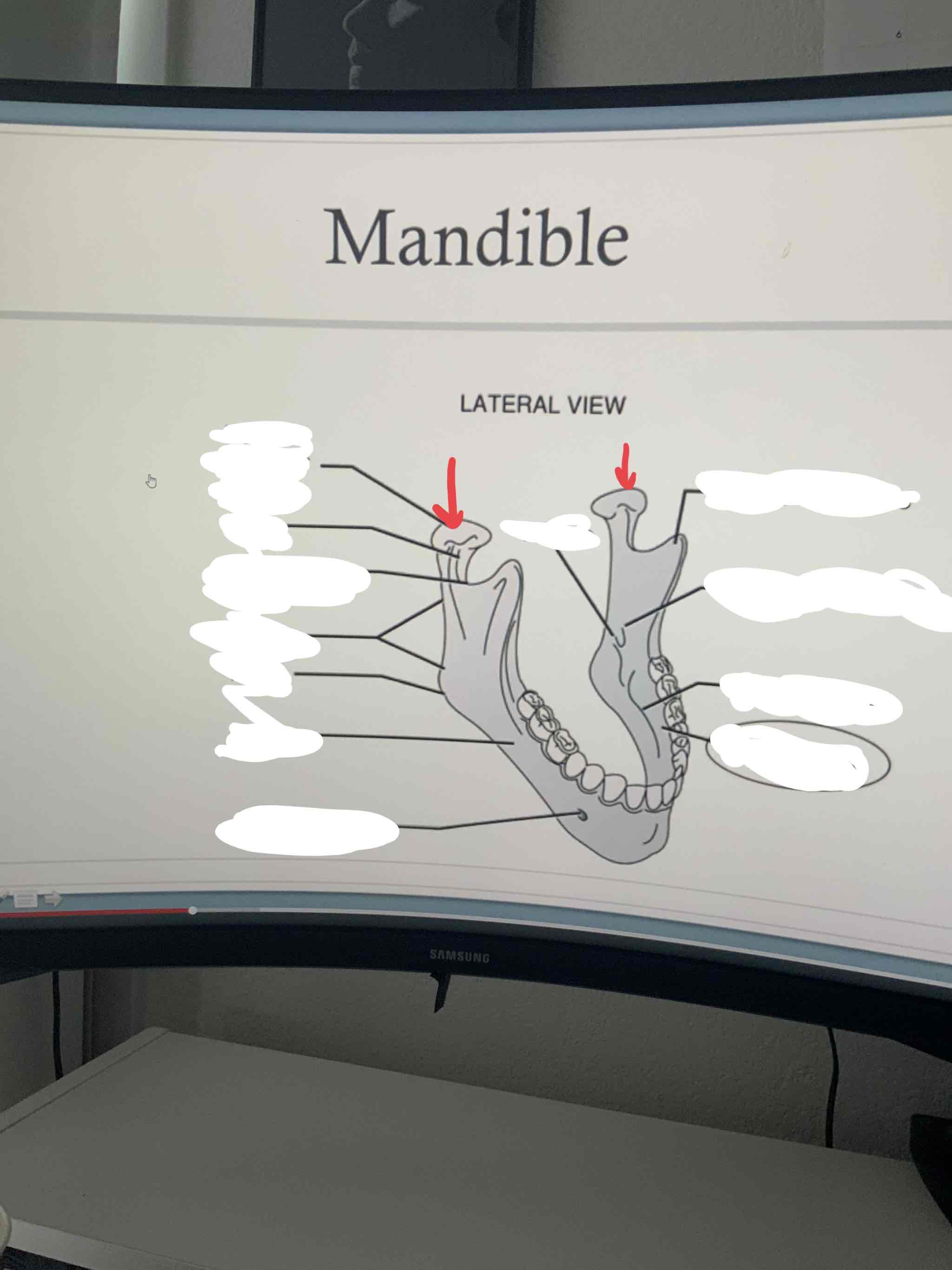
Coronoid Process
point of attachment for temporalis muscle
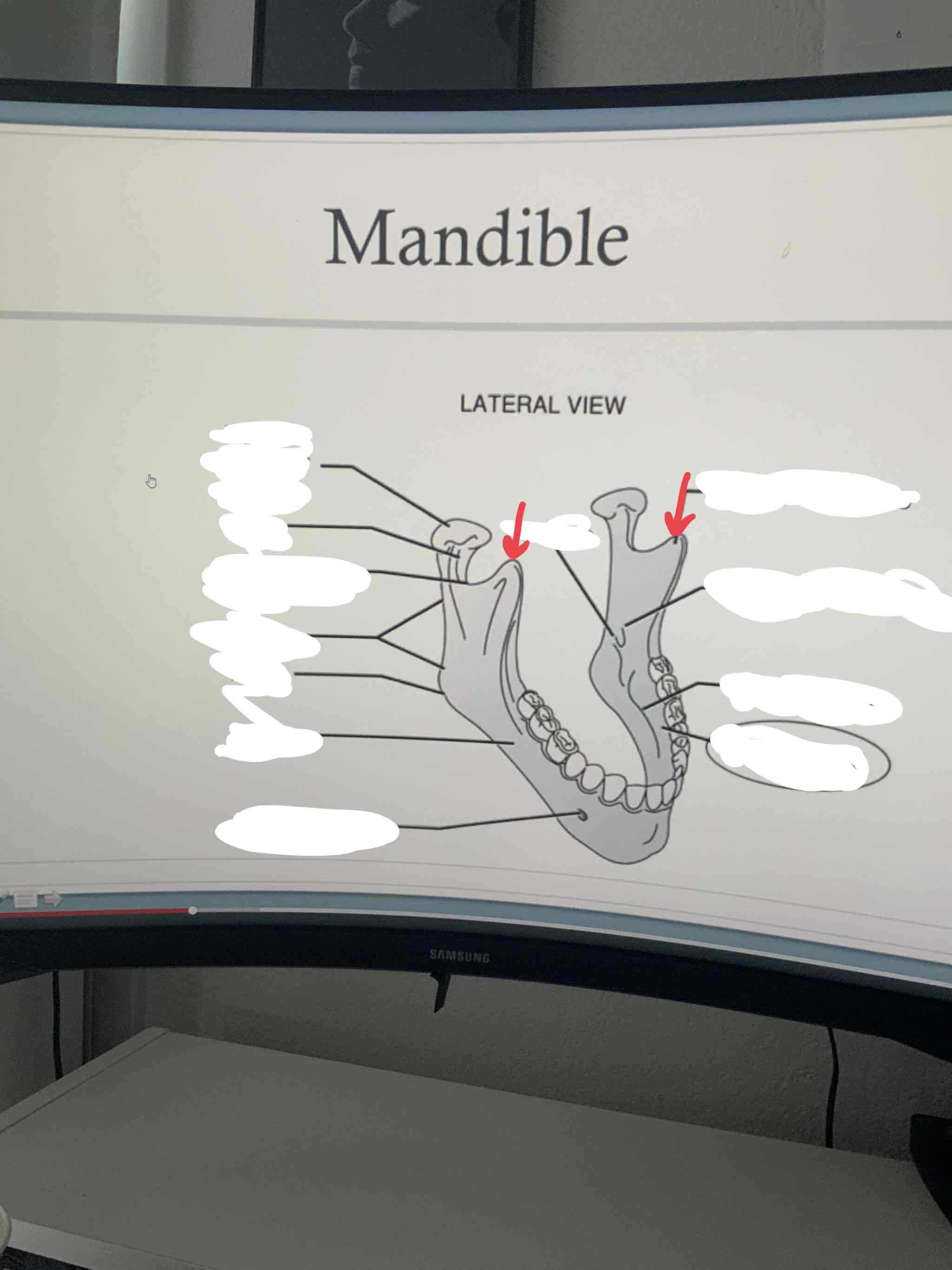
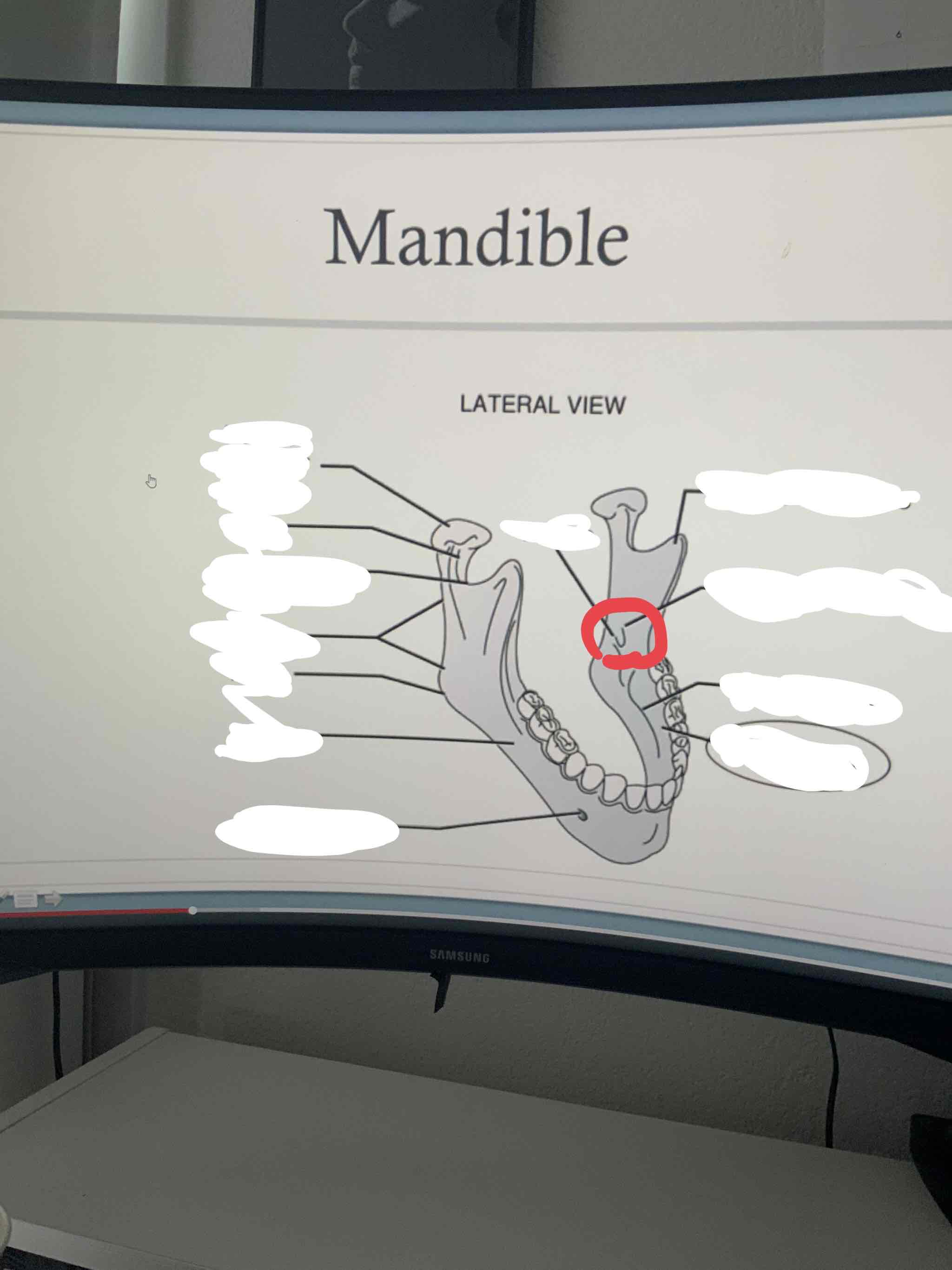
mandibular foramen
allows passage for inferior alveolar nerve (provides sensation for lower teeth)
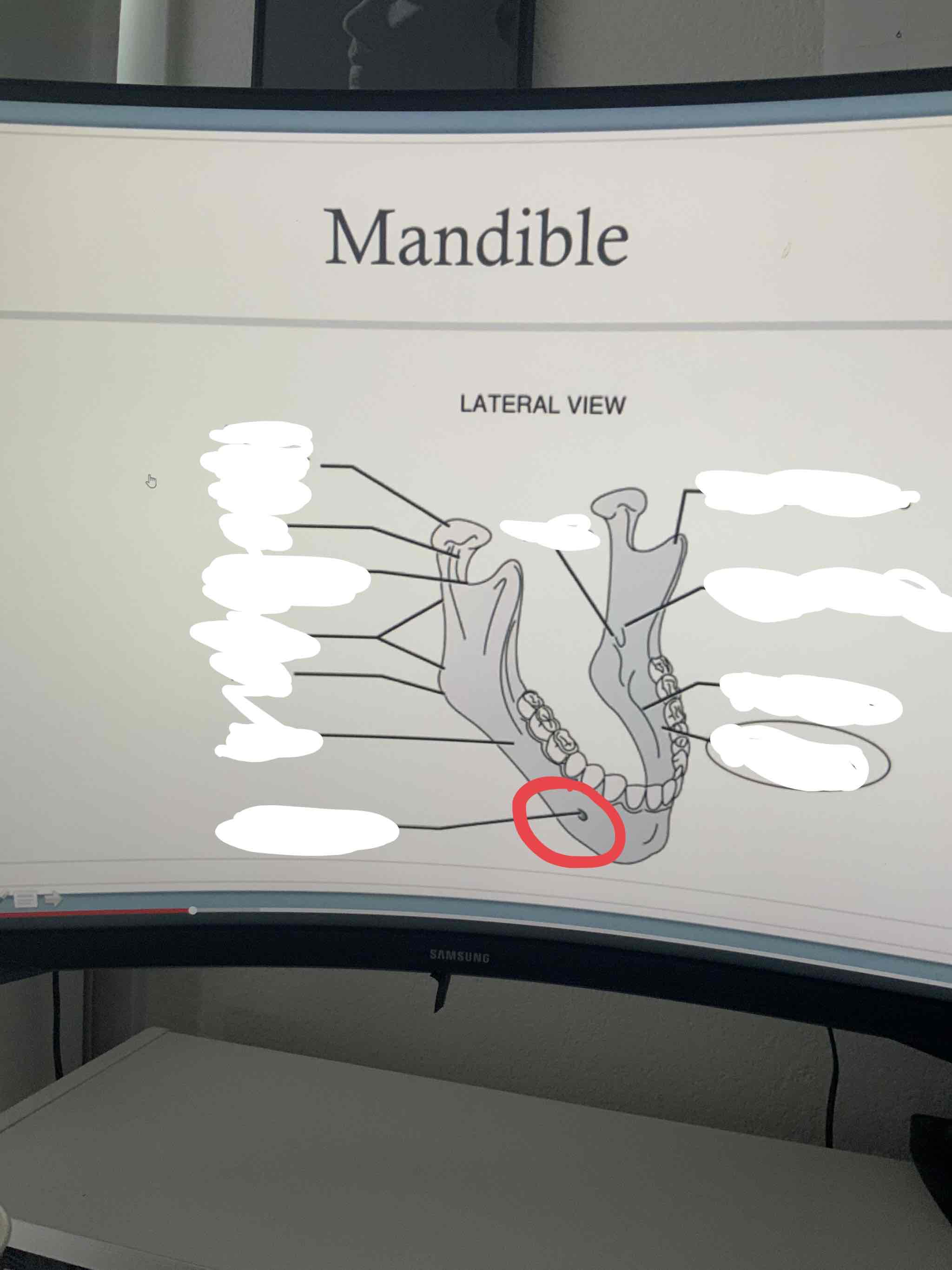
Mental Foramen
allows passage of the mental nerve, which supplies sensation to the chin and lower lip
corpus (mandible)
body of the mandible
angle (mandible)
point between the vertical ramus and horizontal corpus. where the mandible starts to turn

ramus
the vertical branch of the mandible
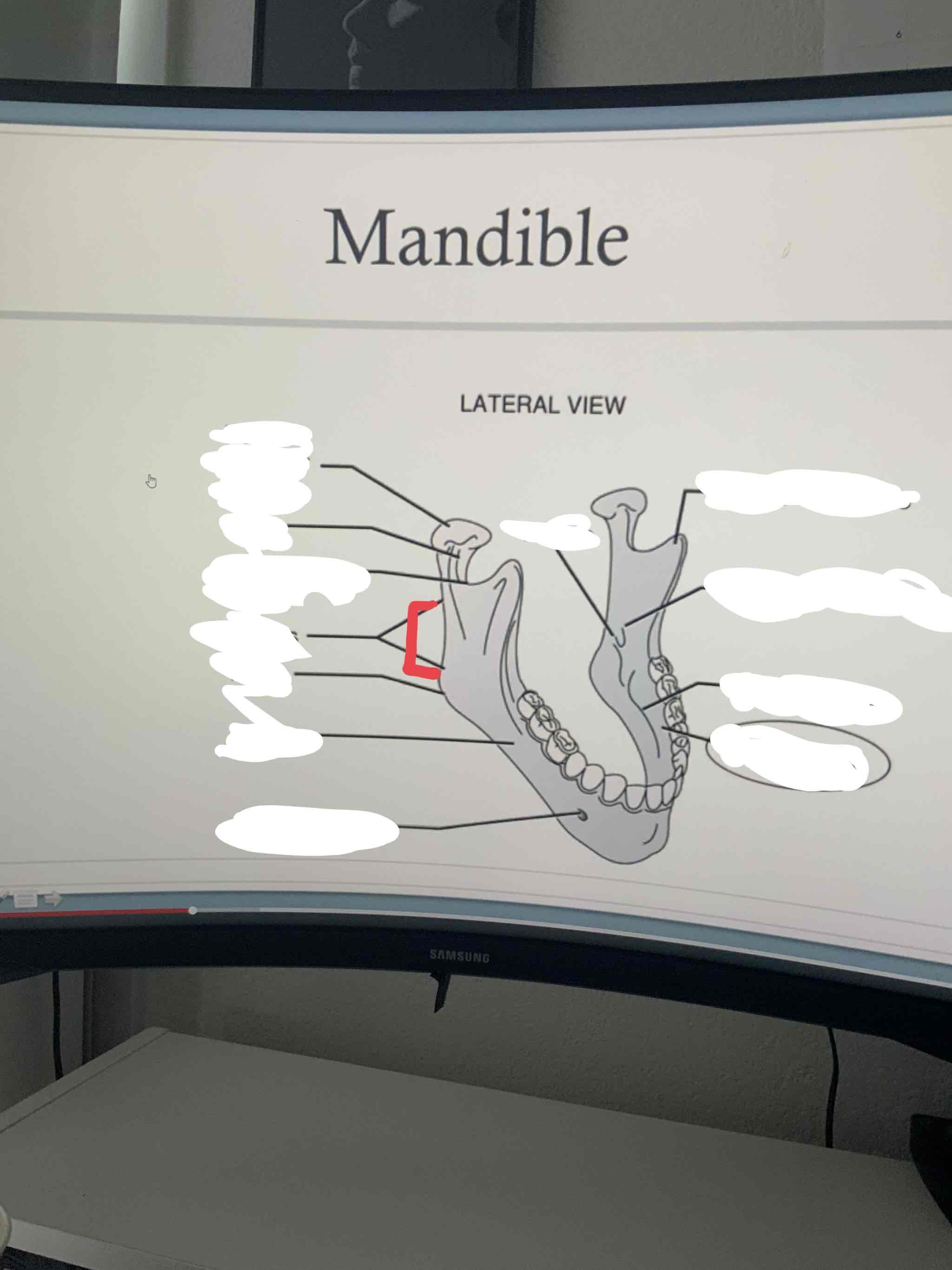
alveolar part (ridge) or (process)
houses the teeth in dental alveoli (sacs)
Maxillae
front of face and upper jaw
frontal process
articulates with the frontal bone
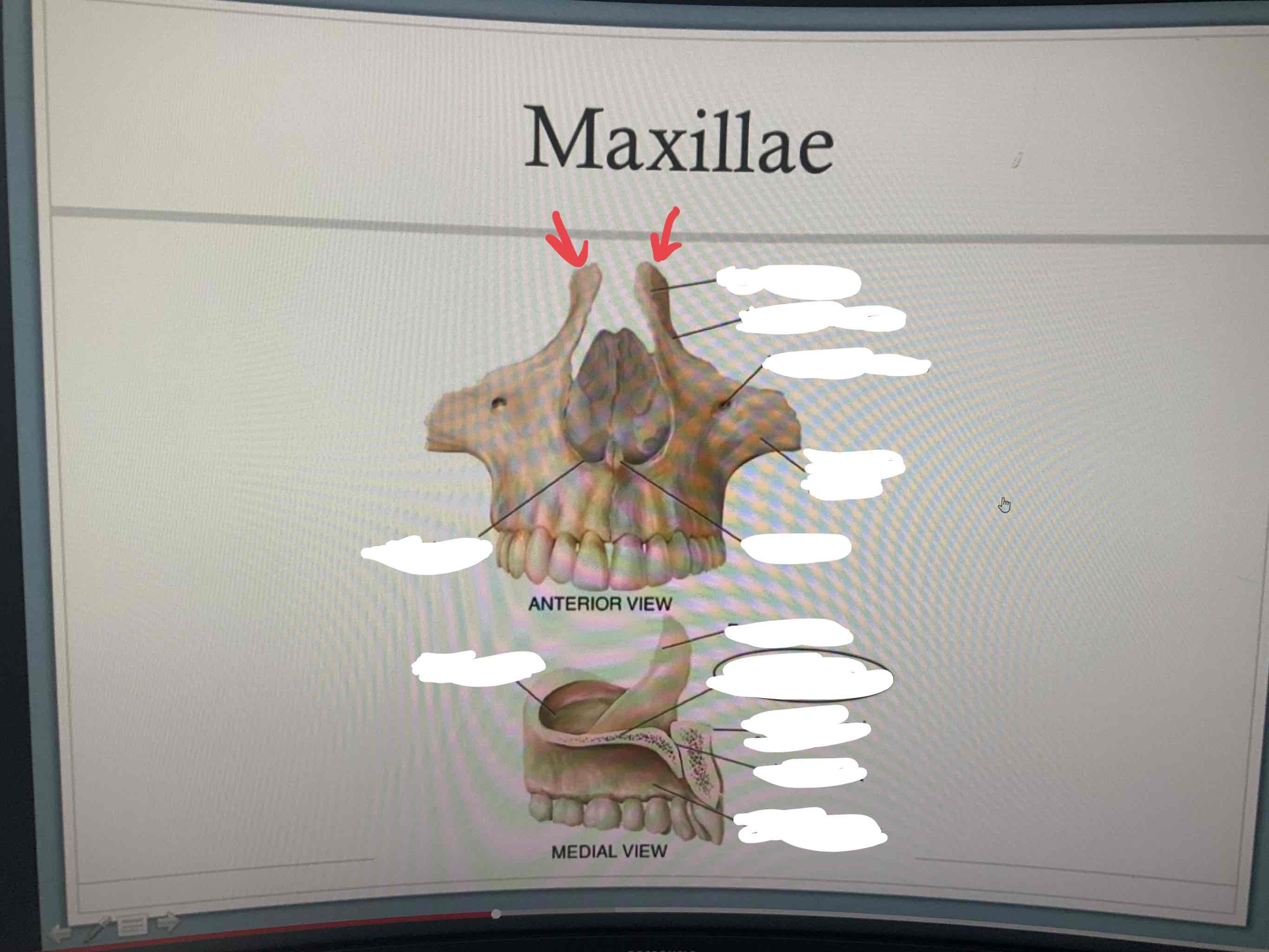
zygomatic process
articulates with the zygomatic bone
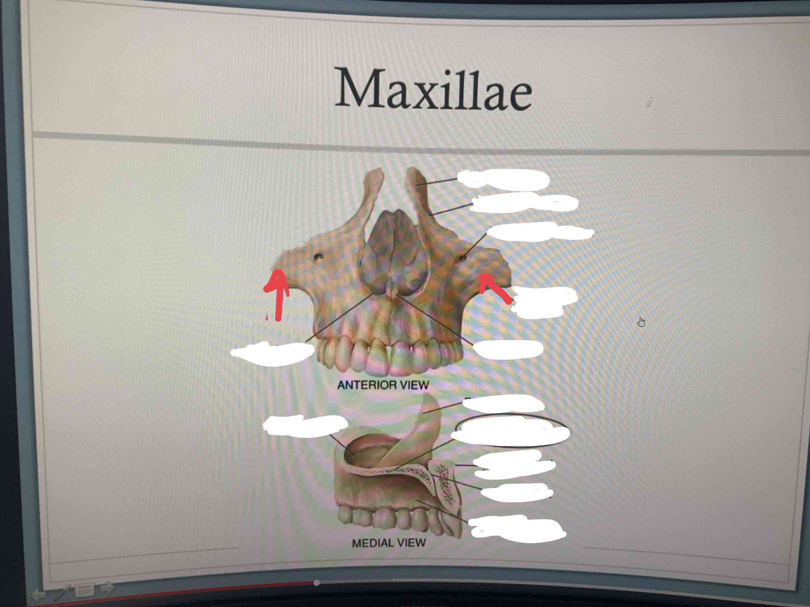
Palatine process
forms the floor of the nasal cavity and the anterior ¾ of the hard palate (roof of mouth
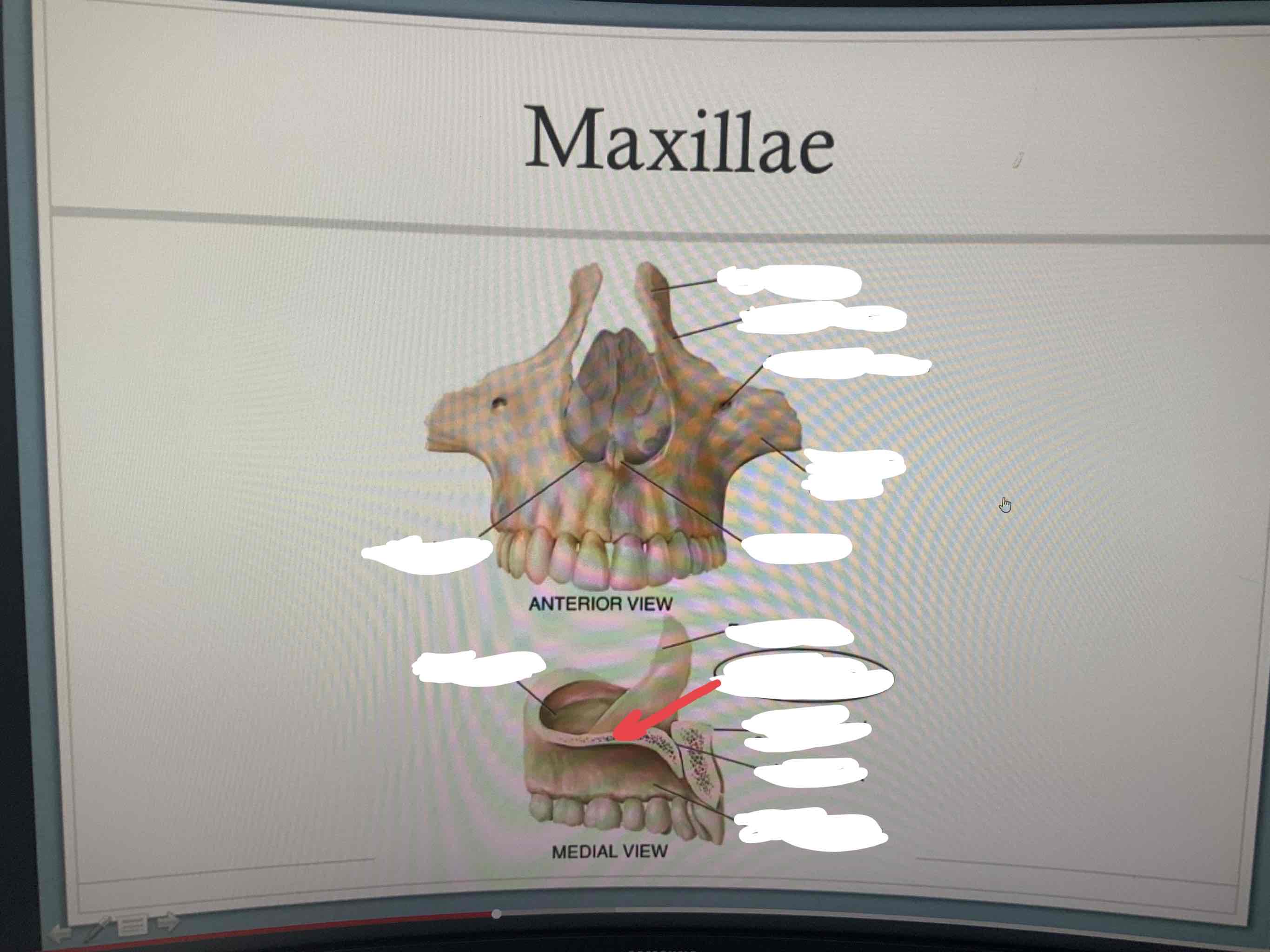
alveolar process
houses teeth

premaxilla
anterior section of the maxillae

Incisive foramen
marks the connection of the premaxilla and both palatine processes

transverse palatine surture
suture between palatine processes of maxillae and horizontal plates of palatine bones
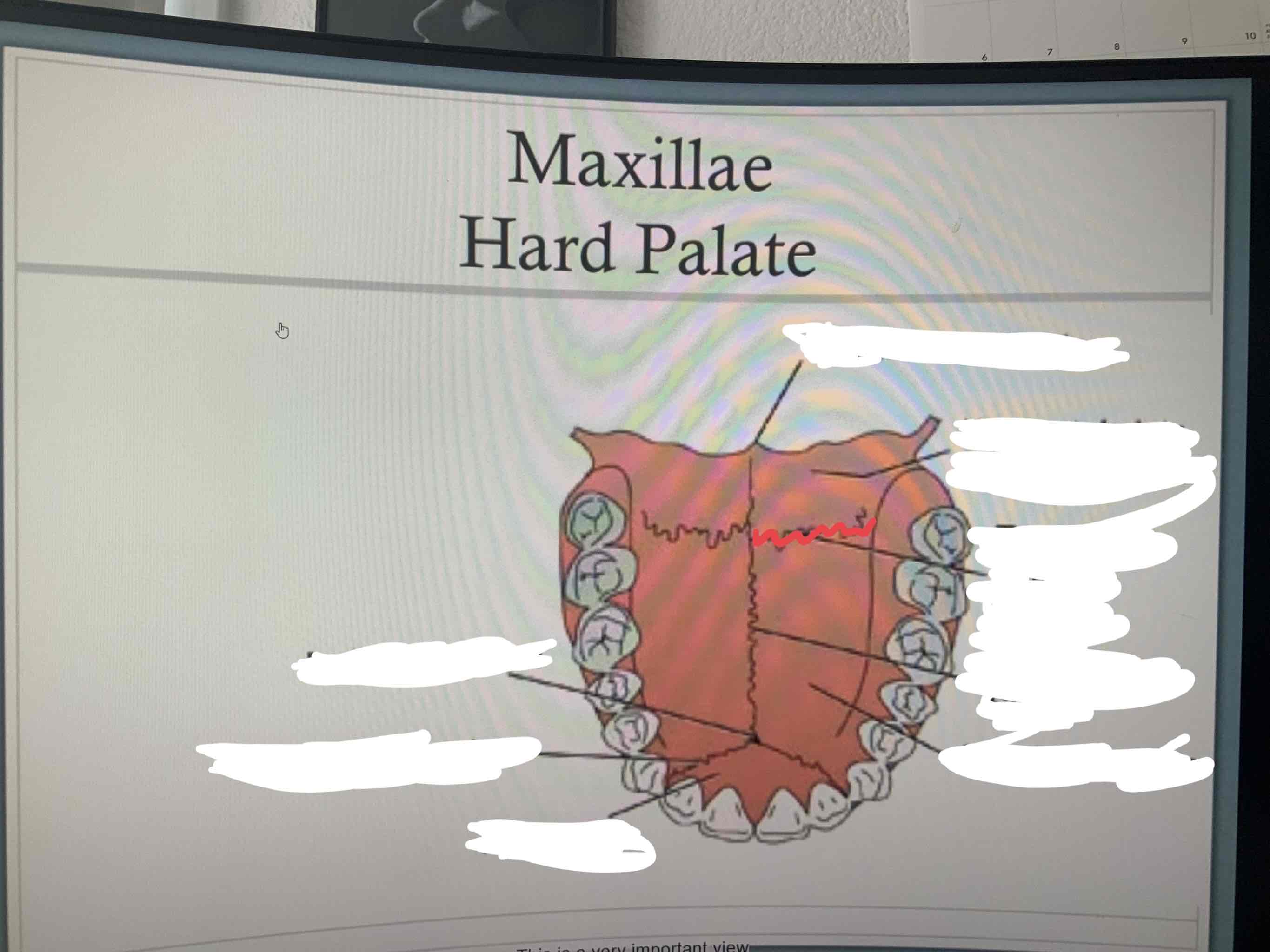
intermaxillary suture
suture between palatine processes. hole for cleft palate happens around here

premaxillary suture
between palatine processes and premaxilla. cleft lip happens around here

palatine bones
horizontal plates — makes up posterior ¼ of hard palate. located posterior to the maxillae
nasal bones
small bones that form the bridge of the nose
inferior nasal conchae
small scroll like bones located on the lateral walls of the nasal cavity. increases the surface area for warming and humidifying air
vomer bone
contributes to the formation of the nasal septum along with the perpendicular plate of the ethmoid bone and septal cartilage
zygomatic bone
cheek bones
lacrimal bones
smallest facial bones, contribute to the formation of the medial wall of the orbital cavity. medial edge of eye sockets
crista galli
extends superiorly from the ethmoid bone into the cranial space
cribriform plate
part of ethmoid bone that separates the cranial cavity from the nasal cavities and allows passage of olfactory nerves
superior nasal concha
part of ethmoid bone that increases surface area to warm and humidify incoming air (superior portion)
middle nasal conchae
part of ethmoid bone that increases surface area to warm and humidify incoming air (middle portion)
perpendicular plate
part of ethmoid bone that contributes to the structure of the nasal septum
hypophyseal fossa (sella tursica)
part of sphenoid bones that houses the pituitary gland
lateral pterygoid plates
part of sphenoid bone. point of attachment for muscles of mastication (lateral portion)
medial pterygoid plates
part of sphenoid bone. point of attachment for muscles of mastication (medial portion)
supraorbital margin
part of the frontal bone. contributes to the orbital cavity
zygomatic process
part of the frontal bone. articulates with the zygomatic bone
coronal suture
suture between the frontal bone and parietal bones
squamosal suture
suture between the temporal and parietal bones
sagittal suture
suture between both parietal bones
lambdoidal suture
suture between occipital and parietal bones
craniosynostosis
when the sutures of the skull become ossified prematurely. can affect one or more of the sutures. often requires surgery to separate the fused bones. may or may not be associated with other genetic syndromes. can affect brain development/learning
foramen magnum
part of the occipital bone. allows passage of the spinal cord to the brainstem
condyles
part of occipital bone. resting point for the first cervical vertebra
external auditory meatus
part of temporal bone. ear canal
zygomatic process (temporal)
articulates with the temporal process of zygomatic bone to form zygomatic arch
Mandibular fossa
part of temporal bone. articulates with mandibular condyle to form temporomandibular joint
styloid process
part of the temporal bone. point of attachment for various muscles
mastoid process
part of the temporal bone. point of attachment for various muscles of the neck
petrous part
part of the temporal bone. houses the organs of hearing (cochlea) and equilibrium (semicircular canals)
deciduous teeth
shedding teeth (baby teeth) 10 in each arch
successional teeth
permanent teeth. 16 in each arch
dental occlusion
relationship of upper dental arch to lower dental arch when teeth come together
class I occlusion
normal
netrocclusion
first mandibular molar is one-half tooth anterior to the first maxillary molar
Class II Occlusion
Malocclusion
First mandibular molar is posterior to normal position
mandible is retracted
Class III Occlusion
Malocclusion
first mandibular molar is anterior to normal position
mandible is protruded
relative micrognathia
condition in which the mandible is small in relation to the maxilla. may be part of other genetic syndromes. may interfere with infant feeding. may cause abnormal alignment of the teeth
open bite
anterior teeth do not occlude (do not touch when you bite down) because of excessive eruption of posterior teeth
closed bite
posterior teeth do not occlude because of excessive eruption of anterior teeth
rugae
folds of tissue on the hard palate
soft palate (velum)
moves posteriorly to seperate oropharynx from nasopharynx
palatine tonsil
located between the two faucial pillars
uvula
aids in velopharyngeal closure. may aid in speech sounds (non-english)
buccal cavity
space between the teeth and cheeks
nasopharynx
space above the soft palate
eustachian tube
provides aeration of the middle ear
pharyngeal tonsil (adenoid)
where the velum reaches to in the nasopharynx
oropharynx
area posterior to the fauces, between the soft palate and hyoid bone
laryngopharynx
area bounded by the hyoid bone, epiglottis and esophagus
nasal cavity
filled with nasal conchae that aid in warming, moistening, and cleaning (filter) the air we breathe
philtrum
vertical groove located above the lips
philtril ridge
location of cleft lip
cleft lip
birth defect that causes a split or opening in the upper lip. leads to difficulties feeding in infancy. surgery will repair the lip. may need orthodontic care when older. mat or may not have speech difficulties
orbicularis oris
closes and protrudes lips. upper and lower. kissing muscle
risoirus muscle
retracts lips at the corners, aids in mastication and smiling. pushes food from cheeks and helps smile
buccinator
compresses the cheeks, moves food onto the surface of molars for mastication
levator labii superioris alaeque nasi
elevates upper lip from wing of nose
levator labii superioris
elevates upper lip
zygomatic minor
elevates upper lip from zygomatic bone
zygomatic major
elevates and retracts the angle of the mouth
levator anguli oris
pulls corners of mouth up and medially
mentalis muscle
depresses lower lip, pulls lower lip out. pout and wrinkle chin
depressor labii inferioris
depresses lower lip. pout
depressor anguli oris
depresses the corners of the mouth and helps compress the upper lip against the lower lip
platysma
depresses mandible
tip
anterior most portion of tongue
dorsum
superior surface. middle tongue
base
part of tongue located in the oropharynx
root
lower area of the tongue where it attaches to mandible
oral or palatine portion
2/3 of the tongue located in the oral cavity