Equine Lameness and Diagnostic Imaging
1/11
There's no tags or description
Looks like no tags are added yet.
Name | Mastery | Learn | Test | Matching | Spaced |
|---|
No study sessions yet.
12 Terms
Basic sequence of orthopaedic lameness examination
Physical examination (before trotting)
Heat
Swelling
Palpable pain
Wounds
Testing pain (manipulative tests)
Care needed → may exacerbate lameness
Not very area specific
Flexion tests
Thoroughbreds → flex carpus to test fractures or synovitis
Wedge/plank test
Hyperextend joint with wedge/plank
Often used for DIP → puts pressure on navicular apparatus
Varus/valgus stress test
Lameness identification
Which limb(s) - trotting/walking for lameness recognition patterns
Origin of discomfort - diagnostic imaging
Cause of pain - nerve blocks
Equine forelimb lameness pattern
Head nod
Sound limb weight bearing → head drops down
Lame limb weight bearing → head raises up
reduces load placed on painful limb
Equine hindlimb lamness pattern
Pelvic hike
Sound limb weight bearing → tuber coxae (ilium) drops down
Lame limb weight bearing → ilium hikes up
Shifting CoM towards sound limb
Additional features for lameness recognition
Not necessarily associated with gait symmetry
Asymmetry of <20-25% difficult to detect
Foot placement → heel → toe strides
Limb flight → affected by joint flexion
Joint angles
Stride length
Stance duration
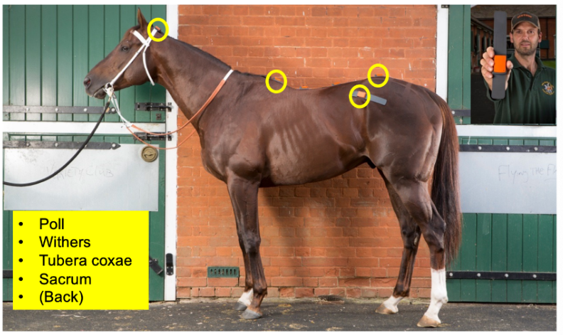
Objective lameness exam- location of inertial motion sensors [5]
Poll
Withers
Tuber coxae
Sacrum
Back
Mixed results from objective scanning
Example:
Sacrum sensor
Right hindlimb pushoff indicates lameness
Measurements show slight asymmetry to the right
Poll and wither sensors indicate contradicting results → mildly right and left forelimb lame
Indicates right forelimb lameness is referred due to right hindlimb lameness
contradiction means not primary lameness
Takeaway → can refer lameness from 1 leg to another
Multi limb lameness → always examine the most lame leg first
Laminitis can impact all 4 legs
Multilimb lameness
Lameness referred: fore → hind:
CONTRALATERAL side
Lameness referred: hind → fore:
IPSILATERAL side
Example presentation:
Asymmetrical hip hike
Extreme head nod
More likely that forelimb is primary
Peripheral nerve blocks (distal → proximal)
Roughly localise source of lameness
Abnormality found with imaging after localisation with block
Palmar digital nerve block (PIP) → removes sensation beyond fetlock
Abaxial sesamoid nerve block (MCP)
Palmar metacarpal/tarsal nerve block → removes sensation beyond metacarpal/tarsal
Palmar nerve block (between suspensory ligaments + ddft)
3 + 4} 4 point blocks - medial and lateral side
Low palmar/plantar digital block → navicular bursa and distal phalanx
Midpastern palmar/plantar digital block → DIP joint + all deep structures EXCEPT lamellar corium
Abaxial sesamoid block → PIP joint, distal sesamoidean ligaments, lamellar corium
Low palmar/plantar block → MCP/MTP joints and proximal sesamoids
4a → distal ends of both splint bones
4b → distal to communicating branch
DP → DIP → PIP → MCP/MTP
Examination - inside the foot
80% of lameness originates from foot
60-70% of these are foot abscesses
Inaccessible to palpation → requires hoof capsule imaging
ACCESSIBLE structures palpated:
Dorsal coffin joint capsule
Proximal coffin joint collateral ligaments
Collateral cartilages
Tendons (+ sheaths) in heel bulbs
Positive response to palmar digital nerve block
Contenders:
Soft tissue
Intra-articular synovial problem
PIP
DIP (dorsal/palmar synovial recesses)
Navicular bursa
All 3 separated by T ligament
DDFT
Additional nerve blocks
Dorsal DIP (coffin) block
Navicular bursa block (palmar/plantar)
Otherwise known as the Hickmans block
Navicular apparatus block between heel bulbs
Imaging
Radiographs
CT → dome
Standing CT
Ultrasonography
MRI
Nuclear scintigraphy
Radiographs can be sufficient for diagnosis → don’t trot horse
fractures → surgery
Acute laminitis → black air in hoof
Severe navicular bone disease
Extensive bone formation
Lytic areas
CT distal limb up to fetlock
Standing CT
Distal limb
also can be used for head and cranial neck
Ultrasonography → flexor tendons
Palmar aspect → look for central area of hypotenicity
Central area core lesion → haemorrhage and granulation tissue
Histology → irregular collagen and haematoma
Breakage or rupture of tendon
Fluid accumulation and irregular contours for bones (e.g. ilium wing stress fracture)
MRI
Do around distal limb
Stimulates tissue with radio frequency
Measures signal + convert to computerised image
Often used for inside hoof
Nuclear scintigraphy
Inject horse with radioactive substance binding to skeleton hydroxyapatite
2-3 hr later → measure radiation and convert to 2D image
Compare L/R for abnormalities