Intro to Physiology of Exercise Exam 2
1/255
There's no tags or description
Looks like no tags are added yet.
Name | Mastery | Learn | Test | Matching | Spaced | Call with Kai |
|---|
No analytics yet
Send a link to your students to track their progress
256 Terms
Functions of the skin
protection
temperature regulation
sensory perception
synthesis of vitamin D
excretion
absorption
Layer of the skin (deep-superficial)
subcutaneous tissue
dermis
epidermis
Epidermis
thinnest, outer layer
composed of epithelium
Dermis
thicker, inner layer
composed of connective tissue
Subcutaneous tissue
located beneath the dermis
it is not considered part of the skin but has very close proximity to the skin
Functions of epidermis
stratified squamous epithelium
avascular
high cell turnover
keratinized
protection
Cells in epidermis
keratinocytes
melanocytes
dendritic cells
tactile (Merkel) cells
Keratinocytes
Produces tough fibrous protein that waterproofs the skin and protects against abrasion
Melanocytes
Produce brown-black pigment
Dendritic cells
derived from monocytes (type of white blood cells)
remove pathogen and alert the system of threat
Tactile (Merkel) cells
Sensory input
Composition of epidermis (deep-superficial)
stratum basale
stratum spinosum
stratum granulosum
stratum luciderm
stratum corneum
Stratum basale
Innermost layer, cell division
Stratum spinosum
Keratinocytes produce keratin, adds strength
Stratum granulosum
Cells contain granules, cells go through apoptosis (programmed cell death)
Stratum luciderm
Only in thick skin and appears lucid (transparent), adds extra protection
Stratum corneum
20-40 layers of dead keratinized cells, protective barrier
Structure of dermis
dense connective tissue
collagen and elastic fibers
Components of dermis
blood vessels
nerves
sweat glands
sebaceous glands
hair follicles
sensory receptors
Structure of hypodermis
loose connective tissue and adipose tissue
fat storage
absorbs shock forces
insulates the body
Compare and contrast thick skin to thin skin
Thick skin: palms of hand/soles of feet, no hair follicles/has stratum lucidum
Thin skin: covers most of the body, contains hair follicles and sebaceous glands/no stratum lucidum
Accessory structures
sweat glands: eccrine & apocrine
oil glands: sebaceous glands
hair
nails
sensory receptors
skin color
temporary changes in skin color
Sweat glands: Eccrine glands
thermoregulation
produce watery sweat
activated during exercise
Sweat glands: Apocrine glands
activated during stress or emotional response
produces a thicker secretion
causes body odor
Oil glands: Sebaceous glands
associated with a hair follicle
secrete sebum into follicle
lubricates hair and skin
antimicrobial properties
Hair
composed of keratinized epidermal cells formed at base of the hair follicle
consists of hair shaft, hair root, hair follicle
associated structures: arrector pili muscle, sebaceous glands, sensory nerve endings
Nails
composed of hard keratin
parts of the nail include nail body, nail root, nail matrix, lunula, cuticle
protection (distal surface of fingers/toes
helps with manipulation
Sensory receptors
haor root plexus
mechanoreceptors
thermoreceptors
nociceptors
Skin color
determined by a combination of pigments, blood flow, and underlying tissue composition
3 pigments in the skin: melanin, carotene, hemoglobin
Erythema
redness due to increased blood flow
common during exercise or inflammation
Pallor
pale appearance from reduced blood flow
seen in shock, cold exposure, or dehydration
Cyanosis
bluish discoloration due to poor oxygen
medical concern
Jaundice
yellowing due to bilirubin buildup
sign of liver dysfunction
Thermoregulation
If body temperature increases:
radiation
conduction
convection
evaporation
If body temperature decreases:
involuntary muscle contractions (shivering)
blood vesicles constrict
During exercise how does the body maintain a stable body temperature?
during exercise muscle contracts and produces heat which causes the body’s core temperature to rise
cooling mechanisms cause vasodilation which activates your body to sweat
body temperature returns to normal
takes place mainly in the hypodermis nut can take place in dermis
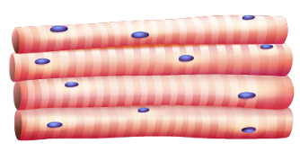
Skeletal muscle
striations
many nuclei/cell
long/parallel shaped cells
voluntary
attached to bones, dermis, ligaments, and other muscles
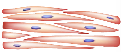
Smooth muscle
no striations
one nucleus/cell
short and tapered cells
involuntary
visceral organs, blood and lymphatic vessels, and skin
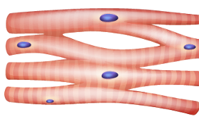
Cardiac muscle
striations
one nucleus/cell
short and branching cells
involuntary
heart
Hierarchy of muscle structure (largest to smallest)
Muscle → muscle fascicle → muscle fiber (cell) → myofibril → myofilament
Muscle fiber (cell) is surrounded by:
Sarcolemma
2 types of myofilaments
actin
myosin
Sarcolemma
Plasma membrane of a muscle fiber maintaining the integrity of the cell
Sarcoplasm
Cytoplasm of a muscle fiber that contains organelles
Nuclei
Contain DNA, which determines cell structure and function
Sarcoplasmic reticulum
Smooth ER in a muscle fiber that stores calcium
Transverse tubules
Extensions of the sarcolemma that penetrate into the sarcoplasm carrying electrical impulses, which trigger the release of the calcium from the sarcoplasmic reticulum
Myofibril
A bundle of myofilaments
Myofilaments
Threadlike contractile proteins that interact to produce contractions
Sarcomere
Contractile unit of the muscle fiber

Sarcomere (z line)
End of sarcomere, actin is attached on both sides

Sarcomere (I band)
Only actin

Sarcomere (A band)
Spans the length of myosin (doesn’t change length)
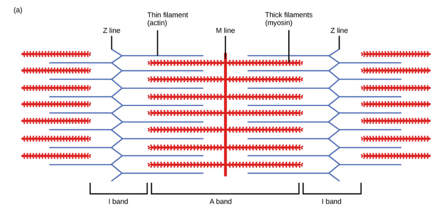
Sarcomere (H band)
Only myosin

Sarcomere (M line)
Center line of sarcomere (doesn’t move)

Actin
2 strands arranged in a double helix
troponin
tropomyosin
thin filament

Myosin
globular heads with a hinge point and fibrous tail
thick filament
moves like a windshield wiper
Motor unit
large motor units: strength
small motor units: precision
Action potential
Electrical stimulation
Neuromuscular junction
The place where a motor neuron meets a muscle fiber and sends a signal to make the muscle contract
Synaptic cleft
The tiny gap between the nerve cell and the muscle cell at the neuromuscular junction
Acetylcholine (ACh)
A neurotransmitter (chemical messenger) that carries the signal from the nerve to the muscle to start contraction
Sliding filament model
The theory that muscles contract because actin and myosin filaments slide past each other, shortening the muscle
All or nothing principle
A muscle fiber either contracts completely or not at all when stimulated — there is no partial contraction of a single fiber
Phases of muscle contraction
latent phase: the short delay between the stimulus and the start of contraction
contraction phase: the muscle shortens and generates force
relaxation phase: the muscle returns to its resting length
Origin
The attachment point of a muscle that stays relatively still during movement
Insertion
The attachment point of a muscle that moves during contraction
Agonist
The main muscle responsible for a movement (prime mover)
Antagonist
The muscle that opposes or reverses the movement of the agonist
Isotonic
A contraction where the muscle changes length and movement occurs.
Concentric = muscle shortens
Eccentric = muscle lengthens
Isometric
A contraction where the muscle produces force but does not change length (no movement)
Flexion
Decrease in the angle of the bones forming the joint
Extension
Increase in the angle of the bones forming the joint
Hyperextension
Increase in the angle of the bones forming the joint beyond the anatomical position
Dorsiflexion
Flexion of the foot at the ankle
Plantar flexion
Extension of the foot at the ankle
Abduction
Movement of a bone away from the midline
Adduction
Movement of a bone toward the midline
Rotation
Movement of a bone around its longitudinal axis
Medial rotation
Rotation of a limb so its anterior surface turns medially
Lateral rotation
Rotation of a limb so its anterior surface turns laterally
Circumduction
Movement of the distal end of a bond in a circle while the proximal end forms the pivot joint
Eversion
Movement of the sole of the foot laterally
Inversion
Movement of the sole of the foot medially
Pronation
Rotation of the forearm when the palm is turned inferiorly or posteriorly
Supination
Rotation of the forearm when the palm is turned superiorly or anteriorly
Protraction
Movement of the body part posteriorly
Retraction
Movement of the body part anteriorly
Elevation
Movement of a body part superiorly
Depression
Movement of a body part inferiorly
Opposition
Movement of the thumb to touch the other 4 fingers
Reposition
Movement of the thumb back to the anatomical position
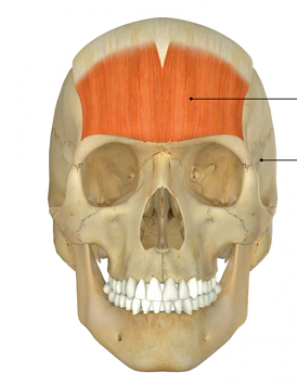
Epicranius frontalis
Epicranius frontalis
Origin: Epicranial aponeurosis
Insertion: Skin and muscles above the eye
Action: Raises eyebrow and wrinkle forehead horizontally
Orbicularis oculi
Origin: frontal bone and maxilla
Insertion: skin around the eye
Action: closes eye
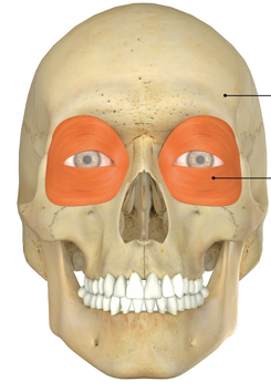
Orbicularis oculi
Orbicularis oris
Origin: muscles around the mouth
Insertion: skin around the lips
Action: closes and puckers lips; shapes lips during speech
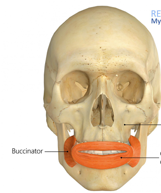
Orbicularis oris
Masseter
Origin: Zygomatic arch
Insertion: Lateral surface of mandible
Action: Raises mandible
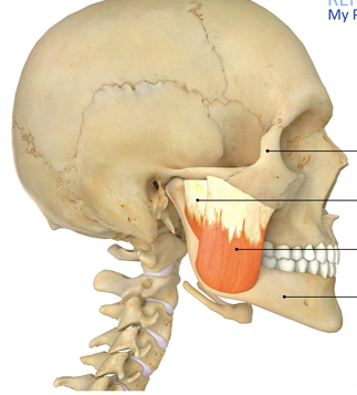
Masseter
Temporalis
Origin: Temporal bone
Insertion: Coronoid process of mandible
Action: Raises mandible