BIOL 319 Lab 9
1/110
There's no tags or description
Looks like no tags are added yet.
Name | Mastery | Learn | Test | Matching | Spaced | Call with Kai |
|---|
No analytics yet
Send a link to your students to track their progress
111 Terms
cerebrum
pons
medulla

cerebellum

spinal cord
thalamus
occipital lobe

pineal gland

fourth ventricle
cerebellum
spinal cord
parietal lobe
corpus callosum

septum pellucidum
frontal lobe
hypothalamus
optic chiasm

pituitary gland
pons
medulla oblongata
cerebrum
largest region of the brain
outer granular layer
mostly made up of stellate cells, axons, and dendrites
outer pyramidal cell layer
mostly made up of pyramidal cells that increase in size as you move deeper into the layer
inner granular layer
mostly made of densely packed stellate cells
inner pyramidal and polymorphic layer
mostly composed of large pyramidal cells in the more superficial portion of the layer and a wide variety of cell morphologies in the deepest parts of the layer
longitudinal fissure
deep groove that divides the cerebrum into left and right hemispheres
Lateralization
specialization of cortical functions between the 2 hemispheres; look exactly alike but have different functions
gray matter
composed of unmyelinated neurons
gyrus
Fold or ridge on the surface of the cerebral cortex
molecular layer
contains mainly dendrites synapsing with cortical neuron axons
sulcus
a shallow groove in the cerebral cortex
corpus callosum
band of white matter that connects the cerebral hemispheres
gray matter forms
the outer convoluted surface of the cerebrum and the foliated surface of the cerebellum
white matter lies
deep to the cerebral and cerebellar cortices
cortical gray matter
made of multipolar neuron cell bodies and attendant dendrites
pyramidal cells
most of the multipolar neurons of the cortex are classified as _____ due to the triangular shape of their cell bodies
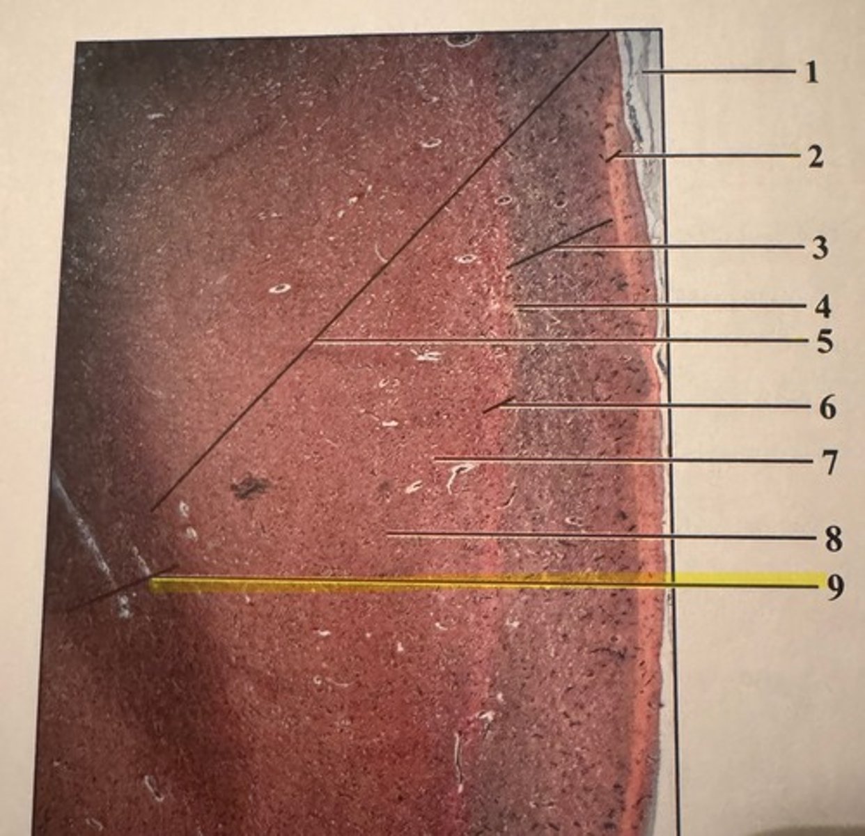
cerebral cortex layers
1. molecular layer
2. outer granular layer
3. outer pyramidal cell layer
4. inner granular layer
5. inner pyramidal and polymorphic layer
meninges

molecular layer

outer granular layer

pyramidal cells

gray matter
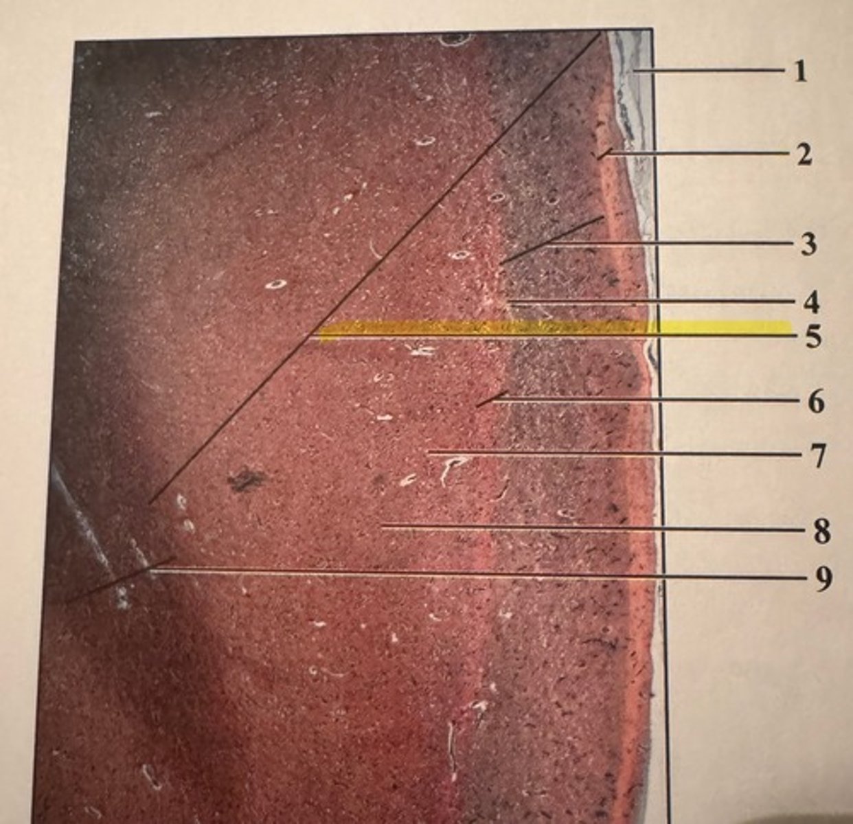
inner granular layer

inner pyramidal cells
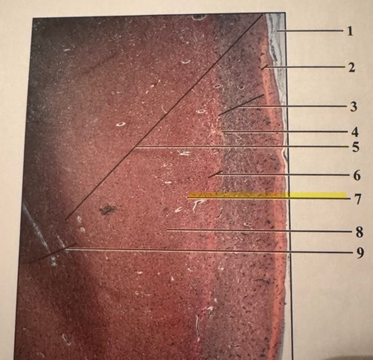
polymorphic cells
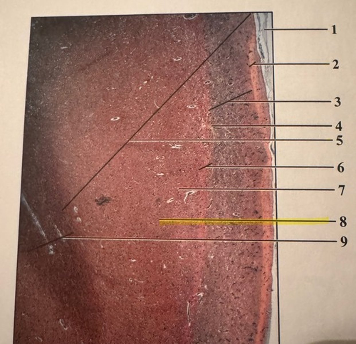
white matter
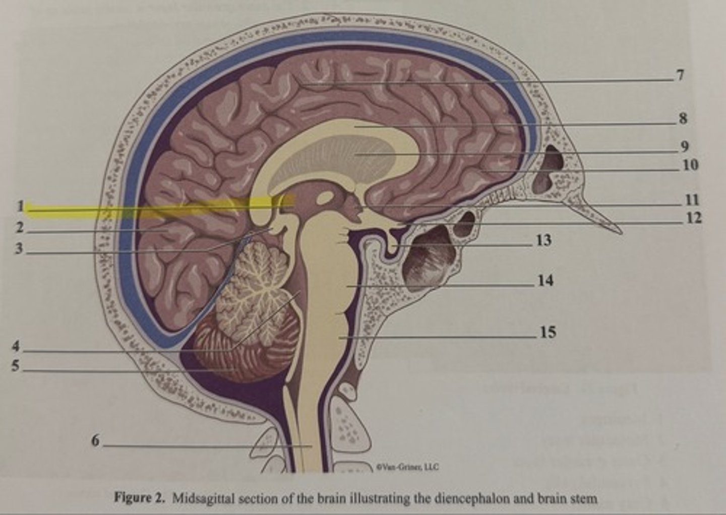
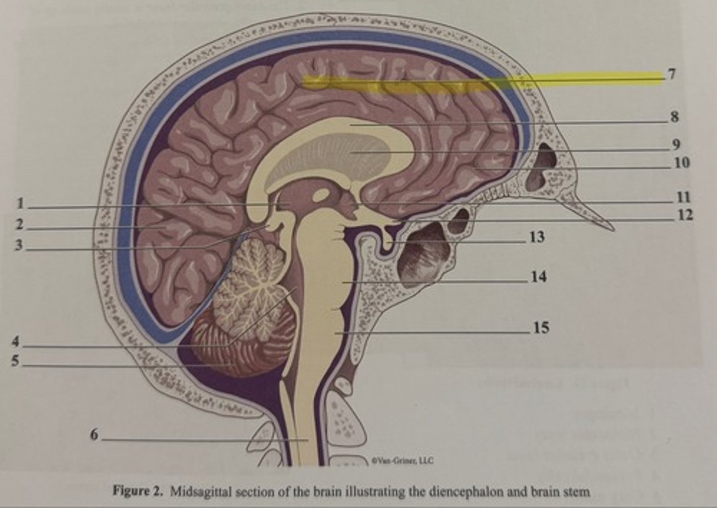
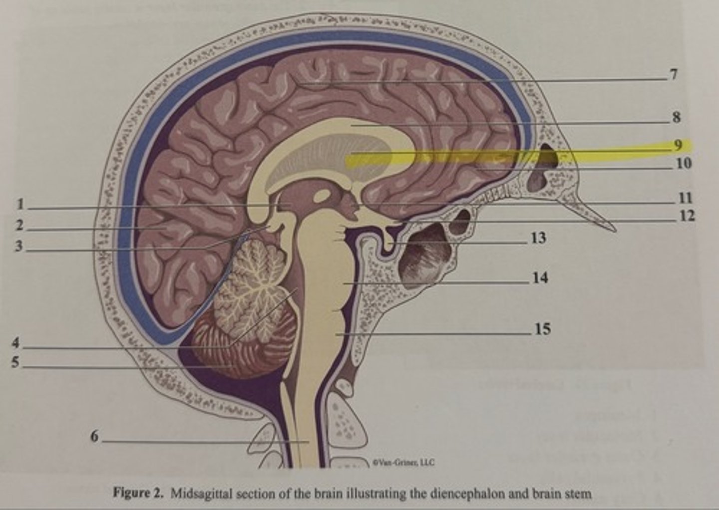
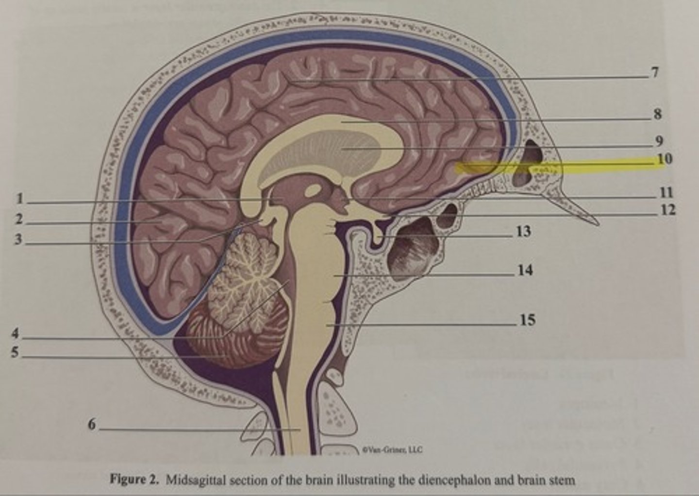
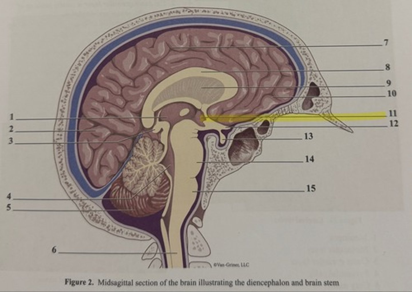
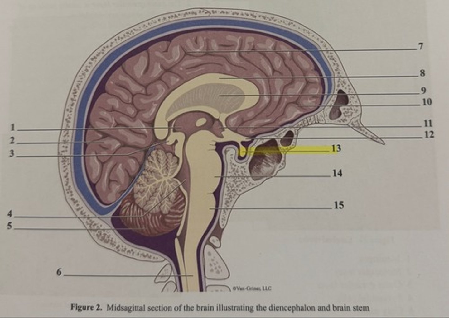
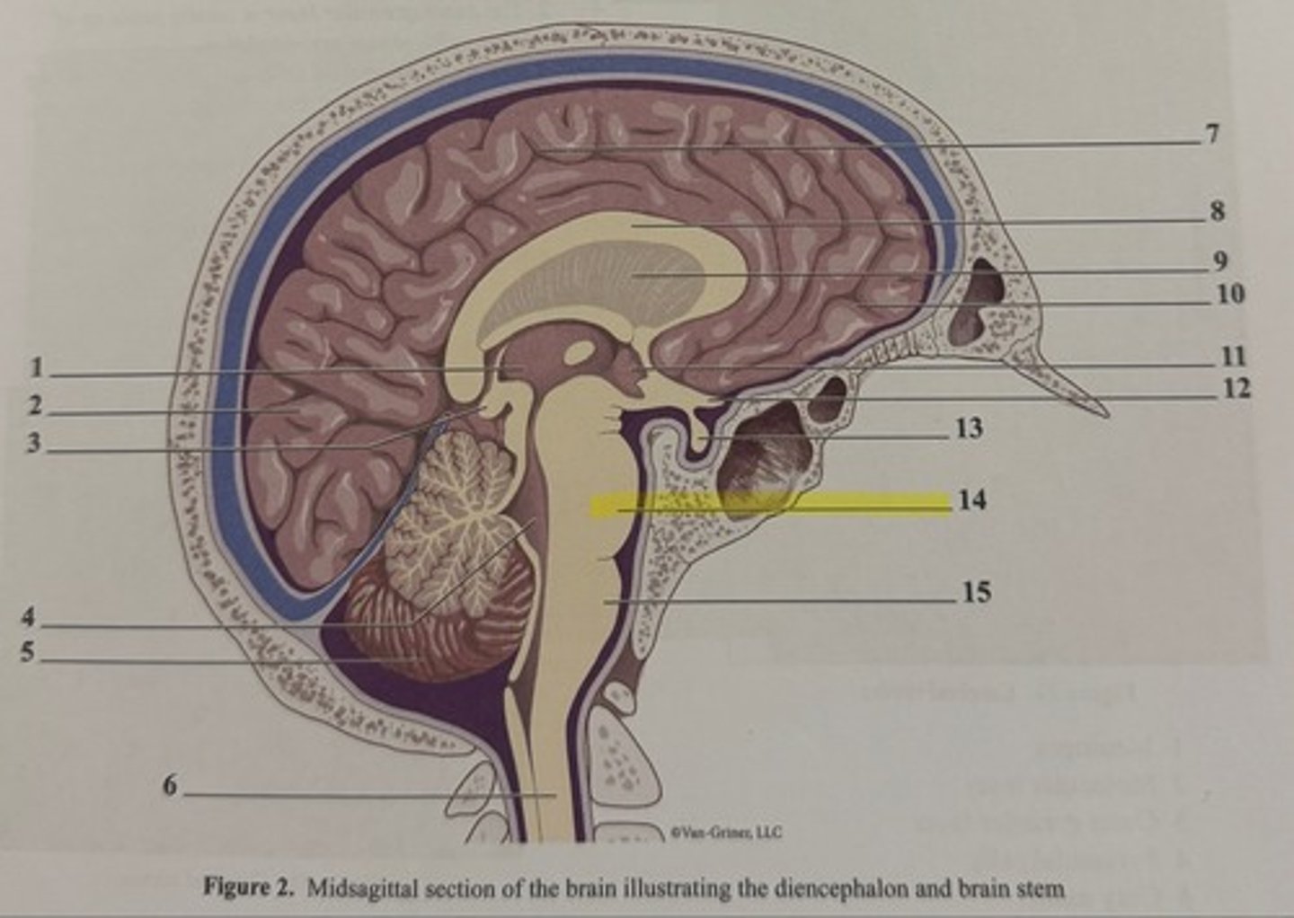
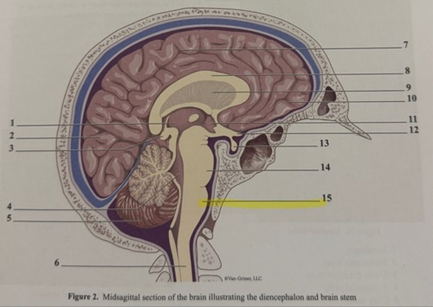
Diencephalon components
thalamus, hypothalamus, epithalamus
-gray matter areas that enclose the 3rd ventricle
thalamus
relay station for incoming sensory or integration information
hypothalamus
autonomic control center, center for emotional response, body temp regulation, regulation of food intake, regulation of water balance and thirst, regulation of sleep-wake cycles, and control of endocrine system functioning
mammillary bodies
relay stations in the olfactory pathways
infundibulum
a stalk of hypothalamic tissue that connects to the pituitary gland
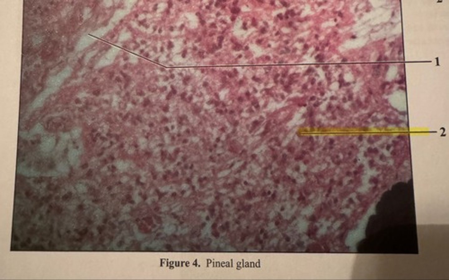
epithalamus
contains the pineal gland
pineal gland
secretes melatonin, which helps regulate the sleep-wake cycles
suprachiasmatic nucleus (SCN)
located in the hypothalamus; controls pineal gland through a complex feedback loop
melatonin
an indoleamin derived from tryptophan that regulates circadian rhythms
neuroglial cells
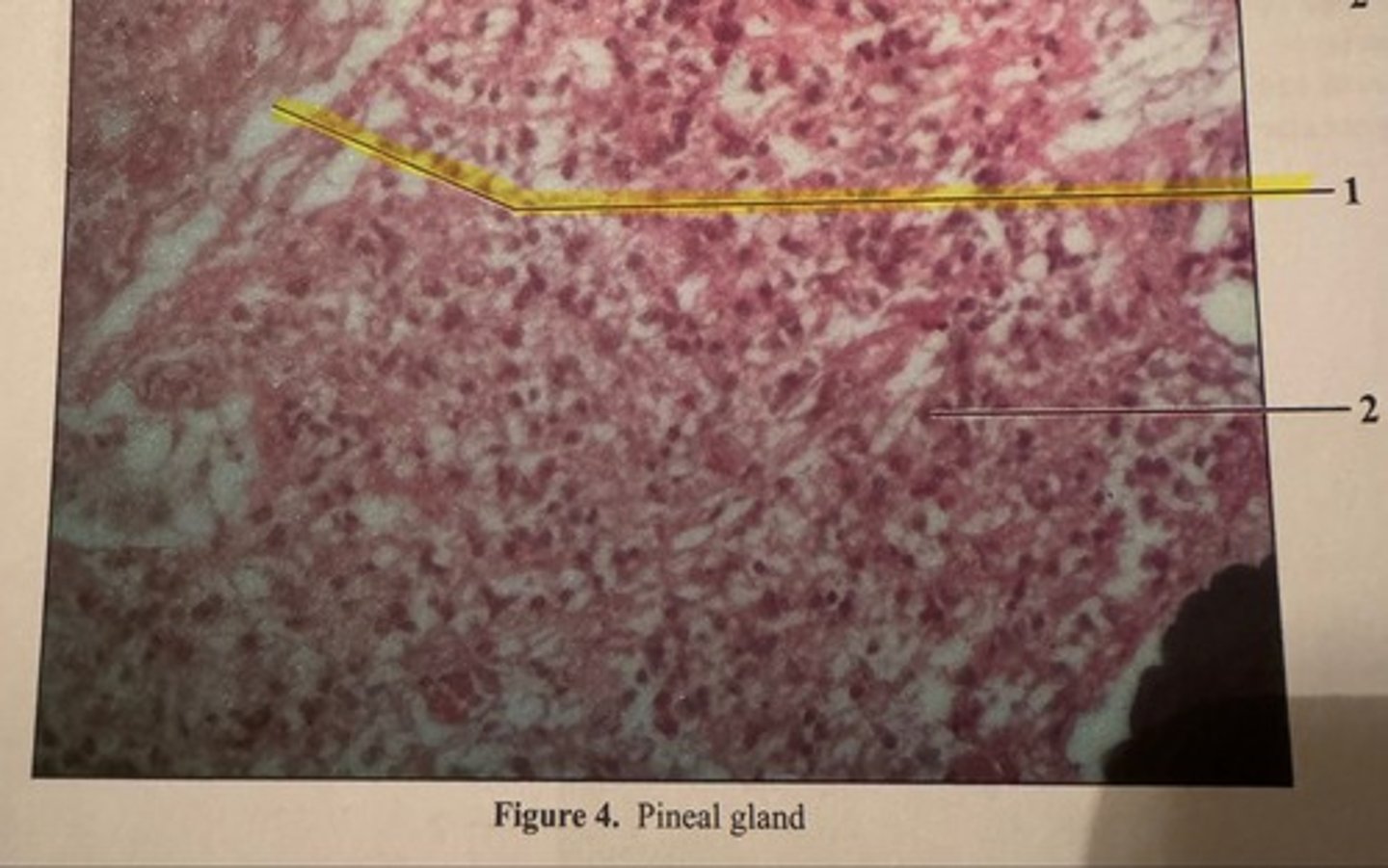
pinealoctyes
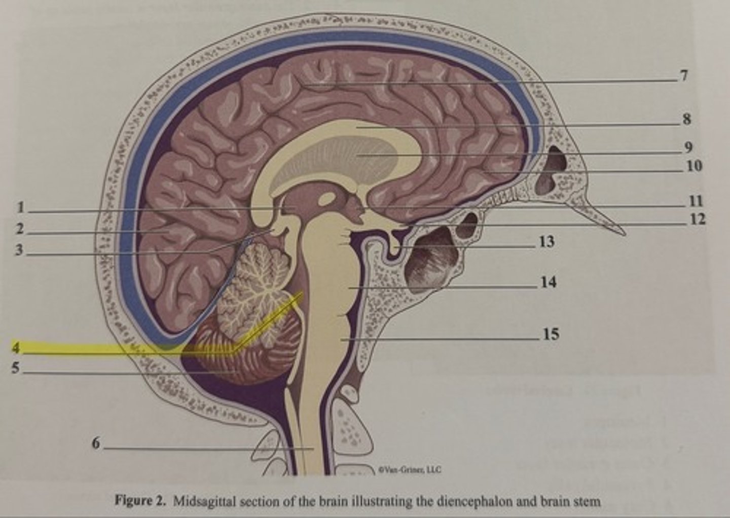
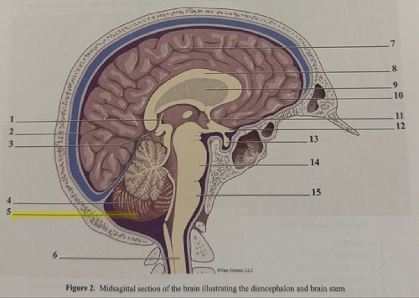
cerebellum function
coordination of complex movements; storage and processing of learned muscle patterns
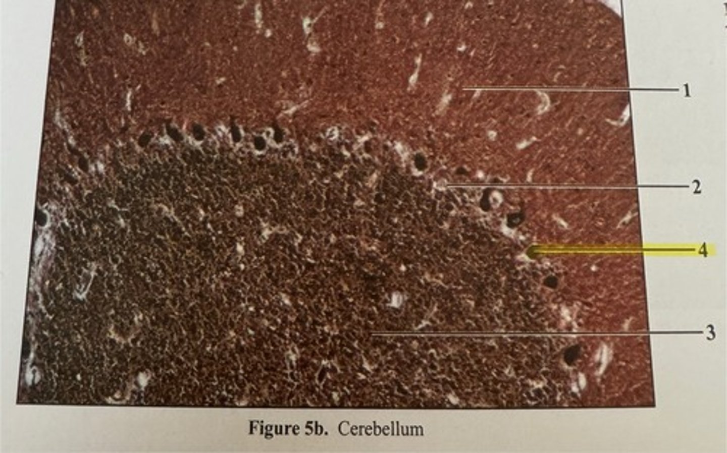
cerebellum layers
molecular, purkinje, granular
molecular layer (cerebellum)
contains stellate and basket cells
molecular layer
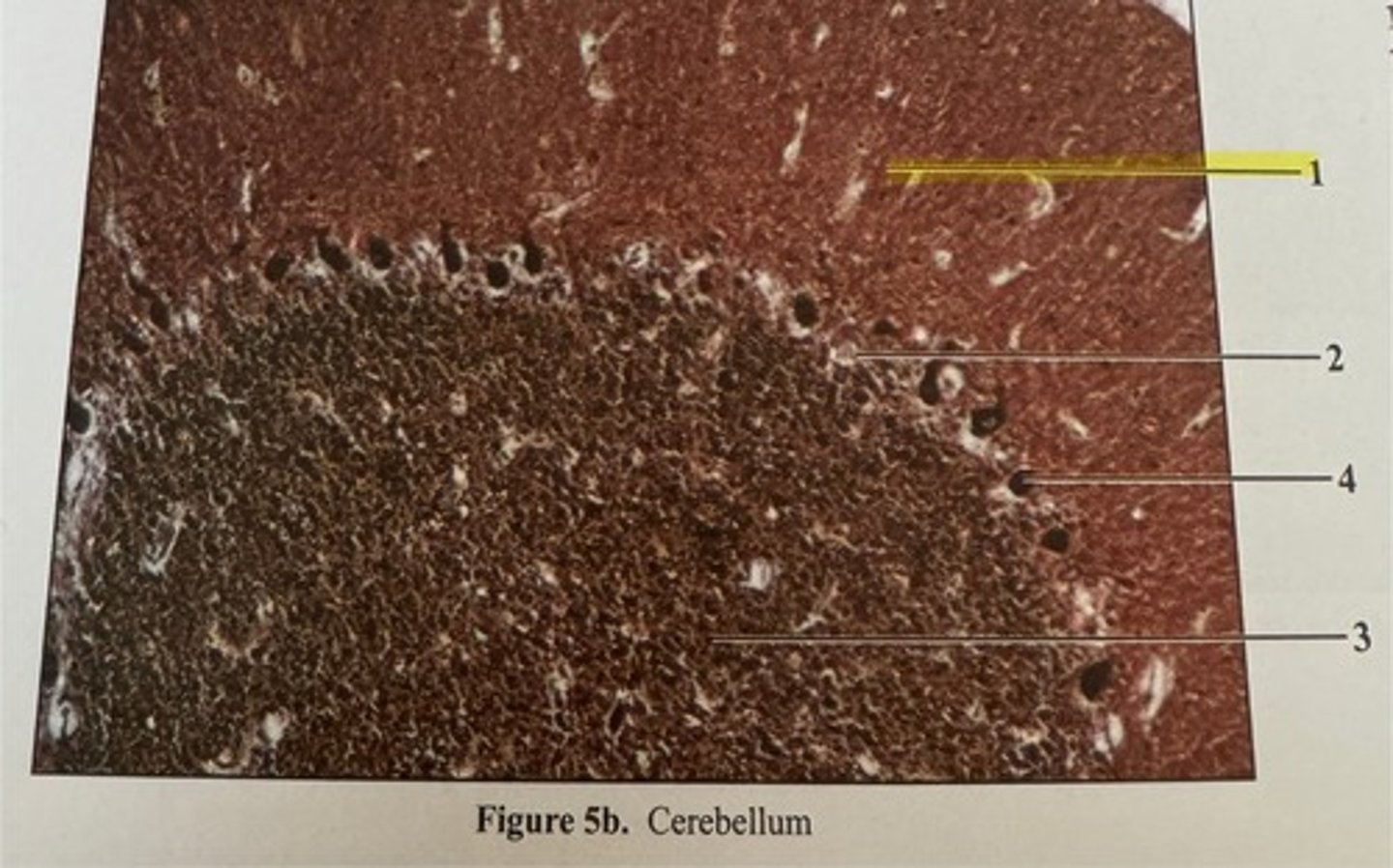
purkinje layer
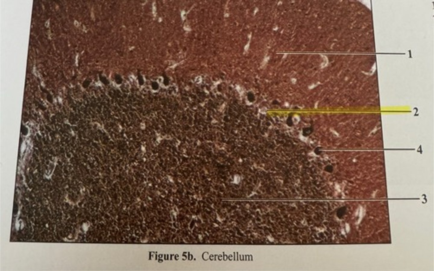
granular layer
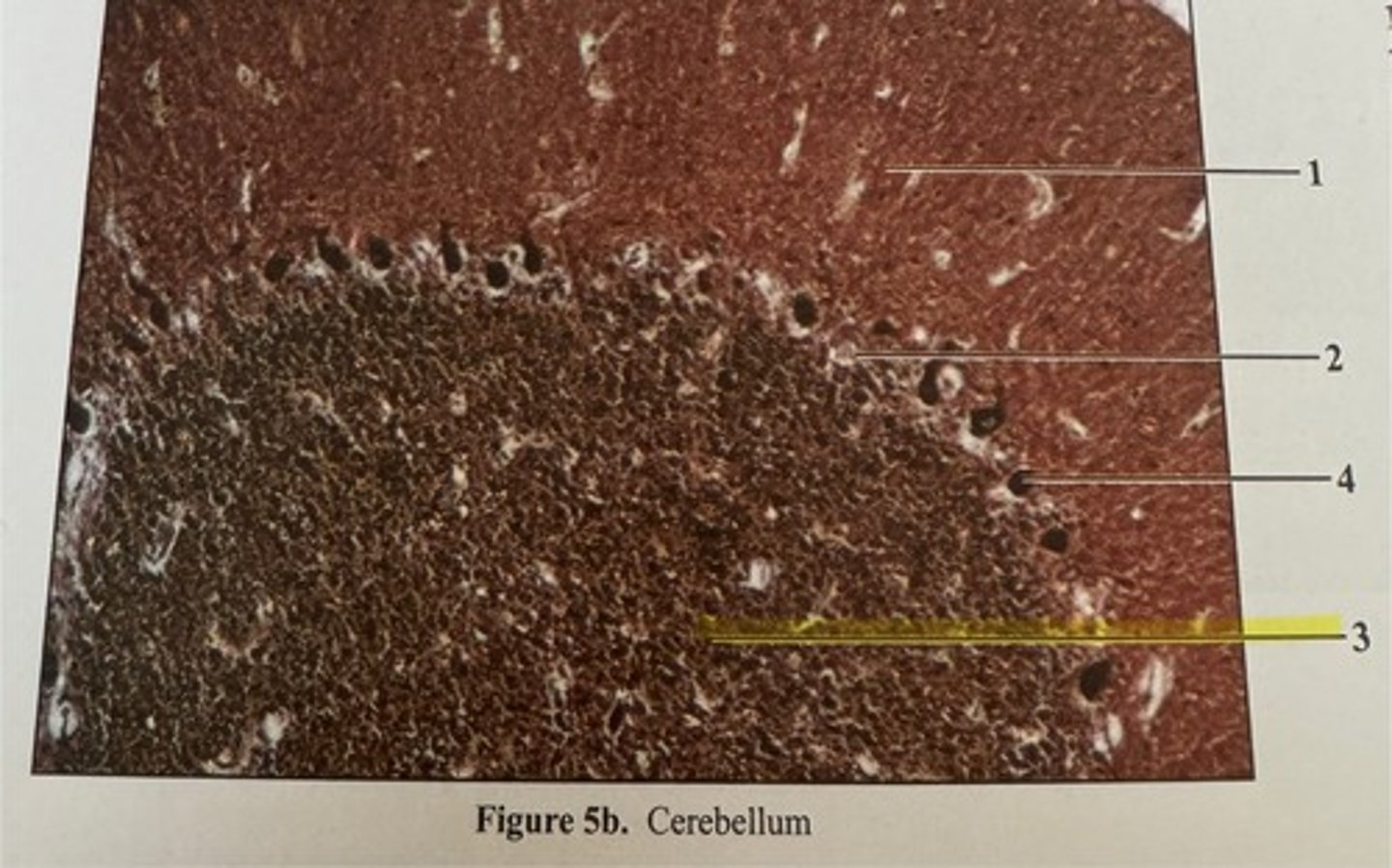
purkinje cell
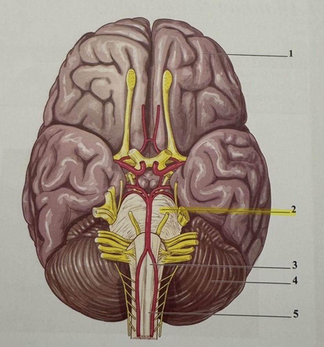
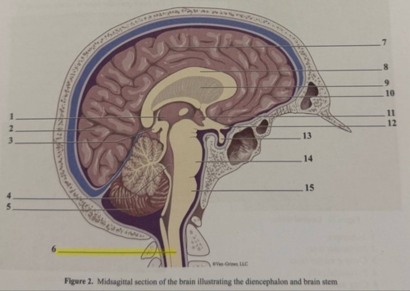
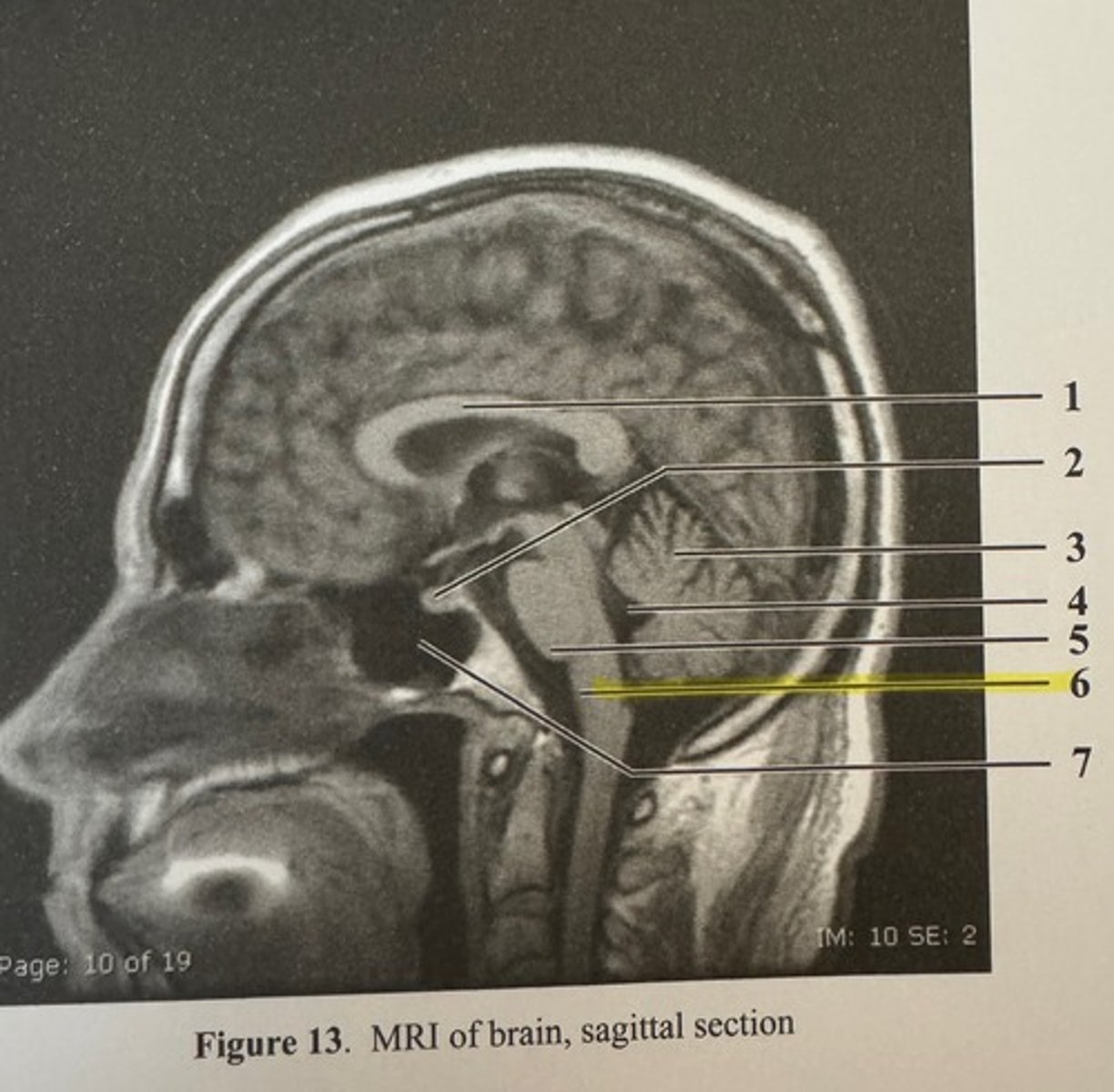
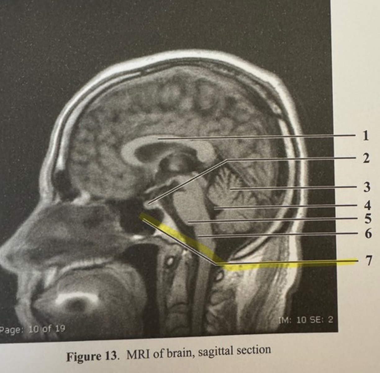
brain stem
consists of midbrain, pons, and medulla oblongata
midbrain
located between the diencephalon and the pons; associated with inhibiting inappropriate muscle movements and dopamine signals to ease inhibition to allow for smooth movements
pons
located between the midbrain and medulla oblongata; chiefly composed of conduction tracts between higher brain centers and the spinal cord or between the motor cortex and cerebellum
medulla oblongata
most inferior part of the brain stem; control over the cardiovascular and respiratory systems
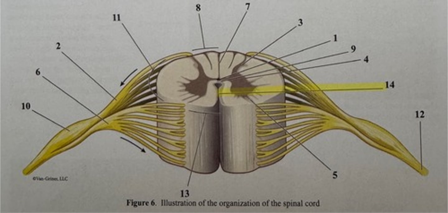
central canal
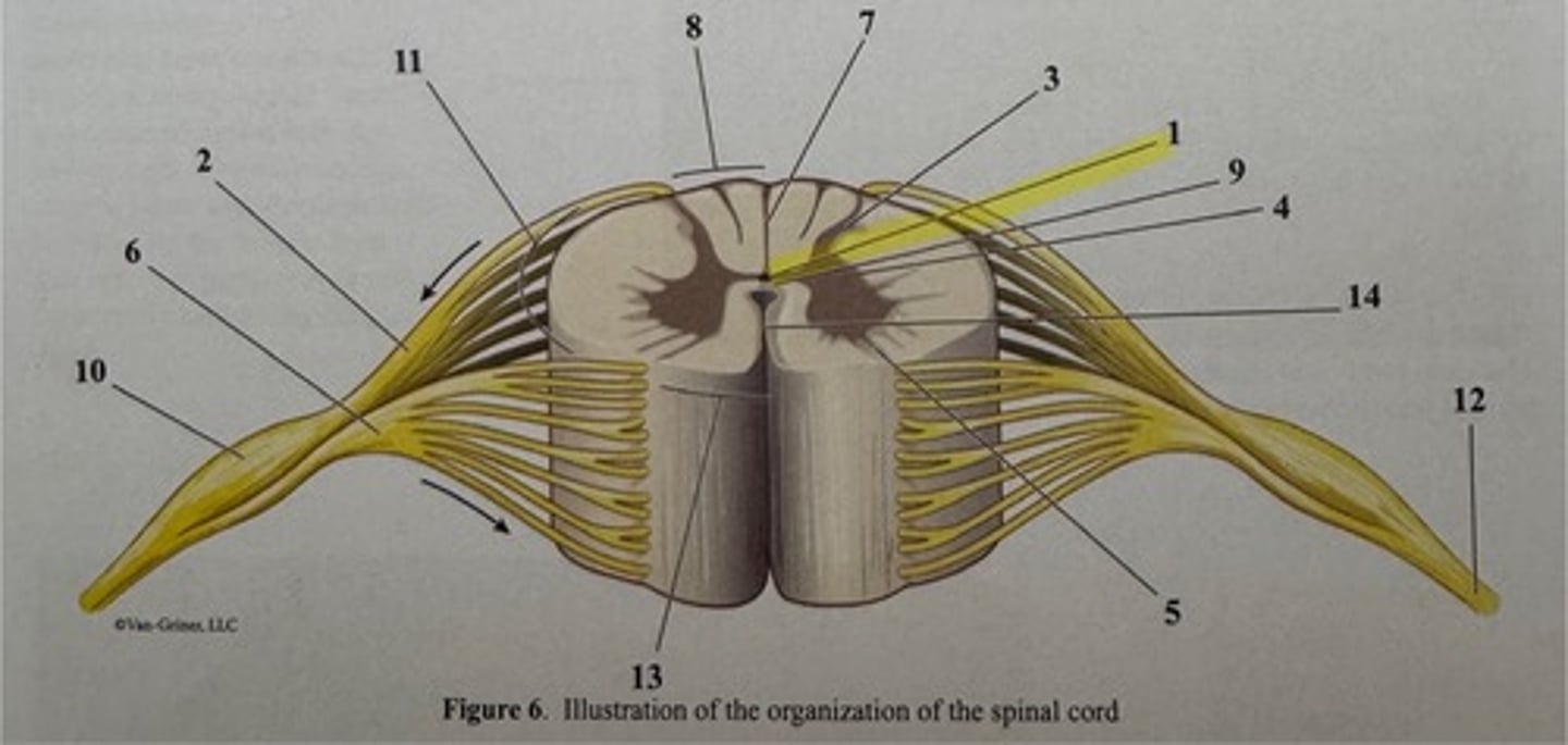
dorsal root
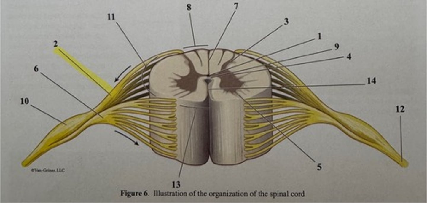
dorsal horn of gray matter
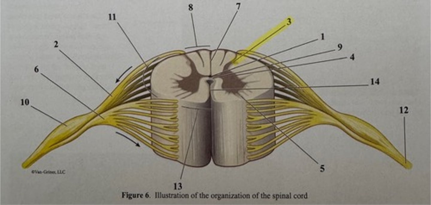
lateral horn of gray matter
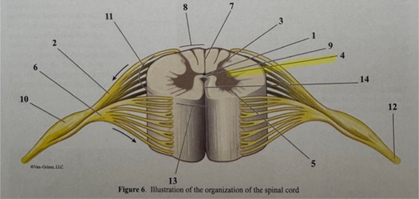
ventral horn of gray matter
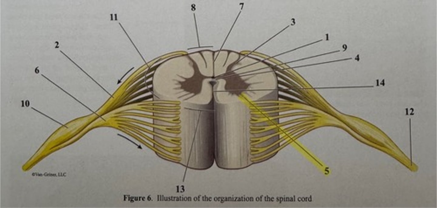
ventral root
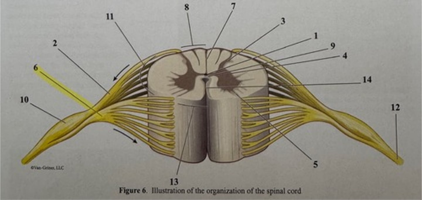
dorsal median sulcus

dorsal funiculus of white matter
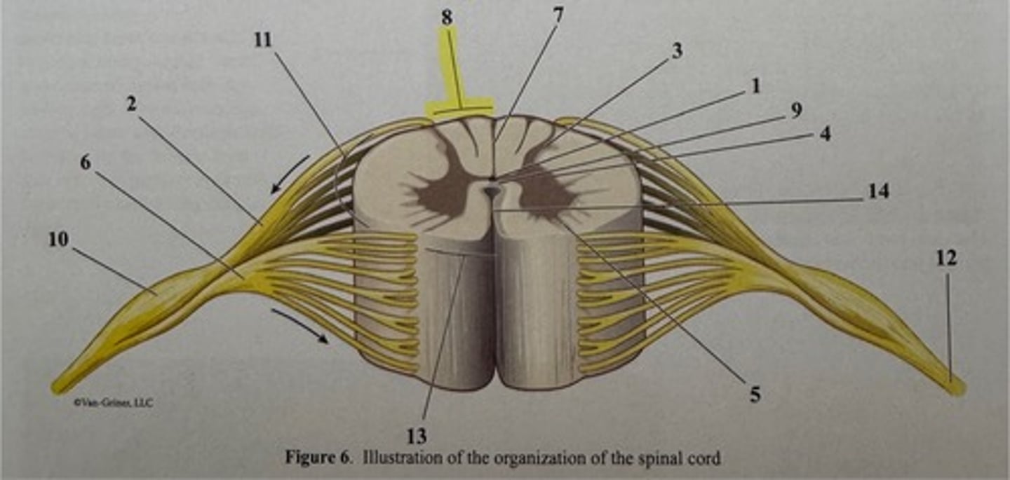
anterior commissure
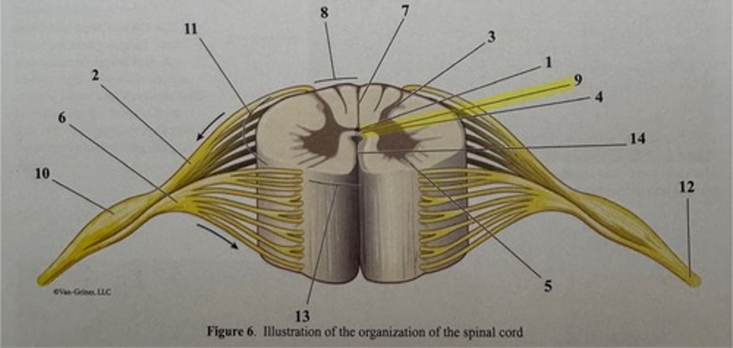
dorsal root ganglion
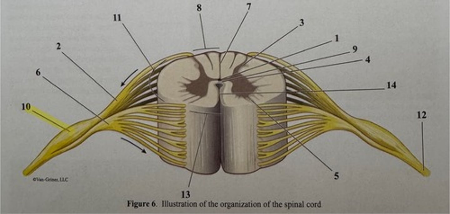
lateral funiculus of white matter
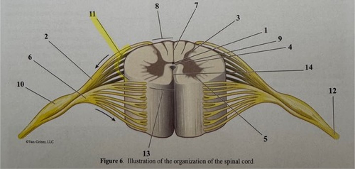
spinal nerve

ventral funiculus of white matter
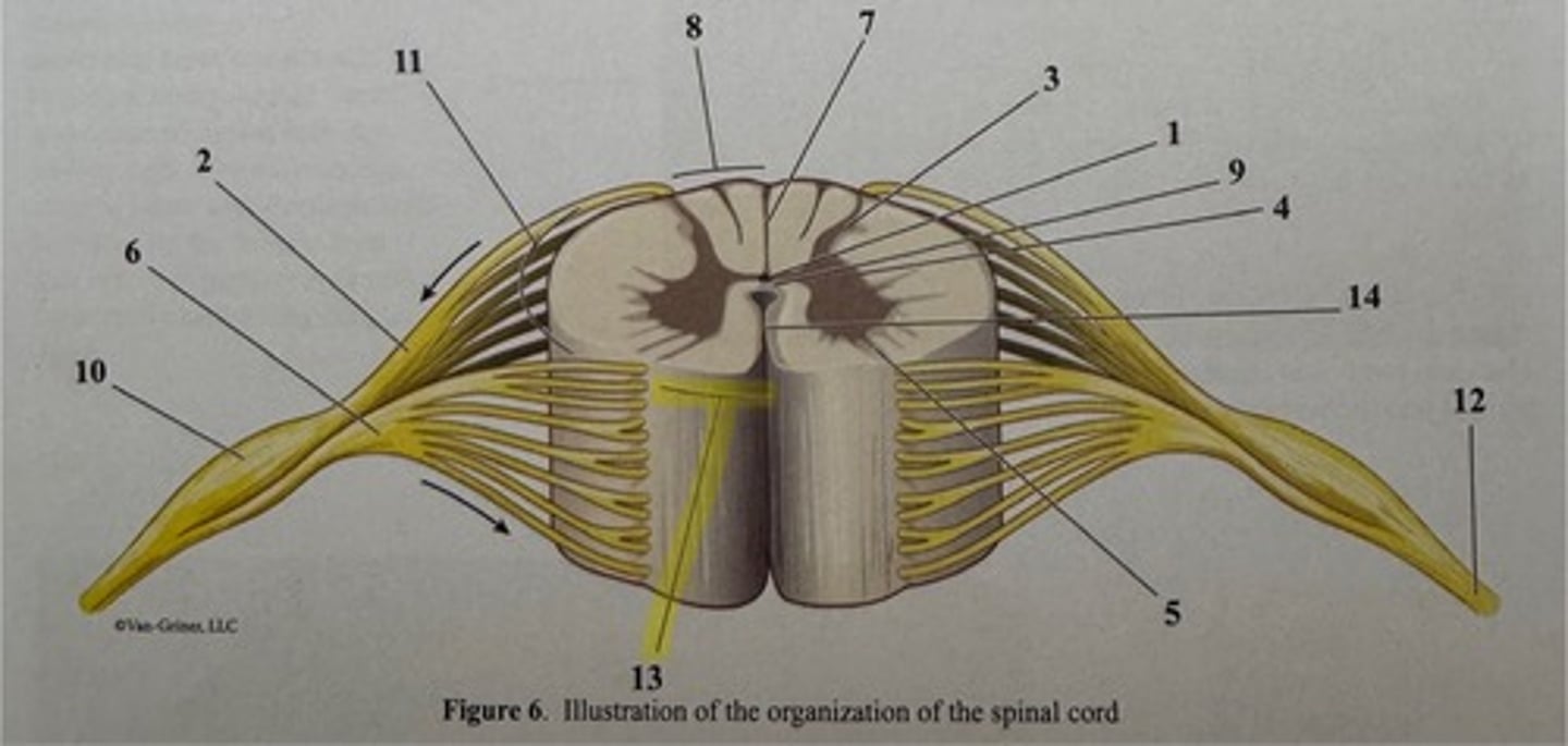
ventral median sulcus
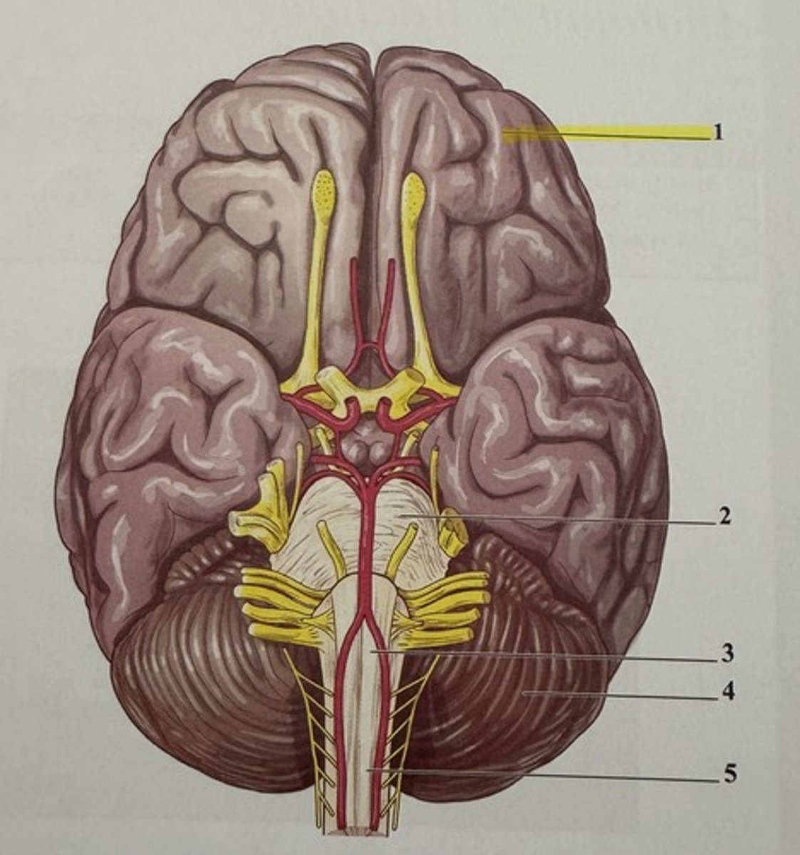
oculomotor nerve
supplies four of the six extrinsic muscles that move the eyeball in the orbit; these muscles are the inferior oblique, medial rectus, inferior rectus, and superior rectus
trochlear nerve
innervates the superior oblique and the trochlea
trigeminal nerve
three branches spring from this, the largest of the cranial nerves. It supplies sensory fibers to the face and motor fibers to the chewing muscles
medulla oblongata
sphenoid sinus
abducens nerve
controls the lateral rectus
facial nerve
a large nerve that innervates muscles of facial expression
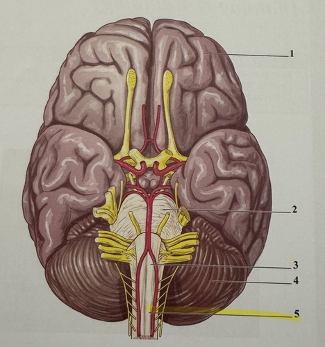
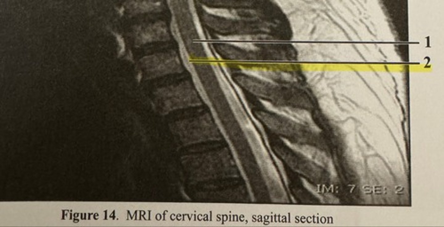
spinal cord
subarchnoid space
vestibulocochlear nerve
a sensory nerve for hearing and balance
glossopharyngeal nerve
innervates the tongue and pharynx, primary sensory to the throat
vagus nerve
only cranial nerve to extend beyond the head and neck to supply motor and sensory fibers to the visceral body organs of the thorax and abdomen
spinal accessory nerve
primarily motor and supplies the trapezius and sternocleidomastoid muscles
hypoglossal nerve
innervates muscles of the tongue
meissner's corpuscles
function as light pressure receptors of the dermis and are located within the dermal papillae just below the epidermal/dermal border
saltatory conduction
the jumping of action potentials from node to node, increases action potential velocity
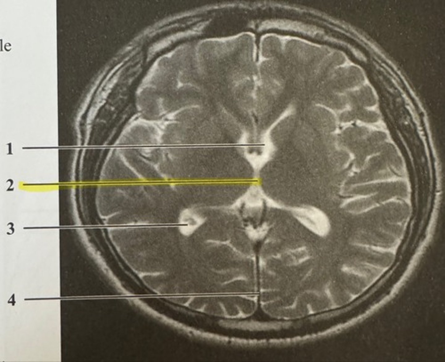
frontal horn of lateral ventricle
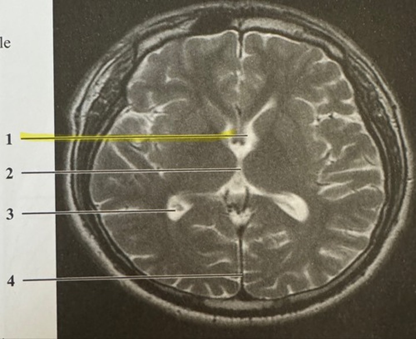
third ventricle