[PO2] Visual Field Tests
1/55
There's no tags or description
Looks like no tags are added yet.
Name | Mastery | Learn | Test | Matching | Spaced | Call with Kai |
|---|
No analytics yet
Send a link to your students to track their progress
56 Terms
Visual fields
🌟 generally assessed to detect the presence of any peripheral vision loss or to assess progession in case of a disease.
🌟 everything you can see around you when you look straight ahead.
Example:
🌟 When you look forward, you can still see things to the sides, up, and down without moving your eyes.
🌟 That whole area is your (BLANK)
Visual field test
🌟 checks how well you can see things in your side vision (peripheral vision).
Clinical importance:
🌟 to determine the magnitude of loss and, thereafter, to equate functional loss with disability.
Use for patients with:
🌟 Glaucoma
🌟 Retinal Diseases
🌟 Neurologic problem (TBI)
Confrontation field test
Automated Static Perimetry
Kinetic Perimetry
Amsler Chart Test
Visual field tests
180 degrees
Binocular field of vision
If you stretch your arms out to your sides, you can see both hands at once. That wide, half-circle view is about
150 degrees
Monocular field of vision
If you close one eye, your view shrinks a little bit
120 degrees
Common Binocular VF
both eyes see the same thing at the same time.
30 degrees
Exclusive VF
only one eye can see.
if something is way over by your left ear, your right eye can't see it because your nose is in the way
45-50 degrees
Upper VF
Your eyebrows block the view a bit, so you see the least in this direction.
60-70 degrees
Down VF
You can see more of your chest and the floor than you can see of the sky.
50-60 degrees
Nasal VF
Your nose is like a "wall" that stops your eye from seeing too far across your face.
80-90 degrees
Temporal VF
This is your best view! There's nothing blocking the side of your face, so your eyes can see almost all the way to your ears.
Factors that limit the VF
• Bones of the orbit
• Size of Palpebral fissure
• Size of the pupil
Tangent Screen Test
What the test is for
The (BLANK) is used to check a person’s visual field, especially the central 30° of vision.
A visual field means everything you can see around you while looking straight ahead.
Doctors use this test to map the areas where the patient can see and areas where they cannot see.
How the test is done
1. Patient position
The patient sits about 3 feet away from a screen.
There is a small target in the center of the screen.
2. Glasses (Rx)
The patient should wear their normal prescription glasses during the test.
If the patient is presbyopic (needs reading help):
A +1.00 diopter lens is placed in a trial frame over their distance prescription.
This is done because the tangent screen is placed about 1 meter away.
Patients should NOT wear multifocal glasses during this test because they can distort the results.
3. Testing one eye at a time
Only one eye is tested at a time.
The other eye is covered.
4. Patient instruction
The patient must look at the center target the whole time and not move their eyes.
5. Moving the object
The examiner moves a small object from outside the visual field toward the center.
The object is moved slowly from different directions.
6. Patient response
The patient tells the examiner when they first see the object.
This shows where their vision begins in that direction.
What the doctor does with the information
Each time the patient sees the object, the examiner marks that point on the screen.
After testing many directions, the examiner can draw a map of the patient's visual field.
This process is called visual field mapping.
Why this test is still used
The (BLANK) is still used because it is:
Inexpensive
Efficient
Fairly accurate for the central 30° of vision
Equipment
The equipment needed is very simple, usually:
A flat screen
A central fixation target
A small moving object
A trial frame with lenses
Because the setup is simple, the test can even be done without standard equipment if necessary.
Perimetry
In this test, you look into the center of a bowl-shaped machine called a perimeter.
The eye that is not being tested is covered with a patch.
The eye being tested has your prescription lenses placed in front of it so you can see as clearly as possible.
During the test, you must keep looking at a small target in the center of the bowl the entire time.
While you are staring at the center, small and dim lights will appear in different places inside the bowl.
Every time you see a light, you press a button.
The machine records:
the lights you saw
and the lights you did not see
Using this information, the machine can find areas of your visual field where vision may be missing.
Kinetic perimetry
A type of visual field in which the boundaries of the visual field are determined by a moving test object of fixed size and intensity while the patient's fixation is held steady.
This can only be done on a Goldmann visual field.
Static perimetry
A type of visual field in which the boundaries are determined by using a test object of fixed size and increasing the intensity until it is seen.
This is a stationary target.
This is done on the Goldmann, Humphrey, or other automatic visual fields.
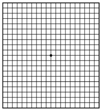
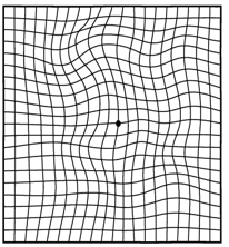
Amsler Grid Test
It is a near point test of central 10 degrees of vision. It is valuable for determining the position and size of central scotomas.
Chart 1
standard chart
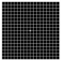
Chart 2
use for px with central scotoma
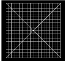
Chart 3
standard chart but red on black
to be used in cases of colour scotoma
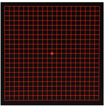
Chart 4
chart with no lines, reveals only scotoma, no form to be disturbed
Chart 5
parallel lines must be looked horizontally and vertically
metamorphopsia
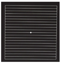
Chart 6
for metamorphopsia, more minue exam of distribution along the reading lines
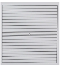
Chart 7
juxta central areas, where the rectangle indicates the limits of fovea
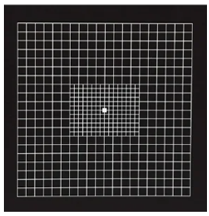
Amsler Grid
Normal (Amsler Grid)
Blind spot (Amsler Grid)
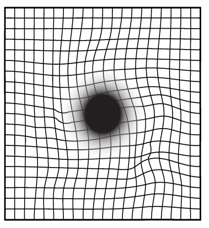
Distortion/wavy lines
Confrontation Field Test
A simple screening test.
The doctor sits in front of you.
You cover one eye.
The doctor moves fingers in different directions.
You say when you see the fingers.
👉 Quickly checks if side vision is normal.
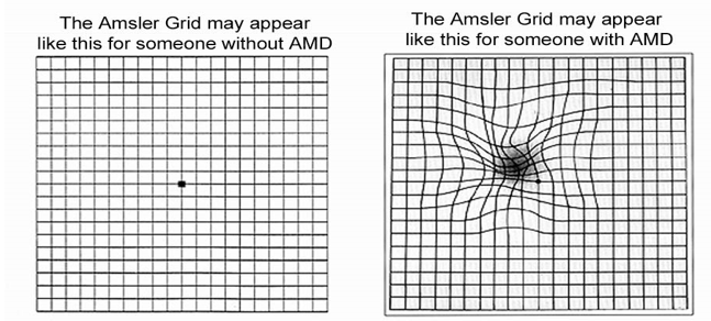
Amsler Chart Test
Used to check central vision.
You look at a grid of straight lines.
If lines appear wavy, missing, or distorted, it may mean a macular problem.
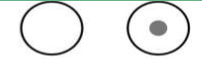
Central scotoma
Monocular vision loss
Bitemporal hemianopia
Contralateral homonymous hemianopia
Contralateral superior quadrantopia
Contralateral inferior quadrantopia
Contralateral homonymous hemianopia with macular sparing
Heteronymous
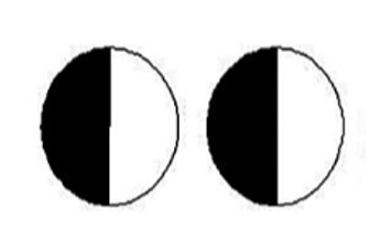
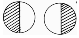
Definition: Loss of different halves of the visual field in each eye.
Example: You lose the left half in one eye and the right half in the other eye.
HINDI PAREHAS
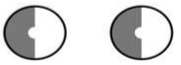
Homonymous
Definition: Loss of the same side of the visual field in both eyes.
Example: You lose the right half in both eyes.
PAREHAS
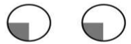
Congruous
defect is identical in both eyes
Incongruous
defects differ between eyes
Left Homonymous Hemianopsia
Left Superior Homonymous Quadrantanopsia
Left Homonymous Hemianopsia with Macular Sparing
Binasal Hemianopsia
Bitemporal Hemianopsia
Left Inferior Homonymous Quadrantanopsia
Inferior Heteronymous Quadrantanopsia
Right Superior Homonymous Quadrantanopsia
1, 4, 6
Contralateral (#)
different side ang defect
2, 3, 5
Ipsilateral (#)
same side ang defect
Superior
Lateral (taas)
Inferior
Medial (baba)
Peripheral
Confrontation field test
Automated static perimetry
Kinetic Perimetry
are type of?
Contralateral
does NOT describe the eyes.
it describes the relationship between the brain lesion and the visual field loss.
opposite side of the brain lesion.
Ipsilateral
(brain)
same side
Example:
Damage in the right optic nerve
Vision loss in the right eye
So:
👉 Right side damage → Right side vision loss