Occipital Bone
1/18
There's no tags or description
Looks like no tags are added yet.
Name | Mastery | Learn | Test | Matching | Spaced |
|---|
No study sessions yet.
19 Terms
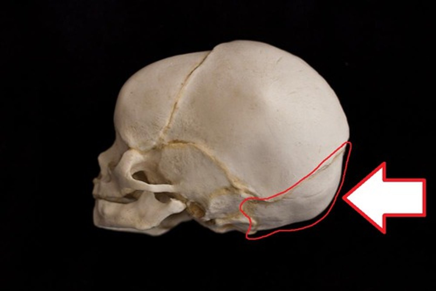
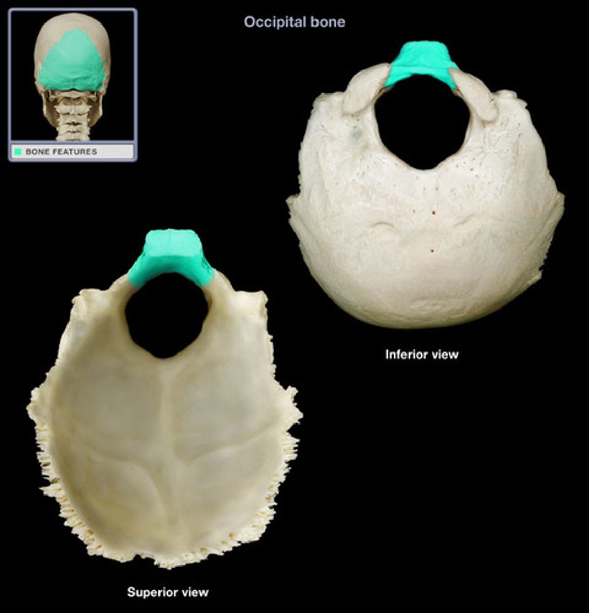
Occipital Bone
The occipital bone is set at the rear of the cranium and articulates with the temporals, sphenoid, parietals, and the uppermost vertebra, the atlas.
(Better pictures:
https://www.bartleby.com/107/31.html)
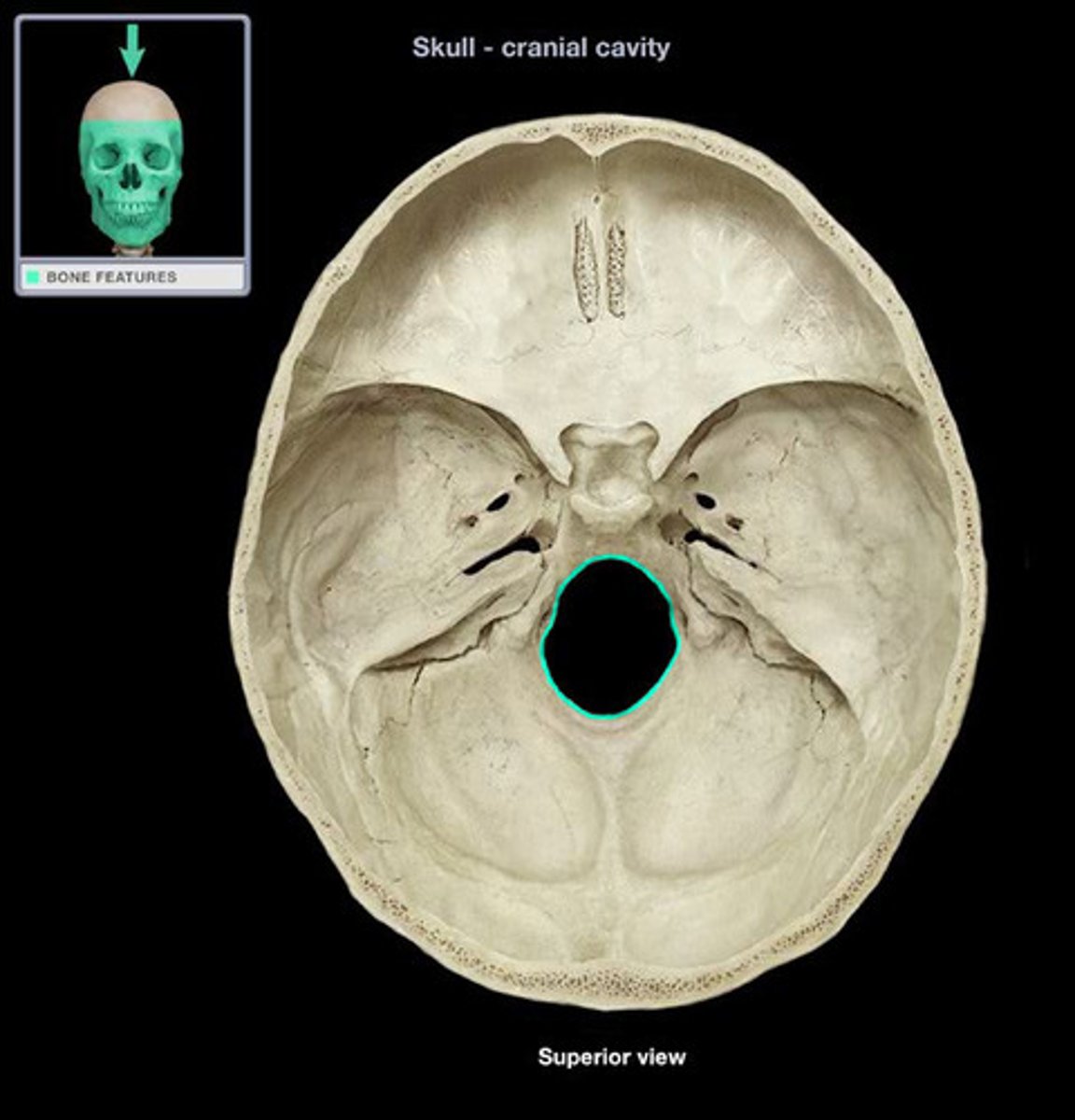
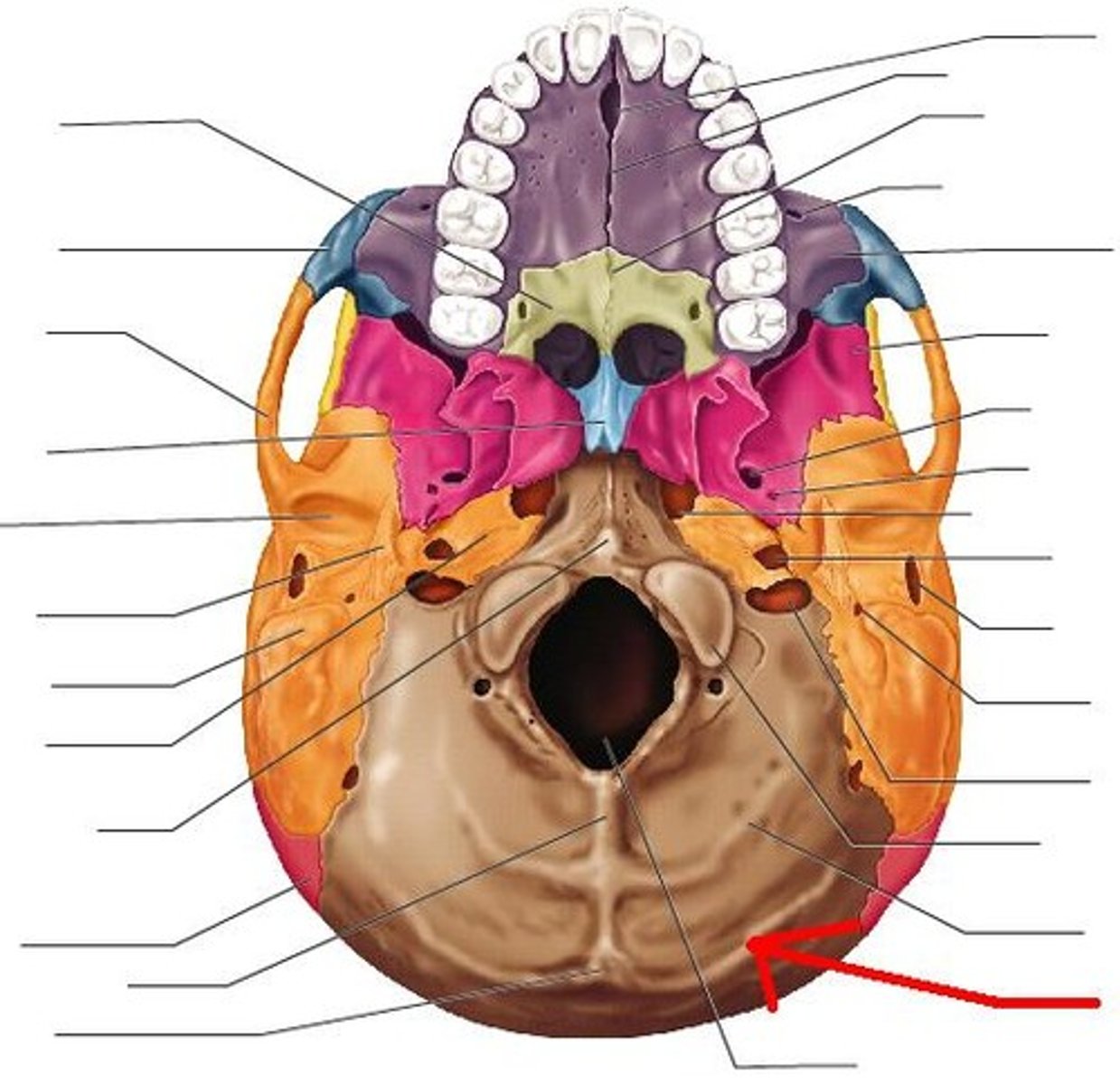
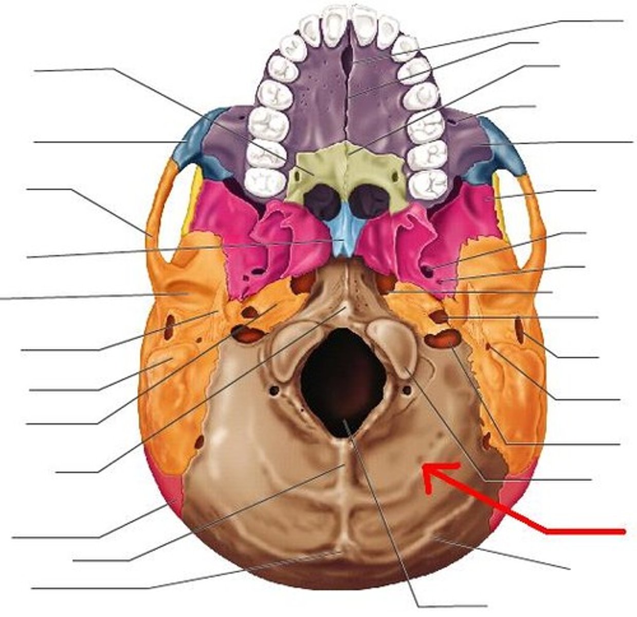
Foramen Magnum
The large hole in the occipital through which the brain stem passes inferiorly into the vertebral canal.
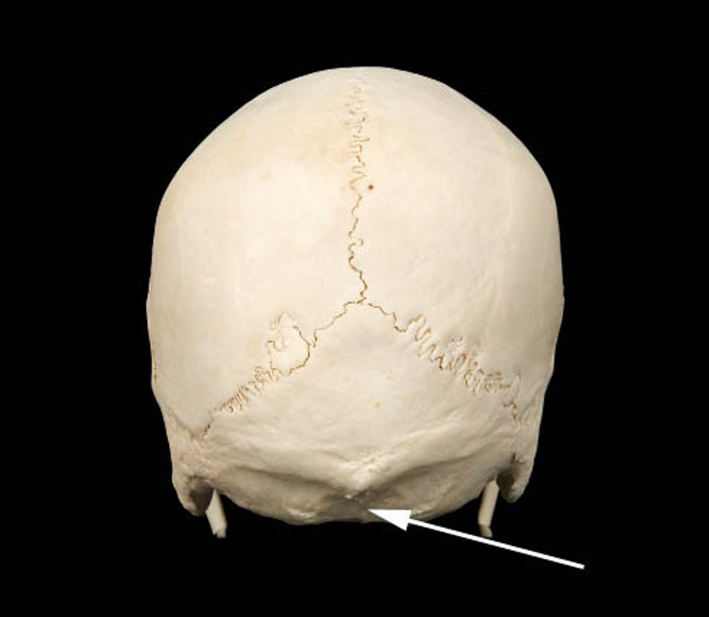
Occipital Squamous
A portion of the occipital bone is by far the largest, constituting the large plate of bone posterior and superior to the foramen magnum.
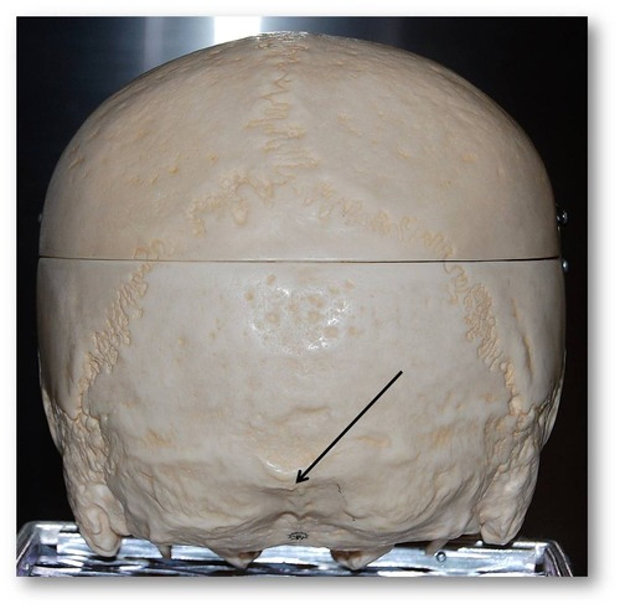
External Occipital Protuberance
This protuberance lies on the ectocranial midline where the occipital and nuchal planes meet. It is highly variable in appearance and heavier and more prominent in male individuals.
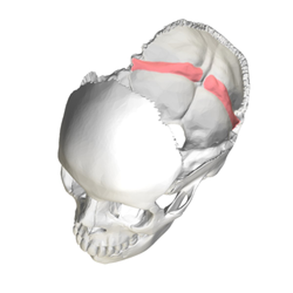
Superior Nuchal Lines
These lines lie to either side of the midline on the ectocranial surface of the squamous portion. The nuchal plane and occipital planes merge at these superiorly convex lines. Several nuchal muscles attach to and below these lines and function to extend and rotate the head.
Inferior Nuchal Lines
These lines are parallel the superior lines but are located about midway on the ectocranial nuchal plane. Fascia separating nuchal muscles attach to the line, whereas additional nuchal muscles attach inferior to this line.
External Occipital Crest
This crest, or median nuchal line, is a highly variable median line or crest that passes between the right and the left nuchal musculature. It stretches from the external occipital protuberance to the rear of the foramen magnum, anchoring the nuchal ligament.
Basilar Part
This part is the thick, square projection anterior to the foramen magnum. It articulates with the petrous portions of both temporals and with the sphenoid via the basilar, or sphenooccipital suture.
Lateral Parts
These parts of the occipital lie to either side of the foramen magnum, articulating with the temporals.
(Picture:
https://en.wikipedia.org/wiki/Lateral_parts_of_occipital_bone)
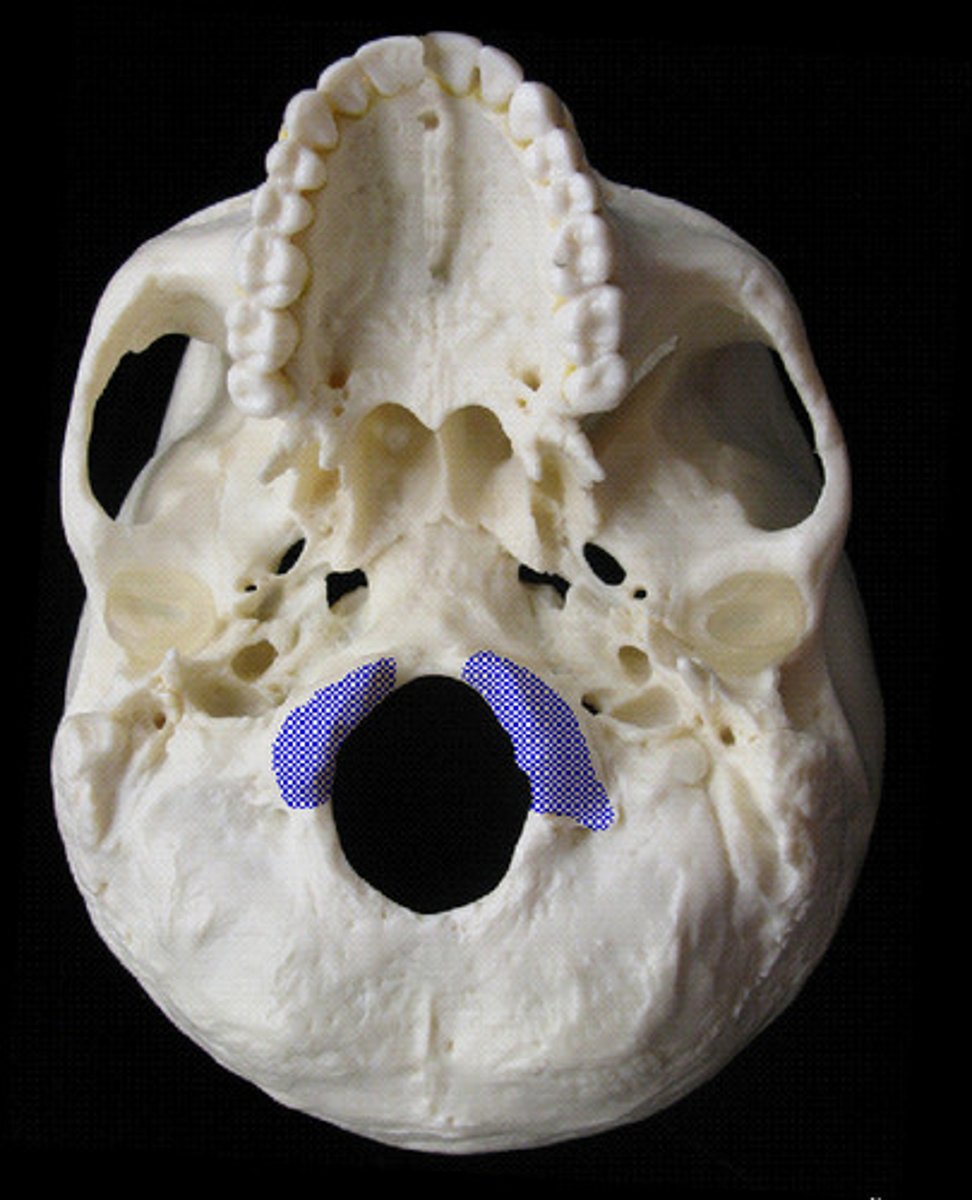
Occipital Condyles
The Condyles are raised oval structures on either side of the foramen magnum. Their inferior surfaces are convex. The articular surfaces of these condyles fit into the concave facets of the atlas vertebra.
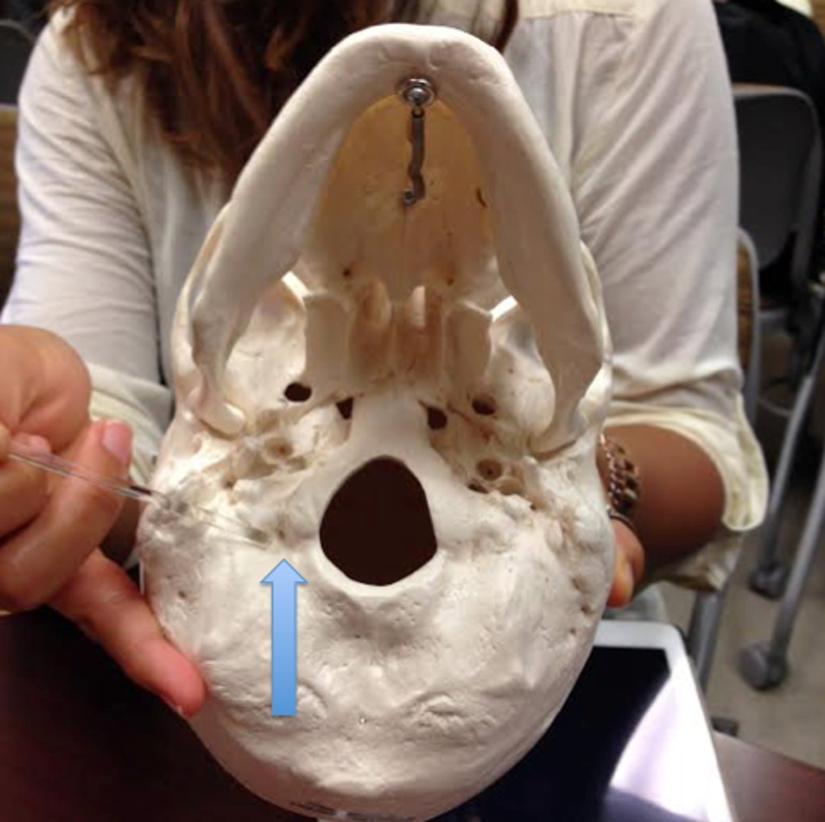
Condyle Fossa
These are ectocranial depressions immediately posterior to the condyles. The fossae receive the posterior margin of the superior facet of the atlas vertebra when the head is extended backward.
(in the picture,it's the tiny hole)
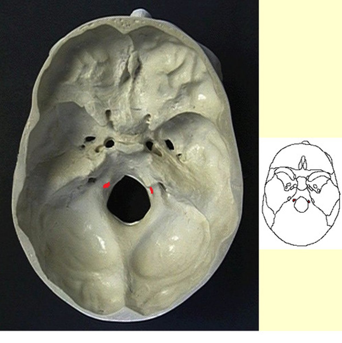
Hypoglossal Canal
Tunnels through the anterior part of the base, therefore superior in placement, of each condyle. These canals give exit to hypoglossal nerves, cranial nerve twelve, and entrance to arteries.
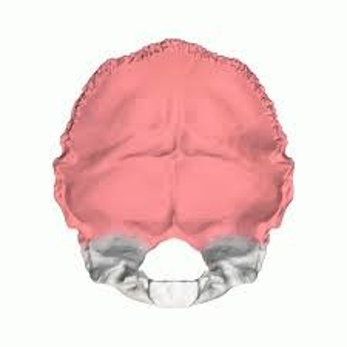
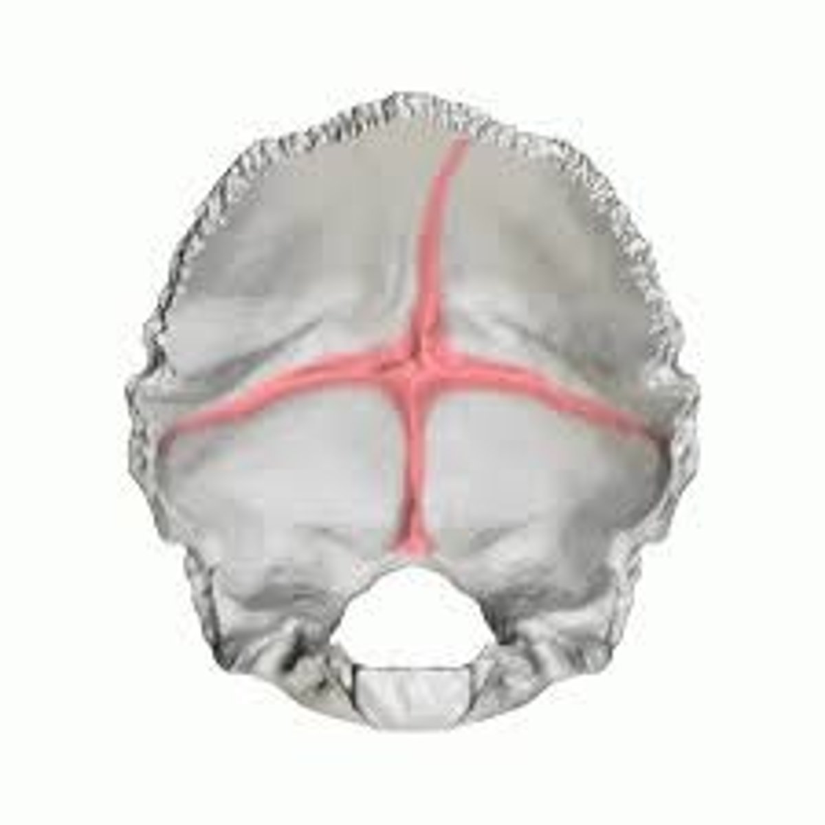
Cruciform Eminence
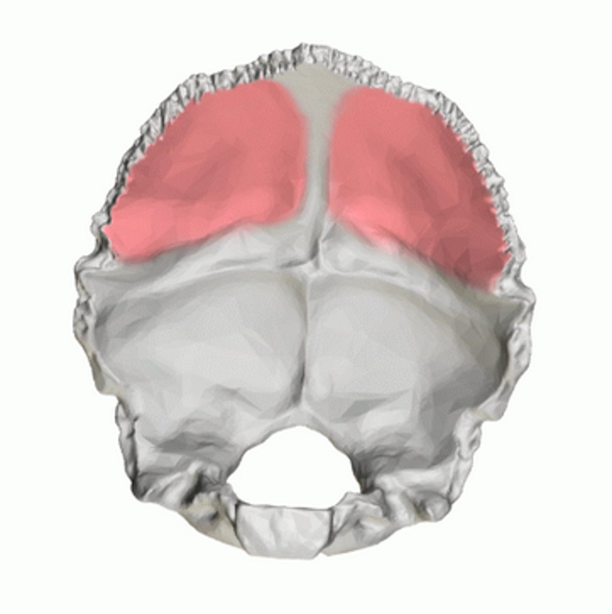
This eminence divides the endocranial surface of the occipital squama into four fossae. It is so named because it is cross-shaped.
Cerebral Fossae
These Fossae are triangular depressions below the lambdoid suture on the endocranial surface of the occipital. They house the occipital lobes of the brain's cerebrum.
(Above the Cerebellar Fossae)
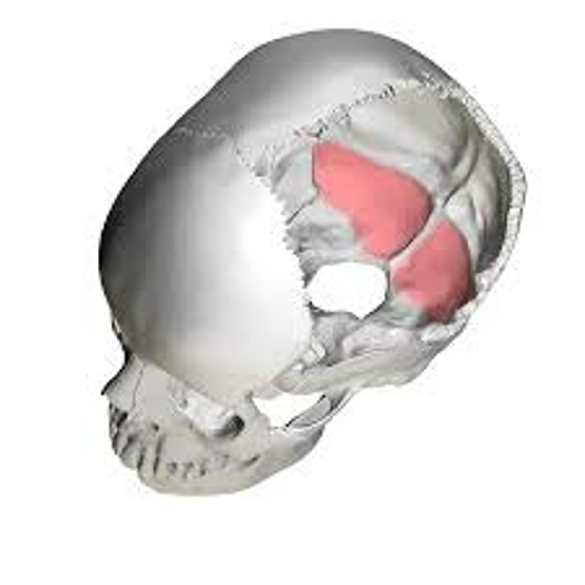
Cerebellar Fossae
These Fossae occupy the inferior part of the endocranial surface of the occipital squama. Therein rest the cerebellar lobes of the brain.
(Below the Cerebral Fossae)
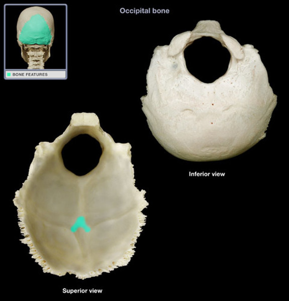
Internal Occipital Protuberance
This protuberance lies at the center of the cruciform eminence.
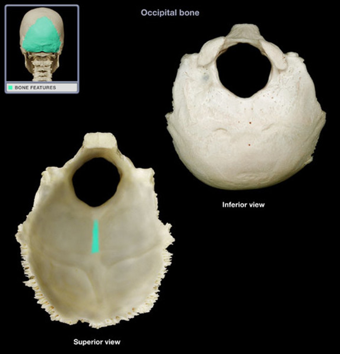
Internal Occipital Crest
This crest is the inferior arm of the cruciform eminence. Sometimes it bears a sulcus that continues on one or both sides of the foramen magnum. Such a sulcus, called an occipitomarginal sulcus, represents an alternative pathway for blood to drain from the brain.
Transverse Sulci
The sulci form the transverse arms of the cruciform eminence. They house the transverse sinuses. The one on the right is usually larger and communicates directly with the sagittal sulcus. However, variations in the soft tissue and bony manifestations of this cranial venous drainage system are common and sometimes pronounced. The transverse sulcus of the occipital connects with the sigmoid sulcus of the temporal and endocranial jugular process, often via the transverse (or sigmoid) sulcus on the mastoid angle of the parietal.
Siding the Occipital
Isolated fragments of the occipital are easily sided by locating the lambdoid suture.
-For isolated condyles, the edge of the foramen magnum is medial and somewhat posterior to the condylar body centers.
-The condylar fossa is posterior, and the hypoglossal canals tunnel from anterolateral to posteromedial.