Exam 2: SHOULDER: THE JOINTS, LIG, BURSA, NO MUSCLES + ELBOW + WRIST/HAND
1/63
Earn XP
Description and Tags
All shoulder, elbow, wrist and hand joints as well as ligaments and concepts. No muscles included.
Name | Mastery | Learn | Test | Matching | Spaced | Call with Kai |
|---|
No analytics yet
Send a link to your students to track their progress
64 Terms
What is the shoulder complex?
The shoulder complex consists of the shoulder joint, scapula, clavicle, and associated muscles and ligaments that work together to allow a wide range of motion and stability in the shoulder.
What joints make up the shoulder complex?
The shoulder complex is made up of the sternoclavicular, acromioclavicular, and glenohumeral joints.
What articulates with the sternal end of the clavicle? And what portion of the clavicle is it?
The sternal end located on the medial portion of the clavicle, articulates with the clavicular notch of manubrium of the sternum.
What 3 movements at the clavicle does the sternoclavicular joint allow?
Movement within the anteroposterior plane, the vertical plane, and mild rotation.
What are the 4 ligaments of the sternoclavicular joint?
Anterior SC Ligament, Posterior SC Ligament, Interclavicular ligament, and Costoclavicular ligament
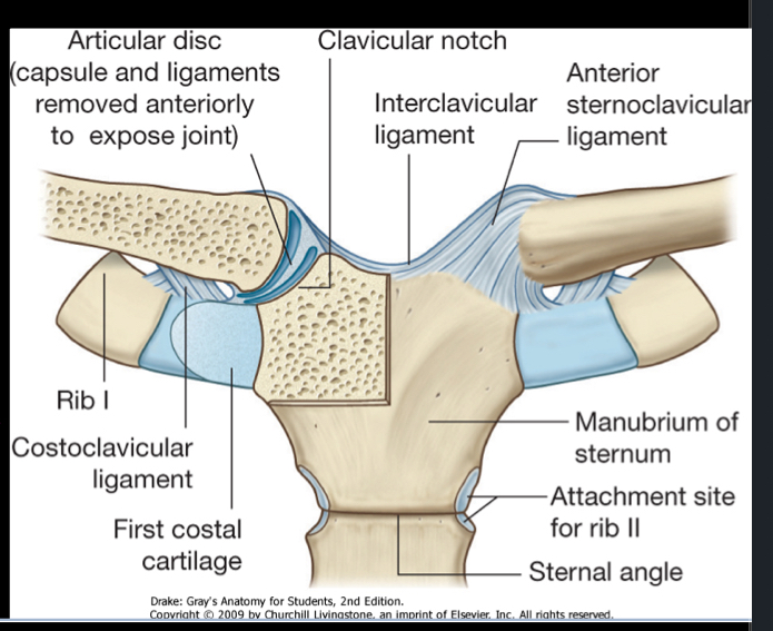
Anterior SC Ligament
This ligament will be between the sternal end of the clavicle and the manubrium of the sternum, providing stability to the joint and limiting anterior movement of the clavicle. The posterior version is the same, and just prevents posterior movement.
Interclavicular Ligament
Think of this ligament as an anchor holding the 2 clavicles together at the manubriums superior surface. It stabilizes the joint by preventing excessive separation of the clavicles during shoulder movements.
Costoclavicular Ligament
This ligament is between the costal cartilage of the 1st rib and the clavicle. It is another important stabilzier that anchors the clavicle down to the costal cartilage to prevent the clavicle from excessively elevating (superior movement).
What is the acromioclavicular joint?
The articulation between the acromial end of the clavicle and the acromion process of the scapula. It is a synovial joint.
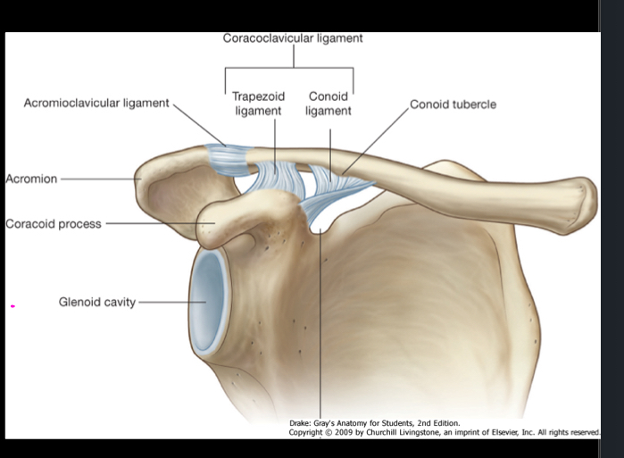
What movement does the AC joint allow?
It allows movement within the A/P plane, vertical plane, and some axial rotation. The same as your sternoclavicular joint.
What are the 2 ligaments that reinforce the AC joint?
The acromioclavicular ligament and the coracoclavicular ligament.
The AC Ligament
Is a ligament that connects the acromion of the scapula to the clavicle and reinforces the acromioclavicular joint, providing stability during shoulder movements.
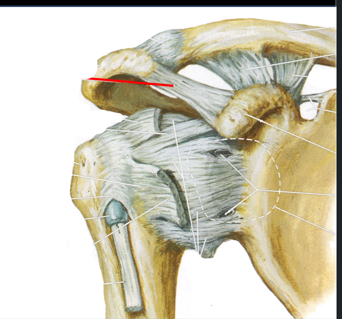
Coracoclavicular ligament
Is between the coracoid process of the scapula and the clavicle. It is an accessory ligament which stabilizes the joint. It does NOT cross over the joint, it only runs from coracoid process to the clavicle.
What is the clinical significance of the AC joint?
The AC joint is the site of shoulder SEPARATION. Which is when the acromion separates from the clavicle. This is different than dislocation which typically involves the humerus. AC joint injuries are common in contact sports and are caused from intense impact to the lateral portion of the shoulder.
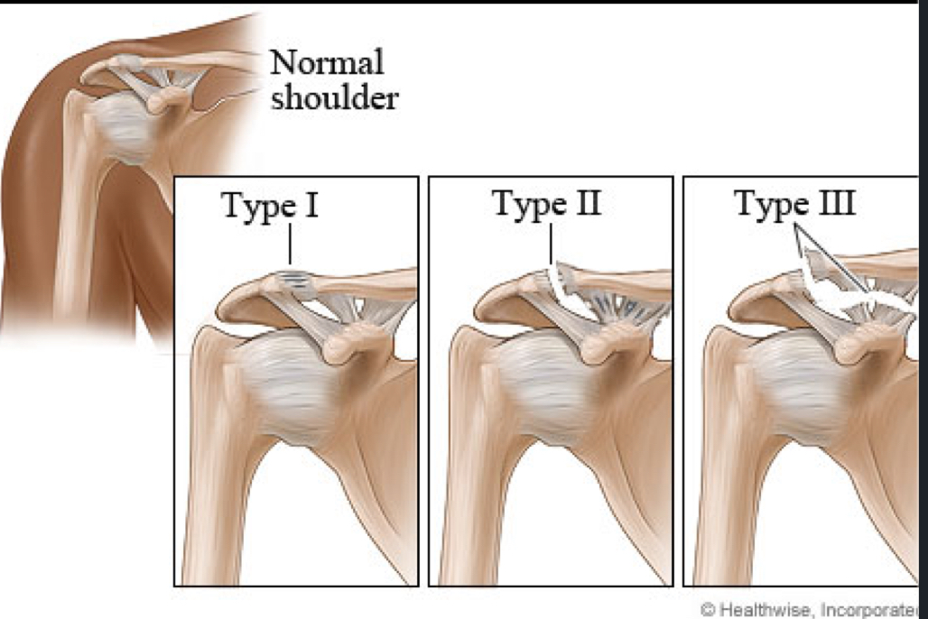
Shoulder Separation
Caused by hard blow to the lateral side of the shoulder or a direct fall on to the shoulder. It’s important to remember that there are different grades of shoulder separation, which indicate the severity of the injury. Grade I is a sprain, Grade II involves partial tear, and Grade III is a complete tear of the ligaments.
Shoulder Separation Grade I
Usually identified as a sprain, which means that there is no tears of either your AC Ligament or your Coracoclavicular ligament. Bones will still be aligned, and it will be mainly damage to the joint capsule around your AC joint.
Shoulder Separation Grade 2
Involves a tear of your acromioclavicular ligament, however, your coracoclavicular ligament will be intact. The AC joint may exhibit mild displacement, and patients typically experience pain and swelling around the shoulder.
Shoulder Separation Grade 3
Involves complete tears of both the acromioclavicular ligament and the coracoclavicular ligament. The clavicle will be pushed out of alignement with the acromion. This results in significant shoulder instability and typically requires surgical intervention for proper healing.
What is the glenohumeral joint?
Is composed of the head of the humerus and the glenoid fossa of the scapula. This joint is the primary shoulder joint, allowing for a wide range of motion and stability. It is a synovial joint, which is characteristic of a ball and socket joint.
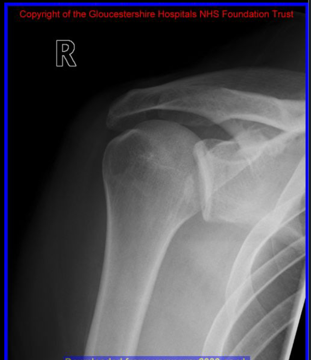
What is clinical diagnosis of this dislocation?
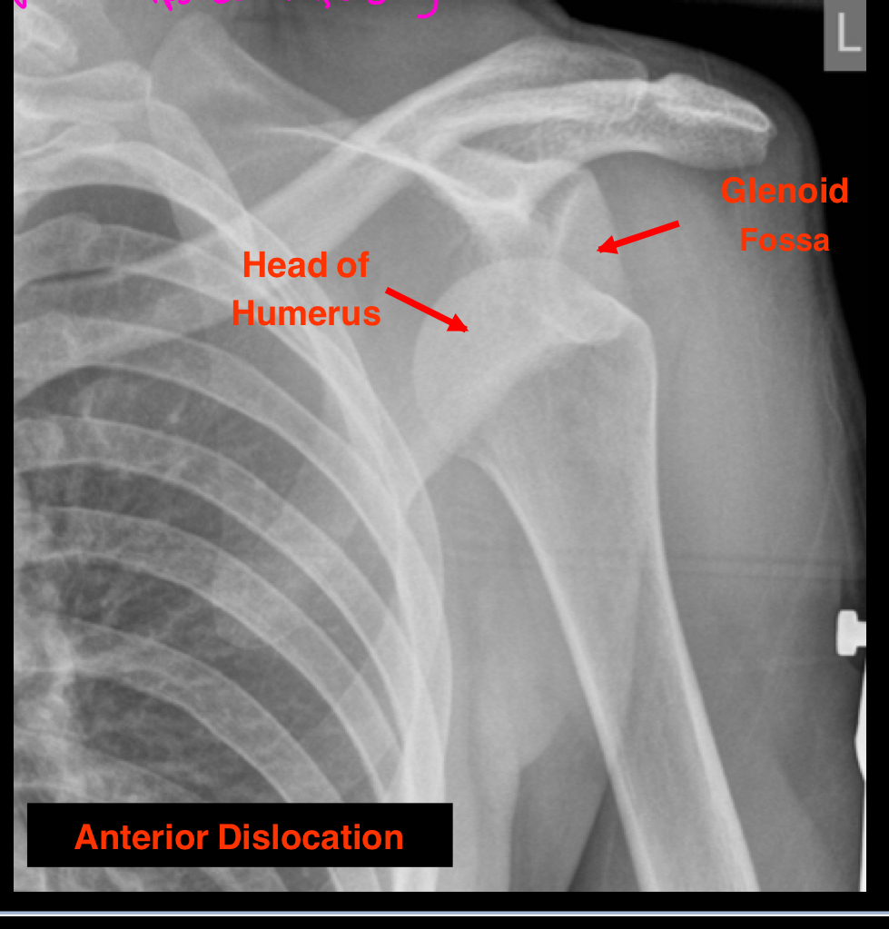
This is an example of a subcoracoid dislocation where the humeral head is displaced underneath the coracoid process of the scapula. This is an anterior dislocation, the most common type of shoulder dislocation. You can see a strong overlap of the head of the humerus with the glenoid fossa, showing that the head dislocated anteriorly.
What is the clinical diagnosis of this dislocation?
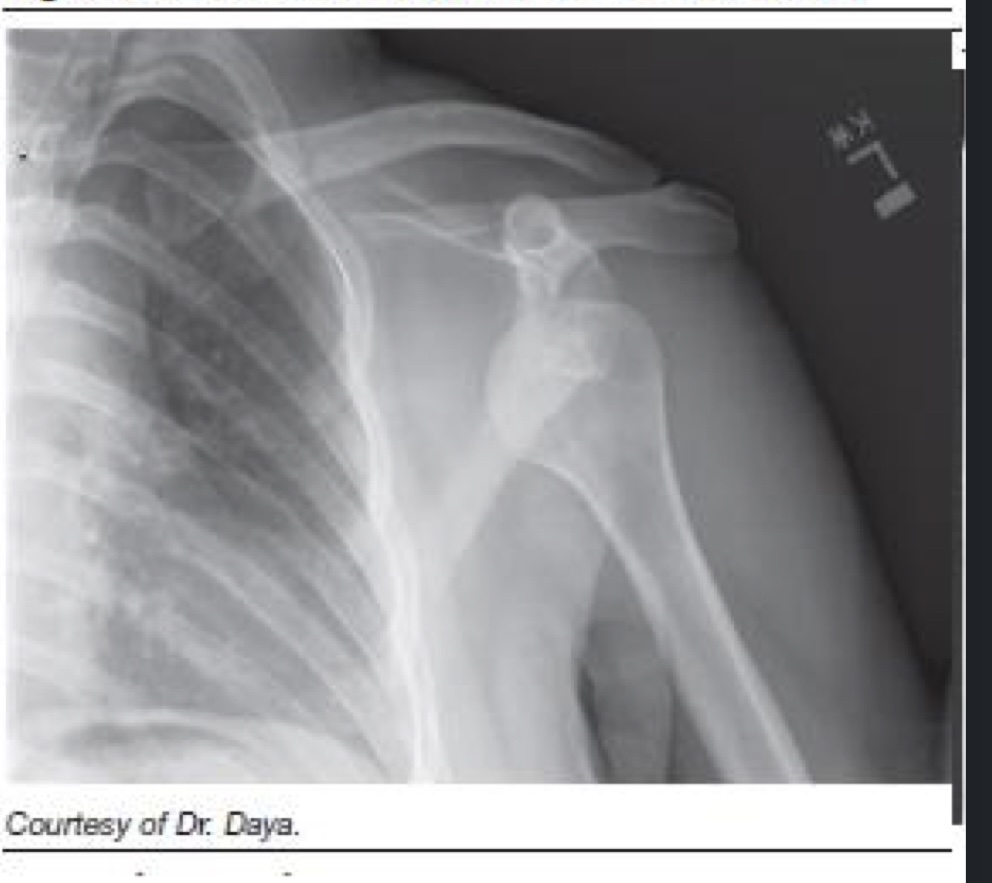
This is another example of a subcoracoid dislocation. It occurs when the humeral head is displaced underneath the coracoid process, typically presenting as an anterior dislocation, which is the most common type.
What is the clinical diagnosis of this dislocation?
This is an example of a subclavicular dislocation, which is less common. This is identified when the head of the humerus is displaced anteriorly and rests below the clavicle. A good indicator of this is when the humerus is close to the ribs and directly below the clavicle, making the coracoid process difficult to identify.
What is the clinical diagnosis of this dislocation?
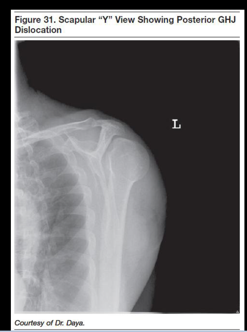
This is an example of posterior dislocation of the humerus. Note the positioning of the humerus, and how it is displaced posteriorly relative to the glenoid fossa. This type of dislocation often results from trauma or seizure activity and may present with a characteristic "square shoulder" appearance. Note: the Y
What is the clinical diagnosis of this dislocation?
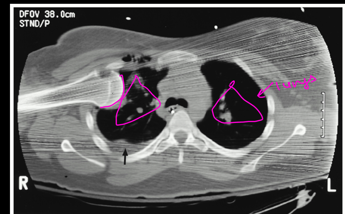
This is an example of an intrathorax dislocation, which is a very uncommon yet severe and life threatening dislocation of the humerus. You can see how the head of the humerus enters the thorax between the ribs and applies pressure to the lungs and other nearby organs.
What is the clinical diagnosis of this dislocation?
This is an example of a posterior dislocation looking anteriorly. This x-ray image shows clearly the displacement of the humeral head relative to the glenoid fossa, emphasizing the posterior movement of the humeral head. Note: This dislocation would most likely injure muscles as well since there are many rotator cuff muscles and stabilizers on the posterior aspect of the GH joint.
What is the main concept behind the “weak point” of the glenohumeral joint?
The "weak point" of the glenohumeral joint refers to anatomical areas, such as the inferior region, where the joint capsule is less reinforced by ligaments. This makes it more susceptible to dislocations and instability. Impact typically travels up to the superior muscles and medial/superiorly placed ligaments, and travels back down inferiorly which is why anterior dislocations are the most common.
What are bursa?
Bursa are closed sacs of synovial membrane which are OUTSIDE the joints and are supportive structures limiting friction during movement. They contain synovial fluid and reduce friction between tissues such as bones, tendons, and muscles. Bursa are compromised of synovial membrane, so they can also make their own synovial fluid.
Where are bursa found?
Bursa are typically found between structures such as 1. tendons and bone, 2. tendons and joints, 3. skin and bone.
What are tendon sheaths?
Tendon sheaths are sheaths of synovial membrane surrounding the tendon. They help reduce friction of the tendon moving and sliding over or through a structure.
What are the 3 bursa of the glenohumeral joint?
Subtendinous bursa of subscapularis
Subacromial bursa
Subdeltoid bursa
Subtendinous Bursa of Subscapularis
Sits between the subscapularis and the fibrous joint capsule. It is formed by the synovial membrane from fibrous joint capsule and communicates with the articular cavity. It functions to reduce friction and cushion the movement of the subscapularis tendon.
Subacromial Bursa
Separates the acromion from the tendon of supraspinatus and does not communicate with the articular cavity. It is fused with the subdeltoid bursa and is the smaller of the two.
Subdeltoid Bursa
This bursa sits between the deltoid muscle and joint capsule. It does not communicate with the articular cavity and is fused with the subacromial bursa.
What is bursitis? Causes?
Bursitis is the inflammation of bursa which is irritation and leads to build up of fluid. Common causes include repetitive motion, injury, and infection.
Bursitis treatment…
Anti-Inflammatories, aspiration, activity changes, corticosteroid injections, recurrent surgical removal of bursa
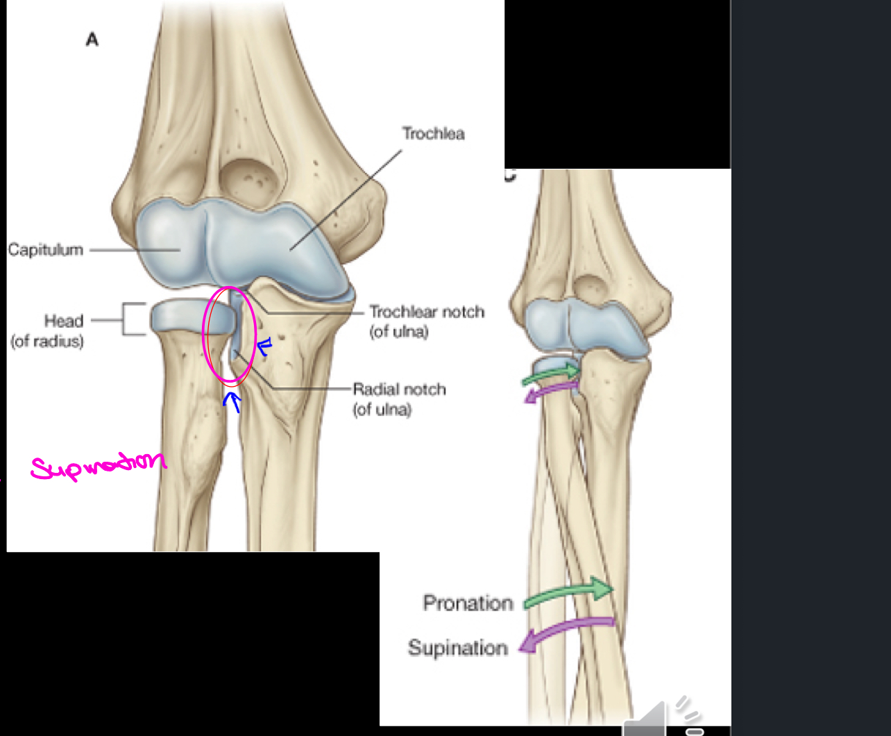
What articulations make up the elbow joint?
Trochlear notch of ulna + trochlea of humerus
Head of radius + capitulum of humerus
Head of radius + radial notch of ulna (proximal radio-ulnar joint)
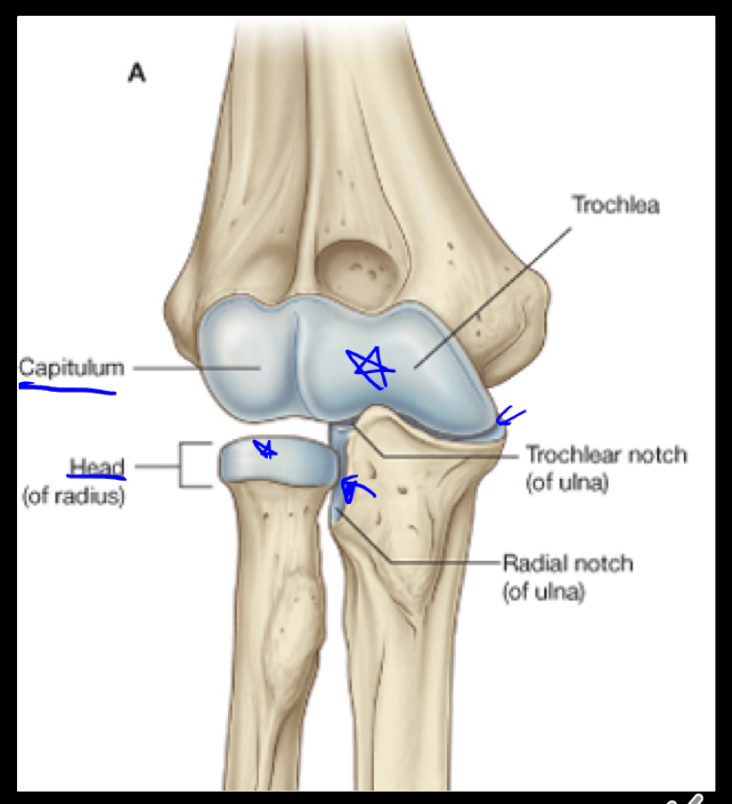
Trochlear notch of ulna and trochlea of humerus articulate to form a ____ joint.
Hinge joint, which allows for extension and flexion.

The head of the radius and capitulum of humerus form a ____ joint.
Hinge joint, which allows for flexion and extension.

The head of the radius and the radial notch of ulna form the ____ joint, which is a ___ joint.
proximal radio-ulnar ; pivot. It helps perform pronation and supination of the forearm.
How many articulations are share the elbow joint synovial membrane?
Three articulations share one synovial membrane cavity.
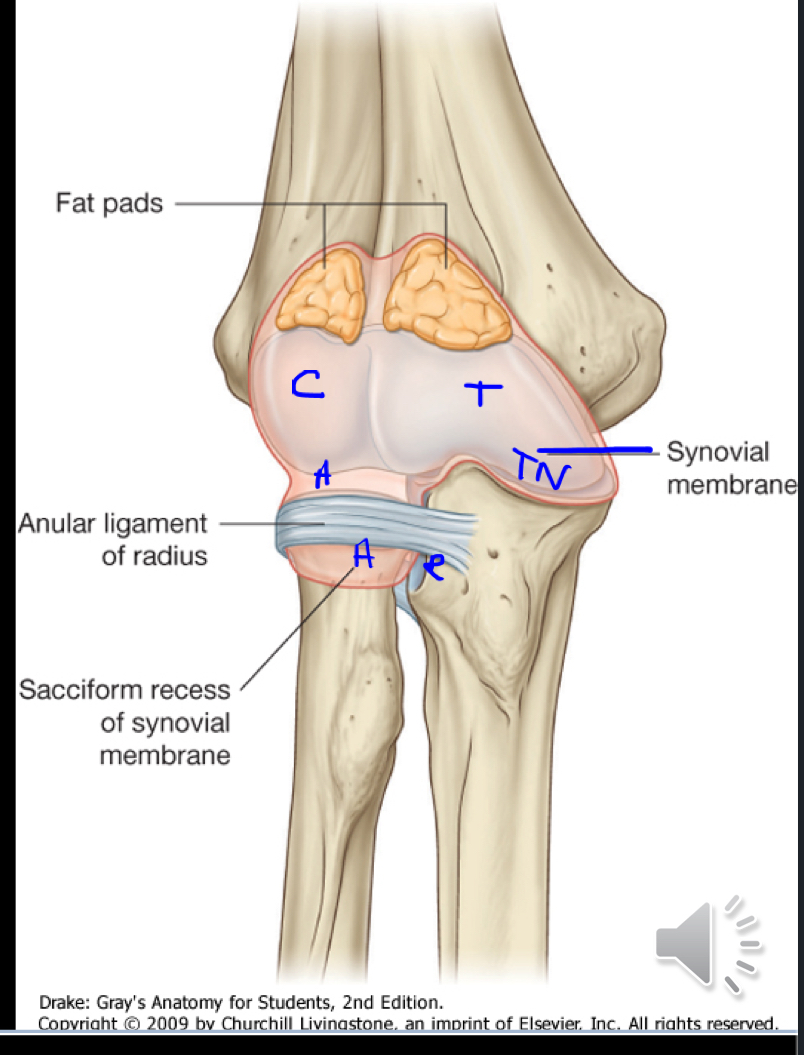
What does the fibrous joint capsule of the elbow attach to?
The fibrous joint capsule of the elbow attaches to the medial epicondyle and the margins of the olecranon, coronoid, and radial fossae of the humerus, as well as to the coronoid process and olecranon of the ulna and the neck of the radius.
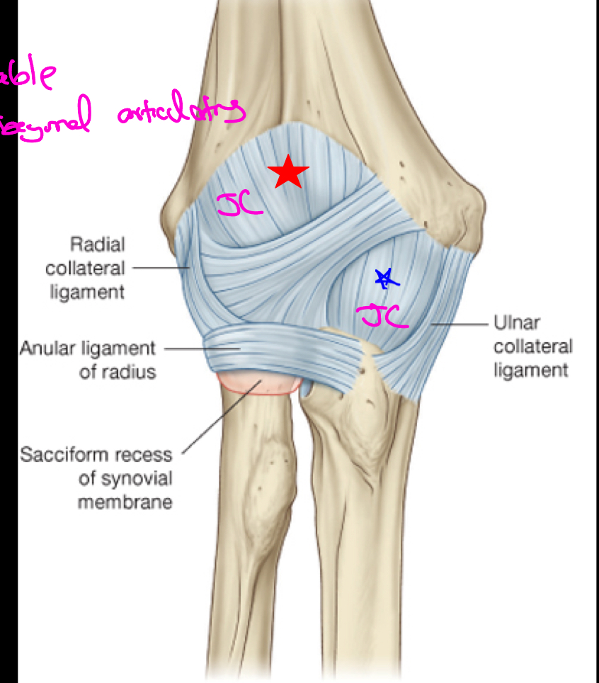
How are ligaments formed?
They are formed from thickened portions of the joint capsule.
What are the ligaments that make up the elbow joint?
Radial collateral ligament: Lateral epicondyle —> Annular ligament (F, E)
Ulnar collateral ligament: Medial epicondyle —> ulna (F, E)
Annular ligament: Around head of radius —> ulna (P, S)
Ulnar Collateral Ligament
It is the main stabilizer of the elbow, from the ulna to the humerus. It is located on the medial side of the elbow and often injured in pitchers.
It keeps the humerus and ulna articulating
Commonly injured with repetitive high speed throwing
What surgery is associated with a tear of the ulnar collateral ligament?
Tommy John Surgery, which involves opening up of the elbows, drilling of holes in the humerus & ulna and the tendon being threaded through them in a figure eight pattern. Over time, the tendon will ligamentize.
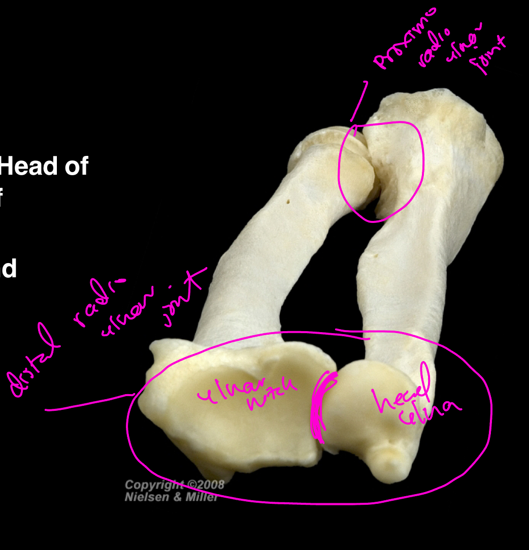
The distal radio-ulnar joint…
Synovial joint; Articulation of the head of ulna and ulnar notch of radius; allows for pronation and supination of forearm.
The Wrist Joint
A synovial joint between the distal end of the radius + triangular disc overlying the distal end of ulna + SLTri

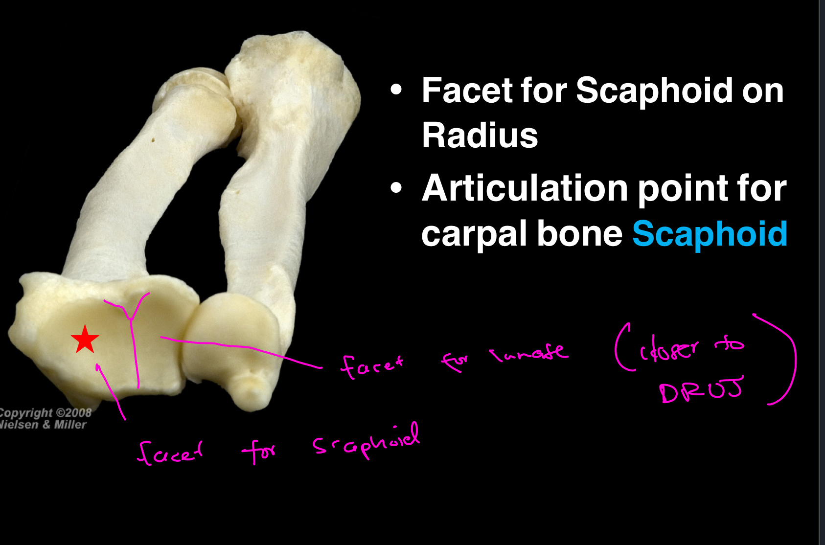
What are the 2 facets associated with the wrist joint?
The facet for scaphoid and the facet for lunate. A good way to remember them is that the facet for lunate is closer to the head of the ulna. BOTH facets are located on the radius.
The main ligaments of the wrist joint are the ____ collateral ligaments and the ___ collateral ligaments.
radial; ulnar
What movements are permitted at the wrist?
Flexion, extension, abduction, adduction, circumduction. VERY mobile joint.
Where are the extensors of the wrist located? Flexors?
The extensors of the wrist are located on the posterior (dorsal) aspect of the forearm, while the flexors are located on the anterior aspect.
All flexors of the wrist insert onto the ____ surface of the hand.
Palmar
All exensors of the wrist insert on to the ____ surface of the hand.
dorsal
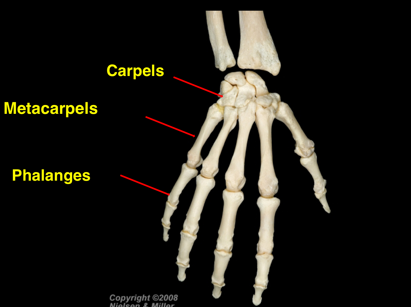
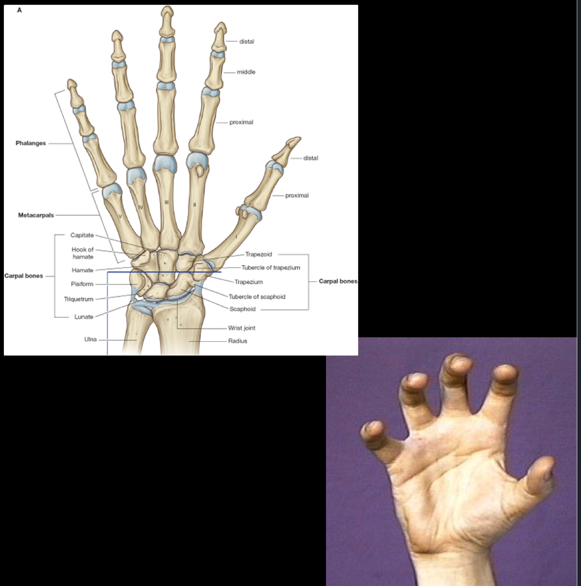
What are the 3 main portions of the hand?
Carpals —> Metacarpals —> Phalanges
The three main portions of the hand are the carpals (wrist bones), metacarpals (bones of the palm), and phalanges (finger bones).
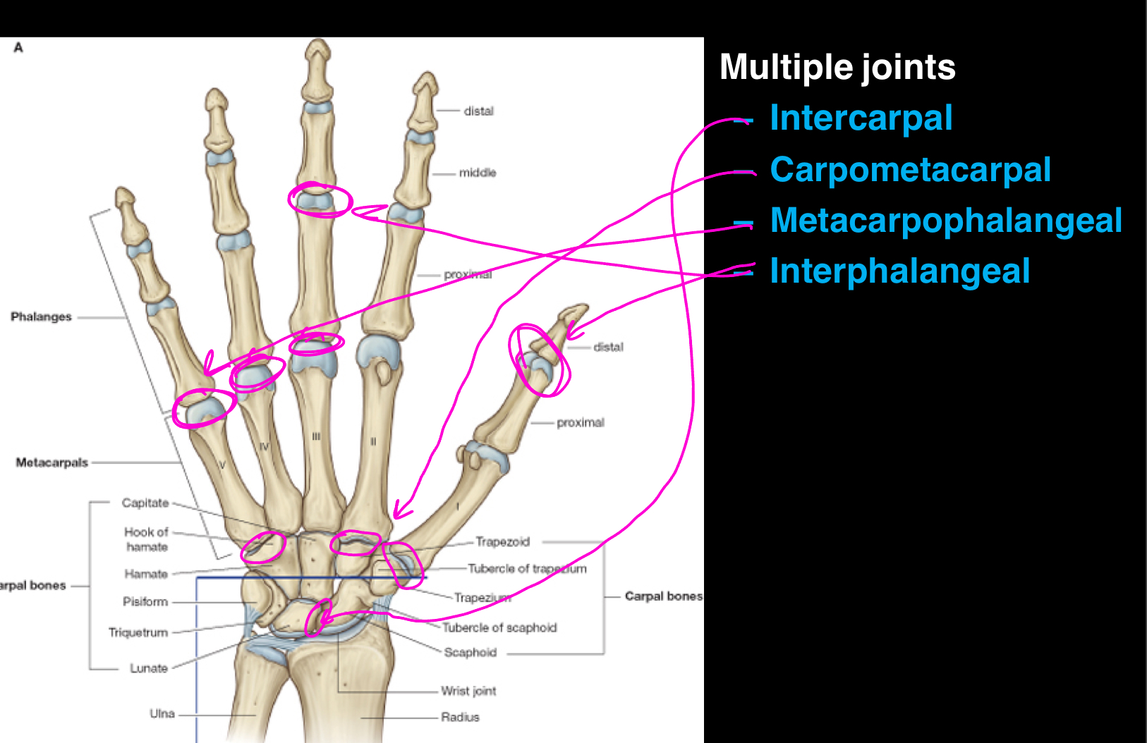
The four types of joints found within the hand are…
Intercarpal (carpal bones to carpal bones),
Carpometacarpal (distal carpal bones to metacarpal base),
Metacarpophalangeal (distal metacarpals to proximal phalanges),
Interphalangeal (between phalanges).
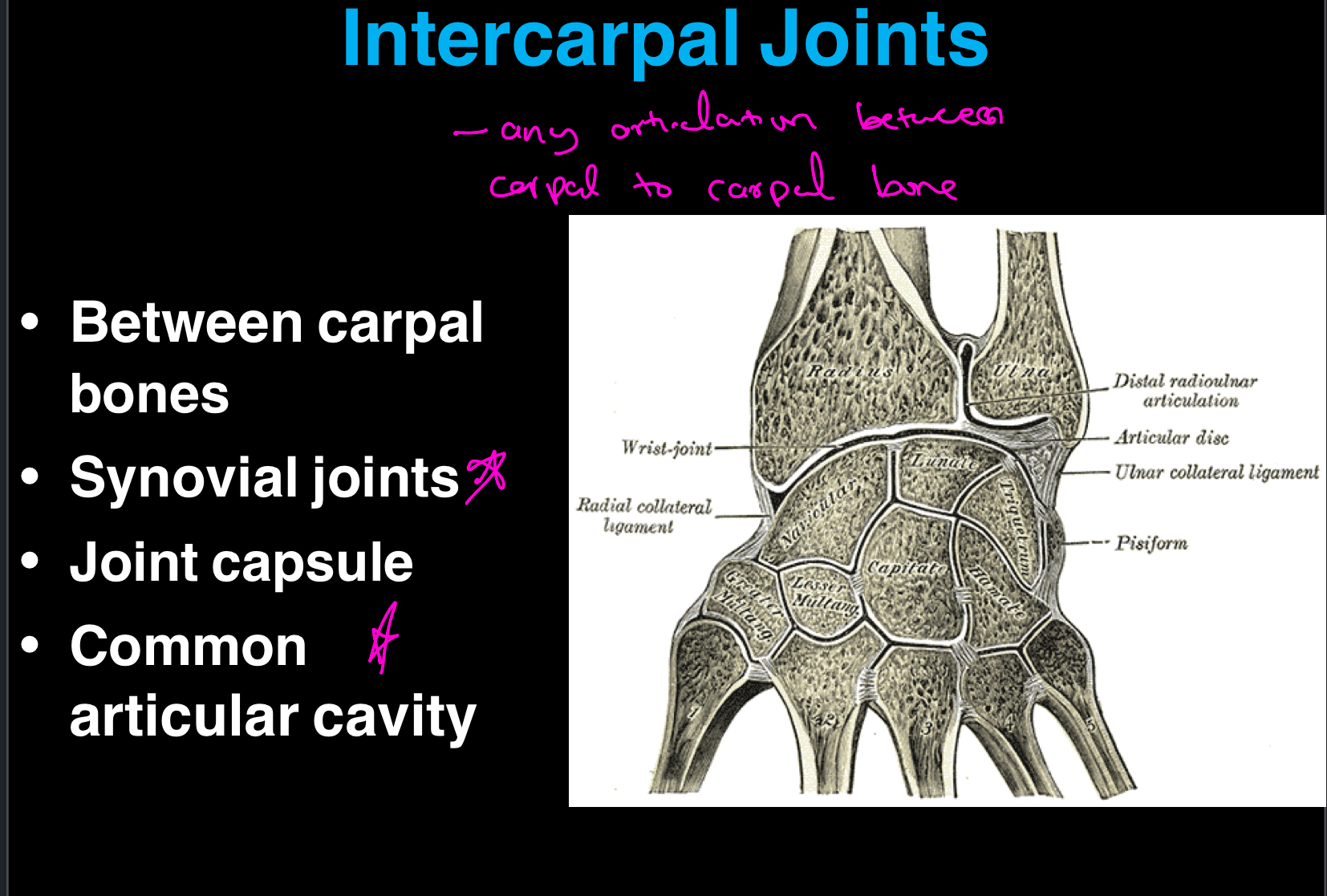
Describe intercarpal joints…
These joints are between carpal bones, ANY articulation between carpal to carpal bone. They are synovial joints, which means they have a joint capsule and a common articular cavity.
It’s important to remember that they have very limited movement and mainly contribute to positioning the hand in abduction, adduction, flexion and extension.
Describe the carpometacarpal joints…
These joints are between the distal row of carpal bones and the base of your metacarpals. There are a total of 5, and they are synovial joints as well. They allow for a range of movements, including opposition in the thumb, and contribute to the overall function and dexterity of the hand.
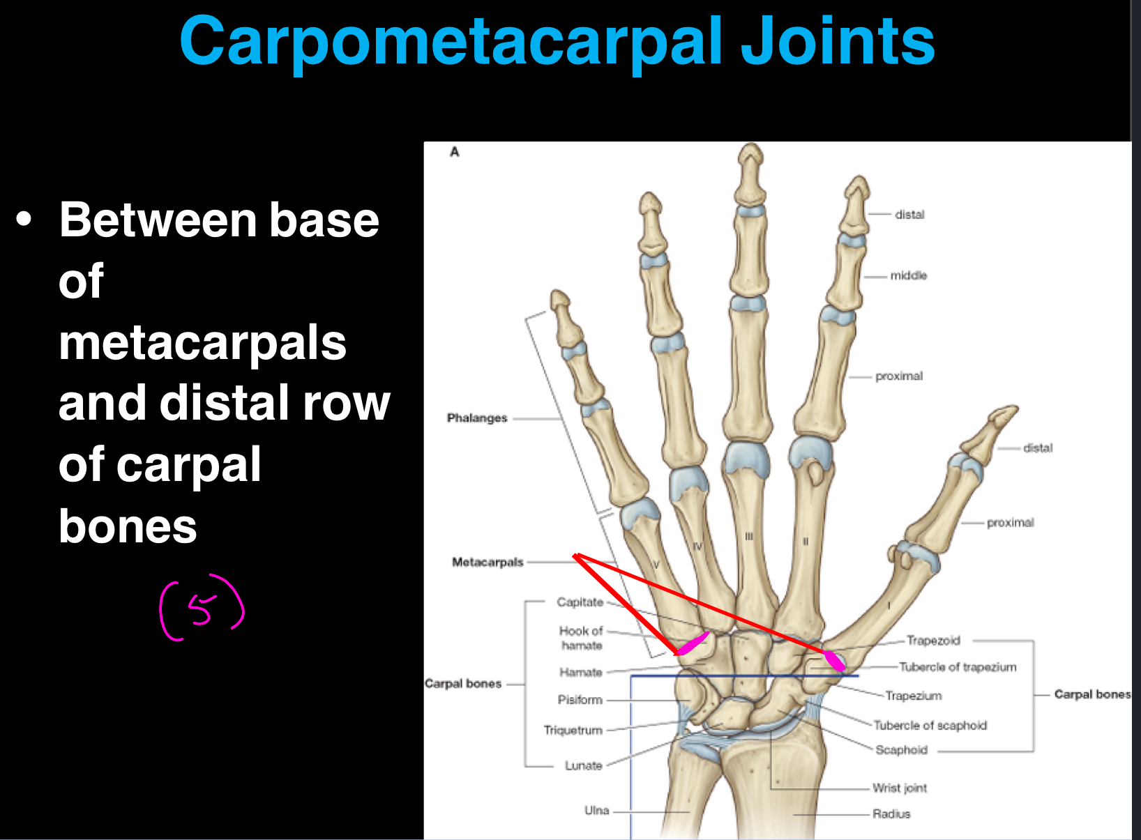
What is the unique carpometacarpal joint?
The unique carpometacarpal joint is the one associated with your thumb (pollox). It is between metacarpal 1 and the trapezium carpal bone and is classified as a saddle joint.
Because of this, you have more movement around this joint compared to others in the hand, allowing for complex functions such as opposition and grasping. (F, E, A, Add, R, Circ)
Carpometacarpal Joints for metacarpals II to V have ___ mobility, ___ gliding movement. However, mobility increases ___.
Less mobility; Limited gliding movement; Mobility increases medially which means metacarpal 5 will glide the most.
Remember: Medial/Lateral is all in reference to anatomical position.
Describe the metacarpophalangeal joints…
These joints are between the distal heads of MCs and the proximal phalanges.
They are classified as condylar joints (type of synovial), and allow for a long range of movements. (F, E, A, Add, C, Limited rotation).
A good way to remember these joints is that they are your first knuckle joints.
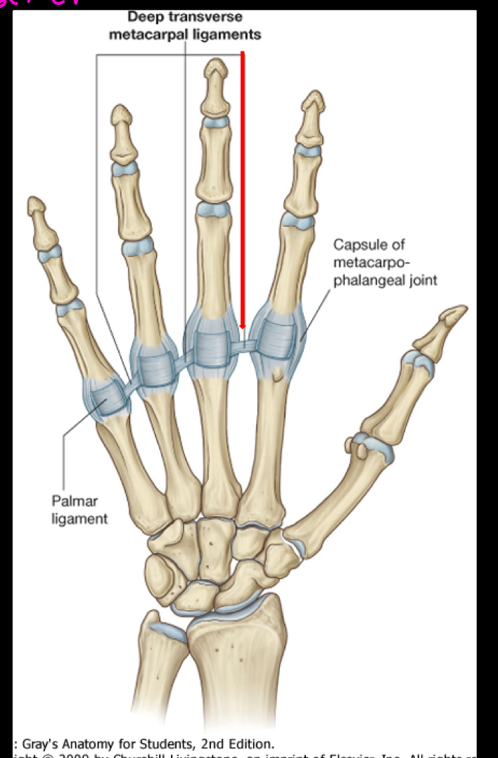
What are the 3 types of ligaments involved in the stability of the metacarpophalangeal joint capsules?
Palmar ligament (Sits right on top of the MCPJ, like a bandaid)
Medial & Lateral Collateral Ligament (Sits on both the medial and lateral side of the MCPJ)
Transverse metacarpal ligaments (Is the tape between each MCPJ that holds everything together going right between the heads of adjacent metacarpals; form the unified palm; absent between digit 2 and thumb)
Describe the interphalangeal joints…
These joints are between the phalanges, so phalange to phalange.
They are hinge joints (again, synovial) and perform flexion and extension.
These joints are supported by your lateral/medial collateral ligaments as well as your palmar ligaments.
The extrinsic extensors of the fingers all share a common innervation which is the….
Posterior interosseous nerve (branch off Radial N)
The muscles included are:
Extensor Digitorum, Indices, Digiti Minimi, Pollicis Longus and Brevis, and Abductor Pollicis Longus.
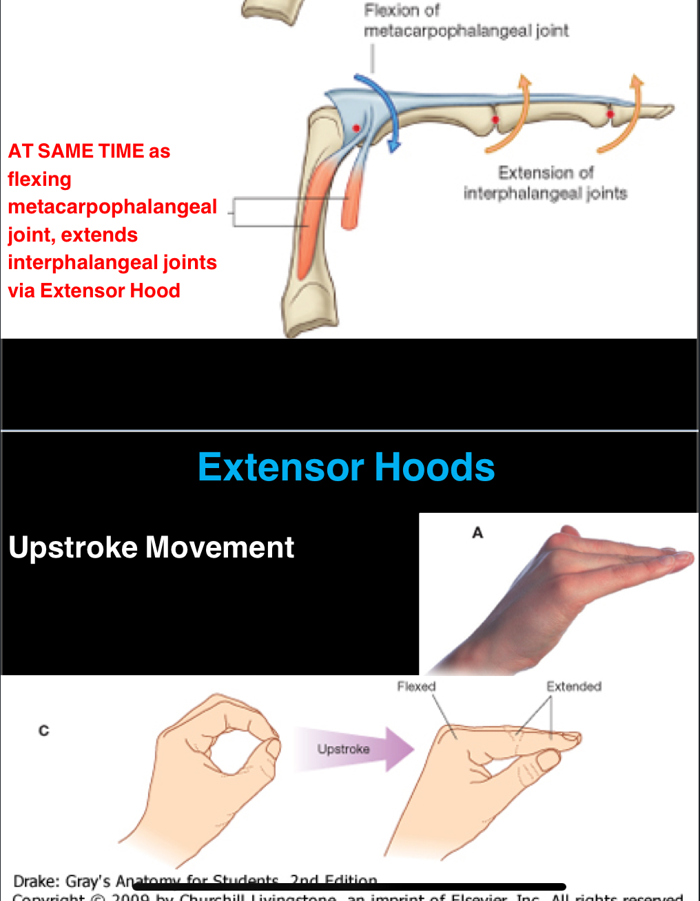
Explain extensor hoods…
Simply put:
The lumbricals attach to two spots, the metacarpophalangeal joint and the extensor hood. The attachment to the MCPJ will do flexion, while the attachment to the extensor hood does extension.
So, the lumbricals are flexing at the MCPJ and extending at the interphalangeal joints.
This allows for an upstroke movement of the fingers during gripping and precise movements. The extensor hood is a structure that spreads across the back of the fingers, allowing coordinated action of muscles for fine motor control.