med anat chapter 10
1/116
There's no tags or description
Looks like no tags are added yet.
Name | Mastery | Learn | Test | Matching | Spaced | Call with Kai |
|---|
No analytics yet
Send a link to your students to track their progress
117 Terms
patella
kneecap
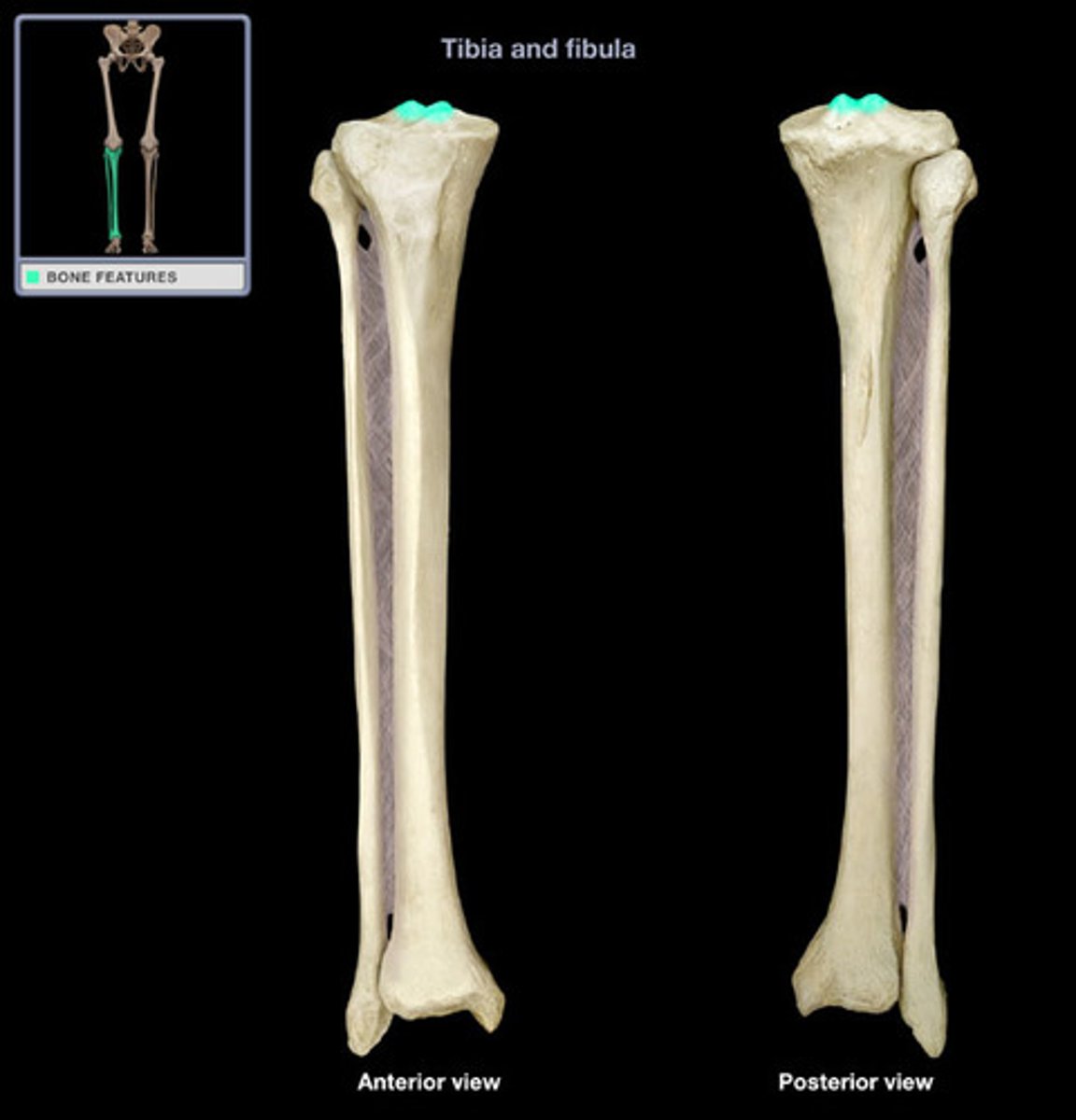
tibia
the medial and larger bone of the lower leg
fibula
The lateral and smaller bone of the lower leg
tibia tuberosity
point where the patellar ligament attaches
tibial condyles
Articulate with the femoral condyles
intercondylar area of tibia
between the condyles
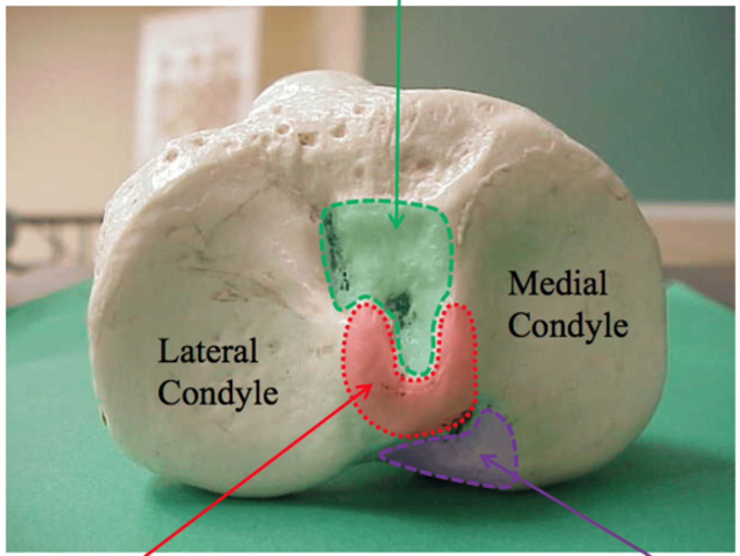
medial intercondylar tubercle
forms the medial part of the intercondylar eminence
anterior facet
for attachment of the medial meniscus
facet under the anterior facet
for attachment of anterior cruciate ligament
small facet
lateral meniscus
anterior on post side
lateral men
posteriormedial facet
medial meniscus
large facet
posterior cruciate ligament
tibial tuberosity
point where the patellar ligament attaches
patellar ligament
connects the tibial tuberosity to the quadriceps tendon
interosseous boarder
along lateral surface of tibia
soleal line
small, diagonally running ridge located on the posterior side of the proximal tibia
attachment sites on fibula
fibular collateral ligament of knee, biceps femoris
styloid process of fibula
Pointed apex of the head of the fibula
anterior of thigh muscles
rectus femoris vastus lateralis vastus medialis, vastus intermedius
medial compartment
gracilis pectineus adductor longus adductor brevi adductor magnus and obturator externus
rectus femoris attachments
AIIS to tibial tuberosity
rectus femoris is innervated by?
femoral nerve
vastus lateralis
extends knee
vastus lateralis attachment
Greater trochanter; lateral lip of linea aspera; "Common tendon"
vastus medialis
extends knee
vastus medialis attachment
Linea Aspera of the Femur to the Tibial Tuberosity via the Patella and the Patellar ligament
vastus medialis is innervated by
femoral nerve
vastus intermedius
extends knee
vastus intermedius attachments
femoral shaft to tibial tuberosity
vastus intermedius innervated
femoral nerve
rectus femoris
extends leg at knee
gracilis
adducts thigh
gracilis attachment
pubis to tibia
gracilis innervated by
obturator nerve
pectineus
adducts, flexes, and medially rotates thigh
pectineus attachments
superior ramus of pubis to pectineal line of femur
pectineus innervated by
femoral nerve
adductor longus
adducts thigh
adductor longus attachment
body and inferior ramus of pubis, linea aspera of femur
adductor longus innervated
obturator nerve
adductor brevis
adducts thigh
adductor brevis attachment
Pubic bone to linea aspera
adductor brevis innervation
obturator nerve
adductor magnus
adducts thigh
adductor magnus attachment
Pubis and Ischium to the Linea Aspera of the Femur
attachment magnus innervation
sciatic nerve
obturator externus
laterally rotates thigh
obturator externus attachment
External surface of obturator membrane; Trochanteric fossa
obturator externus innervated by
obturator nerve
posterior compartment of the thigh
biceps femoris, semitendinosus, semimembranosus
sartorius
Flexes, abducts, and laterally rotates thigh at the hip; flexes knee
sartorius attachment
medial surface of tibia to ASIS
sartorius innervation
femoral nerve
biceps femoris
extends thigh and flexes leg
biceps femoris attachment
long head and head of tibia
biceps femoris innervation
sciatic nerve
semitendinosus
Flexes leg at the knee and extends thigh at the hip; belongs to the hamstring group
semitendinosus attachment
ischial tuberosity and prox tibia
semitendinosus innervation
sciatic nerve
semimembranosus
Flexes leg at the knee and extends thigh at the hip; belongs to the hamstring group
semimembranosus attachment
ischial tuberosity and medial tibial condyle
semimembranosus innervation
tibial division of sciatic nerve
medial meniscus
Cartilage in the knee between the femoral condyle and the medial tibial plateau
lateral meniscus
cartilage in the knee between the lateral femoral condyle and the lateral tibial plateau
patellar ligament
continuation of the quadracepts femoris
collateral ligaments
Ligaments that run along the sides of the knee and limit sideways motion
cruciate ligaments
intercondylar refuon and connects femur and tib
tibiofibular joint
pertaining to the joint between the tibia and fibula
fibular collateral ligament
connects the lateral epicondyle of the femur to the fibula
anterior ligament
Anterior surface of the uterus to the posterior surface of the bladder. Forms vesicouterine pouch.
popliteal fossa
the hollow at the back of the knee
gastrocnemius
plantar flexes foot
gastrocnemius attachments
condyles of femur, calcaneus
gastrocnemius innervation
tibial nerve
plantaris
plantar flexion
plantaris attachments
supracondylar line of femur and oblique pop ligament
plantaris innervation
tibial nerve
soleus
plantar flexes foot
soleus
soleal line line of tibia and calcaneal tendon
soleus innervation
tibial nerve
popliteus
back of knee
popliteus attachments
lateral femoral condyle and proximal tibia
popliteus innervation
tibial nerve
flexor hallucis longus
flexes great toe
flexor hallucis longus attachment
Attachment: distal posterior fibular shaft --> base of distal phalanx of hallux
flexor hallucis longus innervation
tibial nerve
flexor digitorum longus
flexes toes, plantar flexes and inverts foot
flexor digitorum longus attachment
tibia to distal phalanges of 4 toes
flexor digitorum longus innervation
tibial nerve
tibialis posterior
Plantar flexion and inversion of foot
tibialis posterior attachment
originates from the interosseous membrane and adjacent borders of the tibia and fibula. It inserts onto the navicular tuberosity, the medial, intermediate, and lateral cuneiform bones, and the bases of 2nd to 4th metatarsals.
tibialis posterior innervation
tibial nerve
popliteal artery
leg and foot behind the knee
fibularis longus
plantar flexes and everts foot
fibularis longus attachment
lateral surface of fibula
base of 1st metatarsal
medial cuneiform
fibularis longus innervation
superficial fibular nerve
fibularis brevis
plantar flexes and everts foot
fibularis brevis attachments
lateral surface of fibula, tuberosity of 5th metatarsal
fibularis brevis innervation
superficial fibular nerve