Thalamus and Internal Capsule
1/22
There's no tags or description
Looks like no tags are added yet.
Name | Mastery | Learn | Test | Matching | Spaced | Call with Kai |
|---|
No analytics yet
Send a link to your students to track their progress
23 Terms
Thalamus overview
Comprised on the dorsal thalamus and the thalamic reticular formation (ReT)
Conveys input to sensory pathways to cortex (except olfactory)
Info from lower centers to cortex ( ie basal ganglia, cerebellum, hypothalamus)
Gate of info to cortex
Associates info from different sensory modalities and conveys this info to cortical areas involved in attention and executive functions.
Coordinates activity over widespread of cortex for the purpose of cortical arousal.
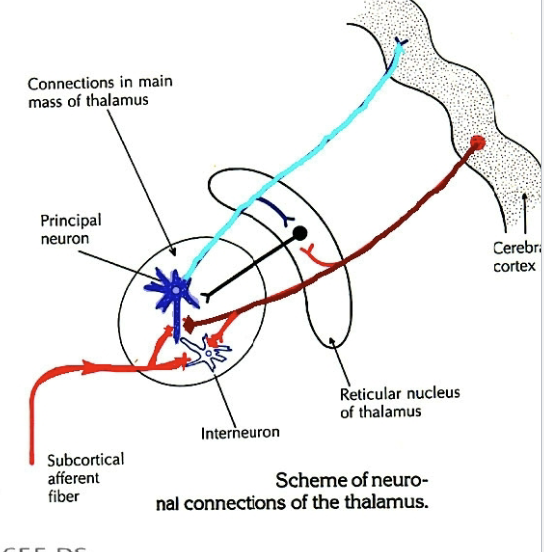
Thalamocortical projections
Nuclei of thalamus (expect fro ReT) reciprocally connected w/ cortex via. excitatory projection neurons.
Cortex sends projections (corticothalamic fibers) back to individual thalamic nuclei.
Corticothalamic projections more numerous than thalamocortical projections.
Corticothalamic projections thought to be modulatory inputs to the thalamus (important for info processing)
Thalamic nuclei characterized based on nature of their thalamocortical projections.
Connection properties
Specific vs no specific
Specific
Efferent projections (thalamocortical fibers) to a particular functional area on the cortex —> relays info to these area
Specific rapid, localized evoked responses in the ipsilateral cortex.
Non Specific
Sends thalamocortical projections to widespread areas of cortex + produce more generalized cortical activation.
Produces widespread activity in both hemispheres with a longer time delay.
Connection properties
Relay vs association nuclei
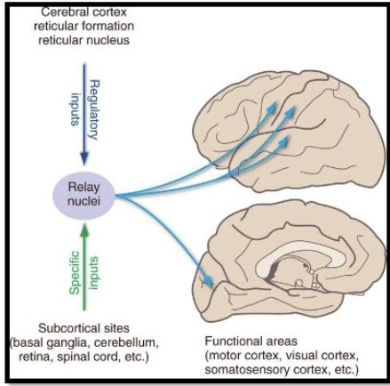
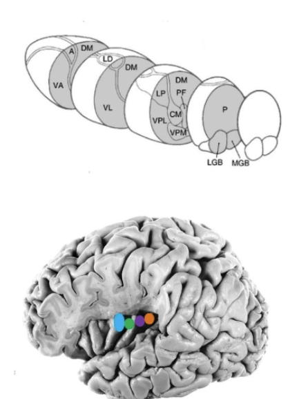
Relay nuclei (first order relay)
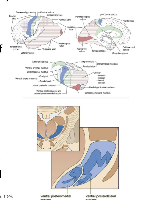
Send projections to primary cortical areas (S1/V1/M1) and covery info from subcortical inputs ( ie. sensory, cerebellum, basal nuclei, mammillary bodies)
Found within the anterior and lateral thalamus.
Sensory relay nuclei
Projections from these nuclei serve as final segment of pathways conveying sensory info from the periphery to the primary sensory fields of the cortex.
Types of nuclei found in this group:
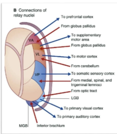
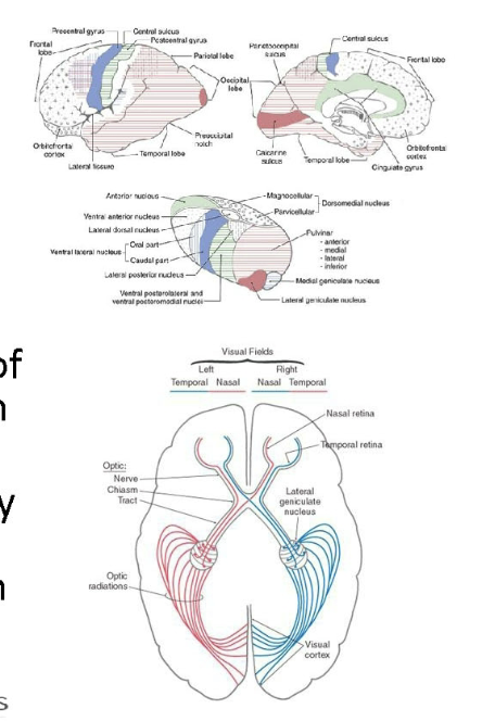
Ventral posterior sulcus - Somatosensory
Lateral geniculate nucleus - Vision
Medial geniculate nucleus - Audition
**** Olfactory input reaches the olf. cortex w/o traveling through thalamus.
Sensory relay nuclei
Ventral posterior complex (VP)
Ventral posteriorlateral nucleus (VPL)
Ventral posteriormedial nucleus (VPM)
Ventral posterior inferior nucleus (VPI)
Contents
Ventral posteriorlateral nucleus (VPL), site of termination of somatosensory pathways representing body
Ventral posteriormedial nucleus (VPM), where trigeminal representing face and oral cavity terminate.
Ventral posterior inferior nucleus (VPI) in a small ares between VPL and VLM
VPI to oral part of VPL receives vestibular info and project to the posterior insular vestibular cortex (PIVC) @ the posterior ends of the inusla and vestibular cortex of the pariteal lobe (in depths of central sulcus(3a) and rostral tip of the intrapariteal sulcus (2v).
VP projects to the primary somatosensory cortex (S1) associated with the postcentral gyrus and posterior paracentral gyrus of the pariteal lobe.
Sensory relay nuclei
medial parvicellular portion of the central posteriormedial nucleus (VPMpc)
Sometimes describes separately as the ventromedial basal (VMb) nucleus
Receives taste fibers directly
General visceral afferent info (via. parabrachial nucleus) from solitary nucleus.
Tase
Conveys to gustatory cortex of the inner frontal operculum and insula.
GVA
Info. conveyed by projections to insula —> GI tract, CV input, and respiratory input represented caudal to the tase representation.
Sensory relay nuclei
Ventromedial posterior thalamic nucleus (VMpo)
More caudal in the ventral medial thalamus lies the ventromedial posterior thalamic nucleus (VMpo)—> receives pain and temperature (+ itch and touch)
Info. from antlat. tract and projects to the dorsal posterior insula (caudal to the viscerosensory representation in insula)
Dorsal insula receiving input from VMb and VMpo considered “interoceptive cortex” representing phys. condition of the body.
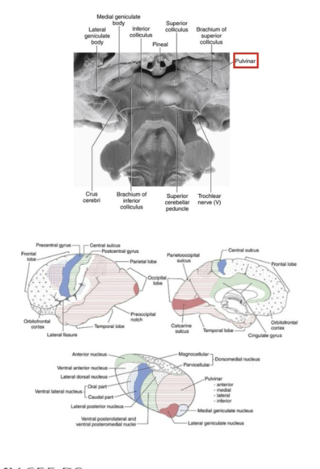
What constitutes the metathalmaus and what is this a continuation of?
Lateral and medial geniculate bodies constitutes the metathalmaus and may be considered as the caudal continuation of the ventral nuclear mass of the thalamus.
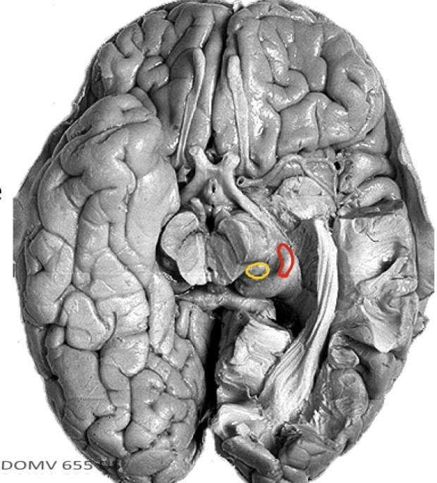
Lateral and medial geniculate nucleus
Lateral geniculate nucleus
LGN is a small, rostrolaterally directed projection from the posterior thalamus.
Laminated, receives input from the retina of both eyes via fiber traveling in the optic tract
Projects to the primary visual cortex (V1) found on the banks of the calcarine sulcus on the medial surface of the occipital lobe.
Medial geniculate nucleus
Recieves auditory input from the inferior colliculus through its brachium.
MGN projects to the primary auditory cortex (A1) associated with the transverse temporal gyri of Heschl
Motor relay nuclei
Projections from these nuclei convey info from the cerebellum and the basal nuclei to the motor cortical fields of the frotnla lobe.
Nuclei within this group:
Ventral anterior nucleus (VA) - Basal Nuclei
Ventral lateral nucleus (VL) - Cerebellum
Ventral anterior nucleus (VA)
Ventral anterior nucleus (VA)
Receives afferent from the output structures of the basal nuclei, the internal segment of the globus pallidus and the pars reticulata of the substantia nigra.
SNrp targets —> more medial magnocelluar part of VA
Parvocellular —> input via GPi
VA sends projections to the frontal eye fields (BA8) and the prefrontal cortex
Contributes to loop circuits of basal nuclei system (Oculomotor, motor, associative)
Involved in motor planning and behavior
Ventral lateral nucleus (VL)
Input from both basal nuclei and. cerebellum
Rostral part of VL (VLo or VLa) receives input from the GPi and projects to premotor cortices (including SMA)
Contributes to motor loop circuit of basal nuclei (motor planning)
Caudal portion (VLp) receives input from the deep nuclei of the cerebellum and projects to motor cortex
Contributes to cerebellar circuits for modulating motor activities (limb movements)
Limbic Nuclei
Contributes to the memory circuit of the limbic system
Nuclei within this group:
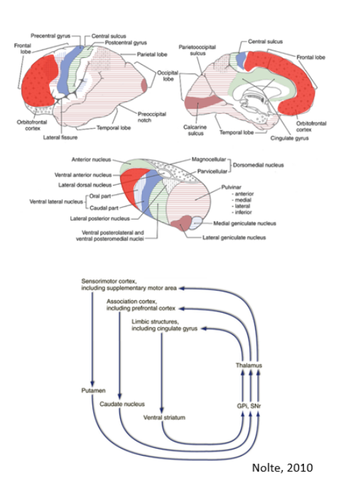
Anterior nuclear group
Lateral dorsal nucleus (LD, can be considered a dorsal extension of the anterior nucleus w/ simular limbic connections)
Anterior nuclei group
Consists of 3 nuclei enclosed by split internal medullary lamina (may be refereed as anterior nucleus)
Caudal lateral to the inter ventricular foramen
Input via hippocampal formation directly via. the fornix and the mammillary bodies (mammilothalamic tract)
Projects primary to cingulate cortex of limbic lobe
Anterior nucleus and its projects are part of the central circuit (memory processing) called circuit of Papez
Connection Properties
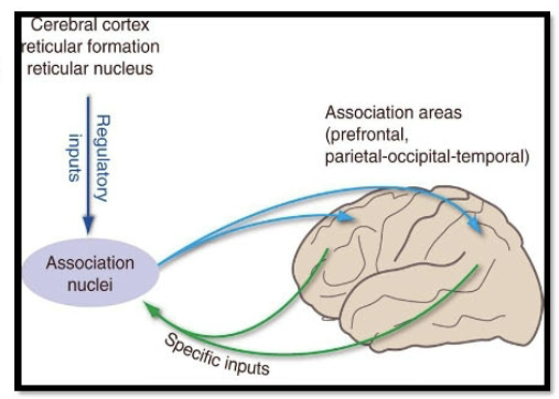
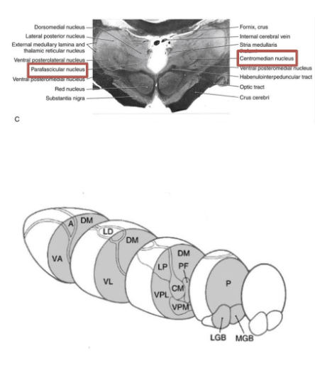
Association nuclei
Higher order relay
Send projections to association cortices (ie pariteo-occiptal, prefrontal) and relays info from primary cortical areas (+ subcortical areas)
Bound within the medial thalamus and posterior part of the dorsal thalamus.
Largest nuclei of the thalamus which has strong reciprocal connections with association areas of the cerebral cortex.
Included nuclei
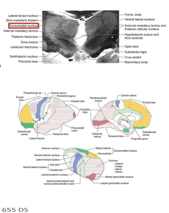
Medial dorsal nucleus (MD)
Pulivinar
Lateral posterior nucleus (LP)
Association nuclei
Medial dorsal nucleus (MD)
Important linking the frontal lobe circuits involved in attention, decision making, and behavioral planning via connections with the amygdala interfaces w/ emotional network of limbic system
Receives input from the amygdala, olfactory cortex (+entrorhinal cortex), substantia nigra, and anterior lateral system
Projects reciprocally to the entire prefrontal cortex (+FEF) and anterior cingulate cortex.
Damage = affected executive functions (judgements, decision making) and affective behaviors.
Association nuclei
Pulivinar and Lateral posterior nucleus (LP)
Pulvinar (L pillow) projects from back of the thalamus dorsal and lateral to the midbrain. Consists of 4 subnuclei.
LP merges with pulvinar (difficult to distinguish caudal boarder)
LP-pulvinar complex receives input from the sup. colliculus, pretectum, and visual cortex of the occipital lobe (vision) + unimodal sensory and associated cortices of the parietal and temporal lobe
Projects to multimodal association cortices of the posterior parietal lobe and lateral temporal lobe.
Major role in spacial attention.
Gate function
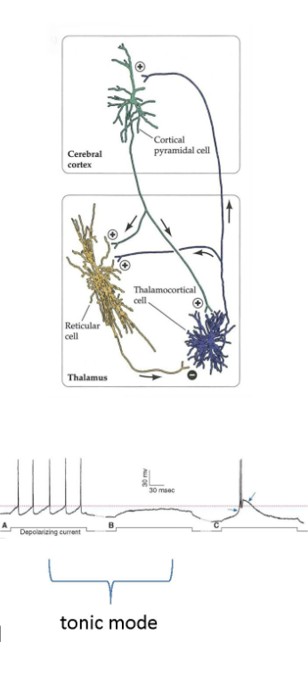
Non specific nuclei
Reticular nucleus (ReT)
Derivative of the ventral thalamus which contains GABAergic neurons that project to all thalamic nuclei
Receives collateral from thalamocortical and corticothalamic projection cortices (unlike nuclei of the dorsal thalamus, these do not project to the cortex)
Gates activity of neurons within thalamic nuclei (ie. related to saliency or focusing on a sensory or motor modality)
Important for synchronization of cortical activity during sleep (non REM) —> sleep spindle and delta wave activity
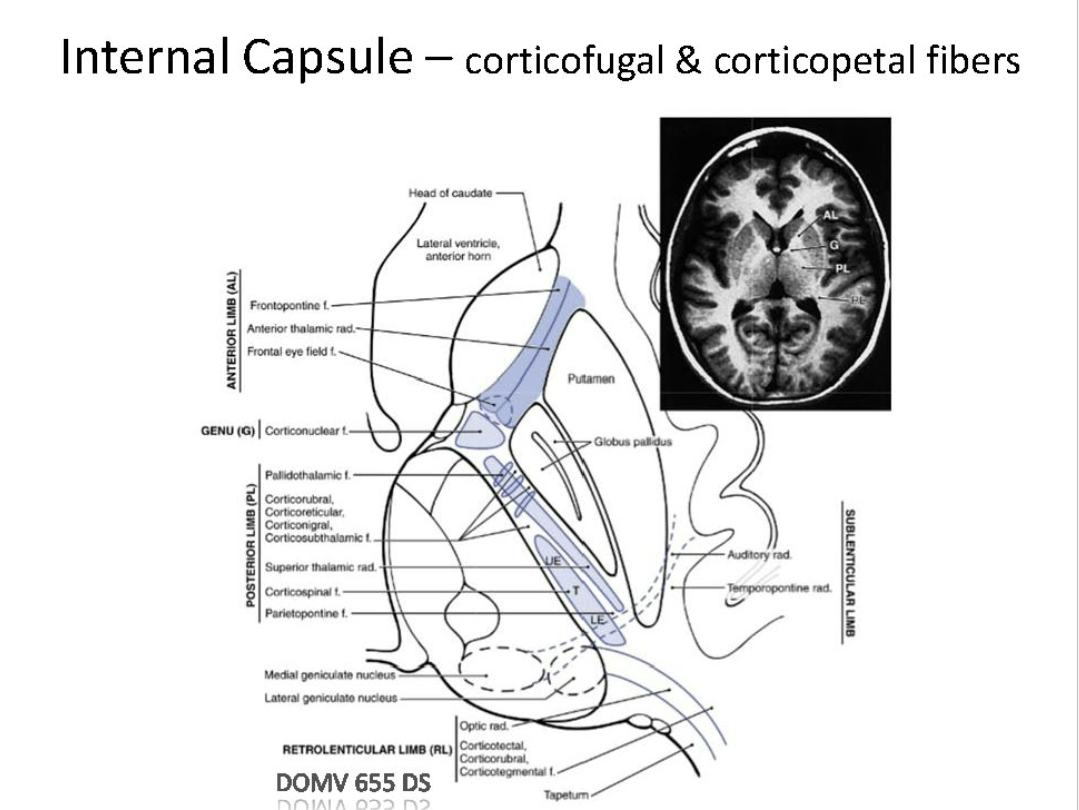
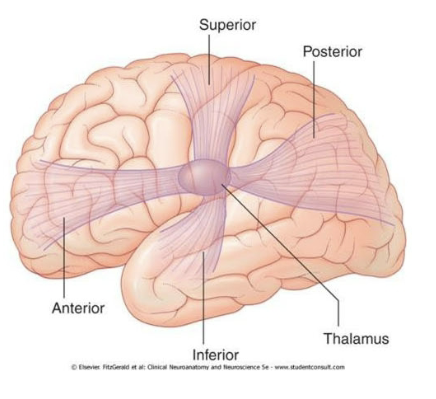
Thalamic radiation
Fibers connecting the thalamus w/ the cortex are grouped into 4 main bundles —>radiations or preduncles —>projects via. internal capsule.
Anterior thalamic radiation
Superior thalamic radiation
Posterior thalamic radiation
Inferior thalamic radiation
Anterioior radiation
Fibers connecting mediodorsal nucleus and anterior nucleus with the frontal lobe and cingulate cortex. (travels via ant. limb of int. capsule)
Superior radiation (aka central thalamic radiation)
Fibers connecting VP w/ parietal lobe and VA-VL w/ frontal lobe (travels via central limb of int. capsule)
Posterior radiation
Fibers connecting LGN w/ occipital lobe (geniculocalcarine tract - optic radiation)
Projects via retrolenticular part of the internal capsule
Also has pulvinar projections —> occipital lobe and posterior parietal lobe.
Inferior radiation
Fibers connecting MGN w/ temporal lobe (auditory radiations)(travels via sublentriuclar part of int. capsule)
Internal capsule
Structural relationships
Thalamocortical projections
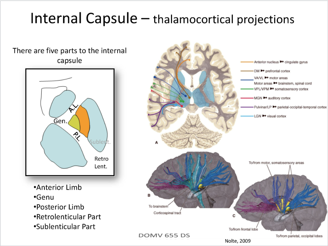
Internal capsule
Corticofugal and corticopetal fibers