Lecture 6 - Neuro 3; AP, Refractory period, Myelin, Synapses, Ca2+
1/21
There's no tags or description
Looks like no tags are added yet.
Name | Mastery | Learn | Test | Matching | Spaced | Call with Kai |
|---|
No analytics yet
Send a link to your students to track their progress
22 Terms
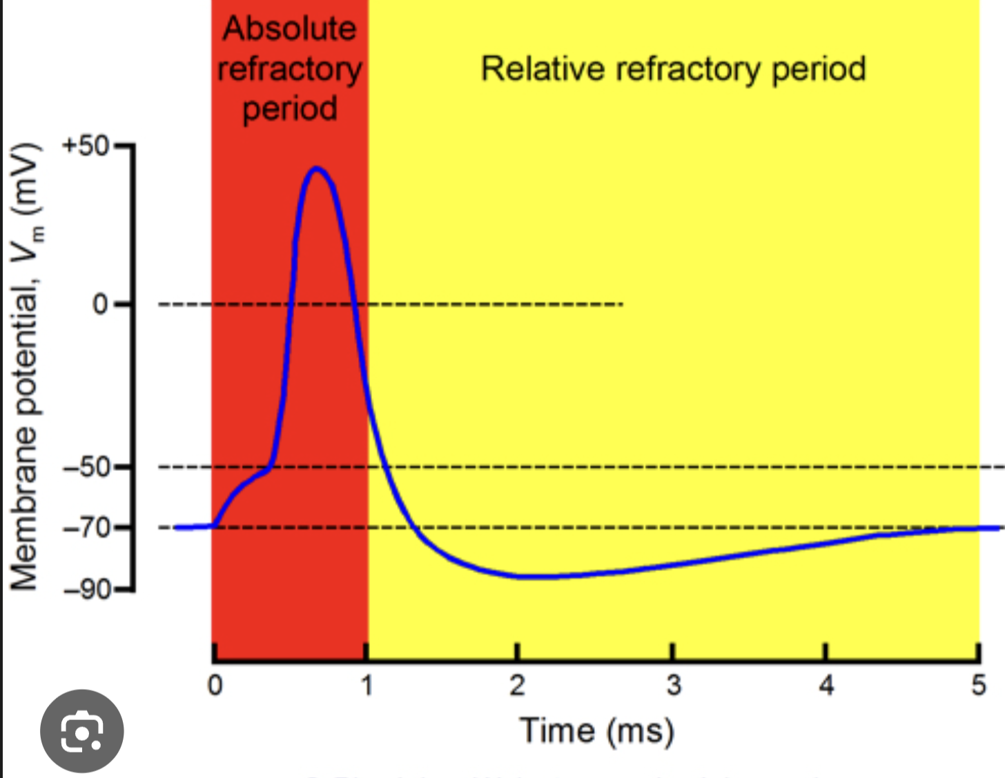
How many refractory periods in an action potential?
2
Absolute and relative
Absolute refractory period
Time during which another AP can’t be triggered
No matter the stimulus strength
Occurs form the onset of AP (start of depolaritzation) until end of Na+ inactivation (end of repolarization)
Why it happens (key mechanism)
Voltage-gated Na⁺ channels are INACTIVATED
The inactivation gate physically blocks the channel
Channel cannot reopen until the membrane repolarizes
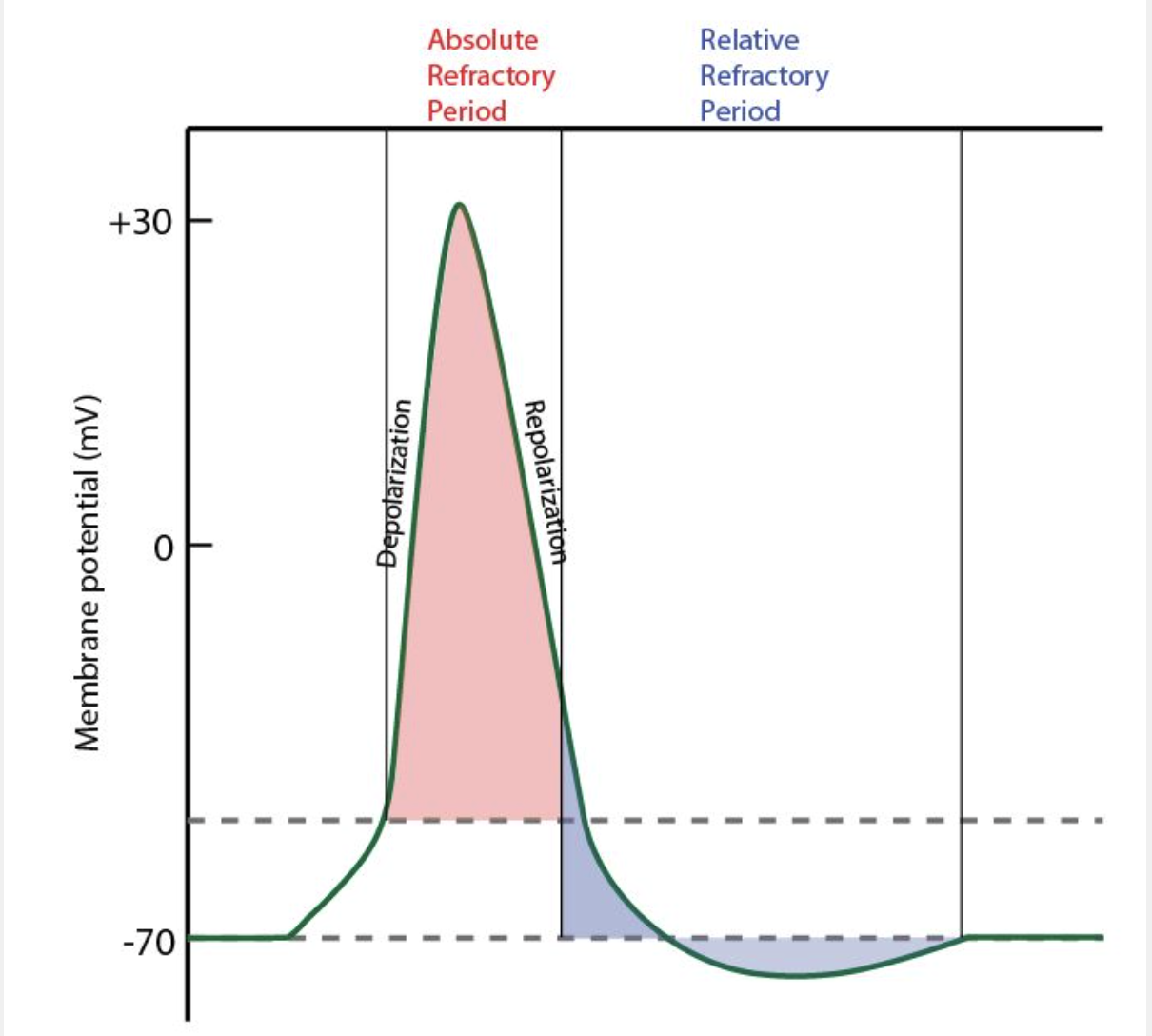
Relative refractory period
Time during which a stronger stimulus is necessary to trigger an AP
More depolarizing current is required to reach threshold
Due to hyperpolarization
If we were to get a stimulus of the same size that triggers a normal action potential → no AP fired, because, we are starting more negative than usual, and therefore need a more positive signal
Two things are happening:
1. Some Na⁺ channels are still inactivated
Fewer available Na⁺ channels
Harder to reach threshold
2. K⁺ channels are still open
High K⁺ permeability
Vm is hyperpolarized (more negative than RMP)
→ Threshold is effectively farther away
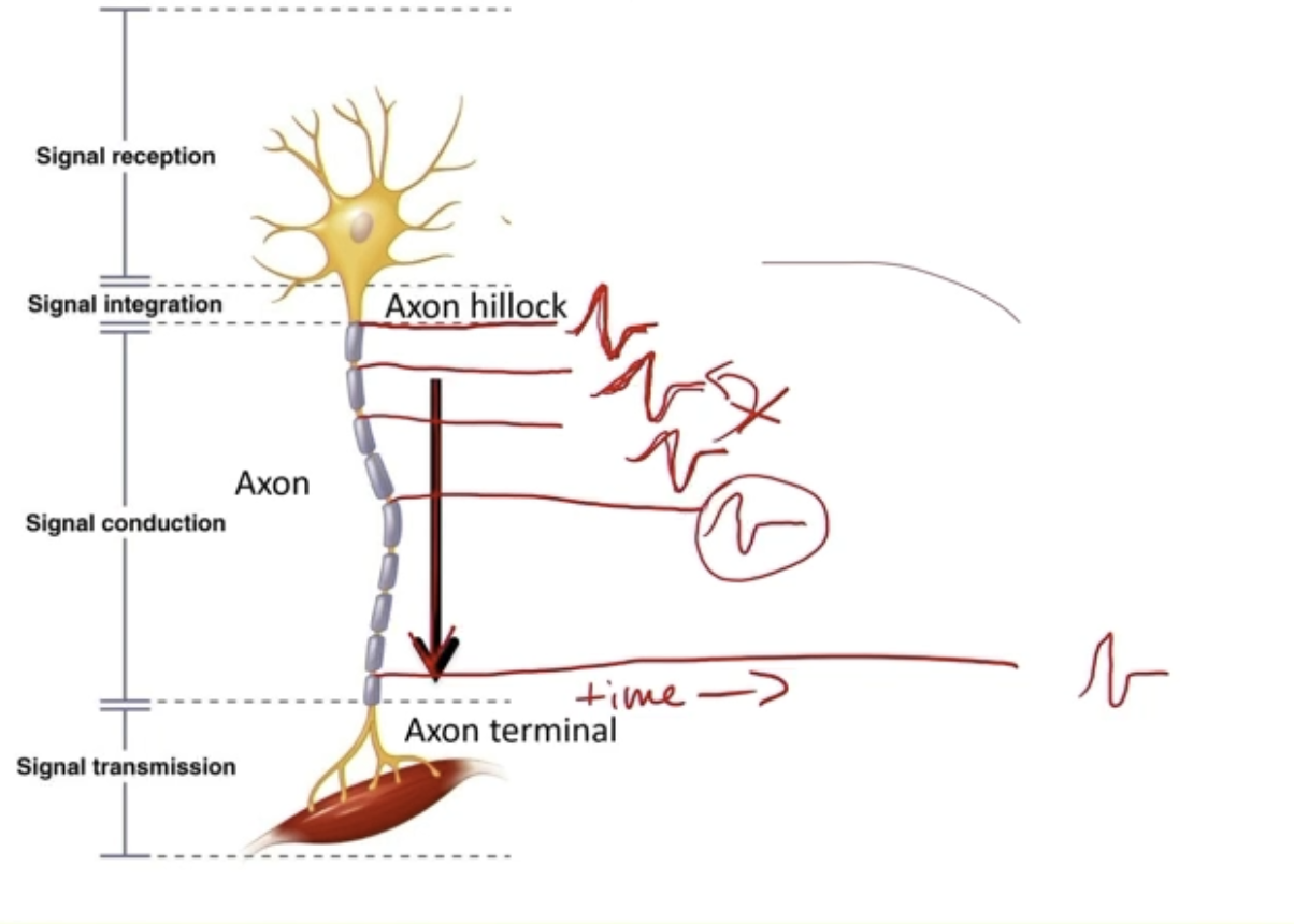
Action potential depolarization spread
AP spread from the Axon hillock to the Axon terminal
Signals spread through myelin sheaths, fire an AP at each Node of Ranvier, where Voltage-gated channels are concentrated at
All AP have same shape because there are the same Voltage gated Na+ and K+ down the axon, which determine its shape
AP spreads DOWN in ONE DIRECTION
Absolute refractory period prevents the AP from going backwards, since Na+ channels behind the AP are inactivated and can’t reopen immediately
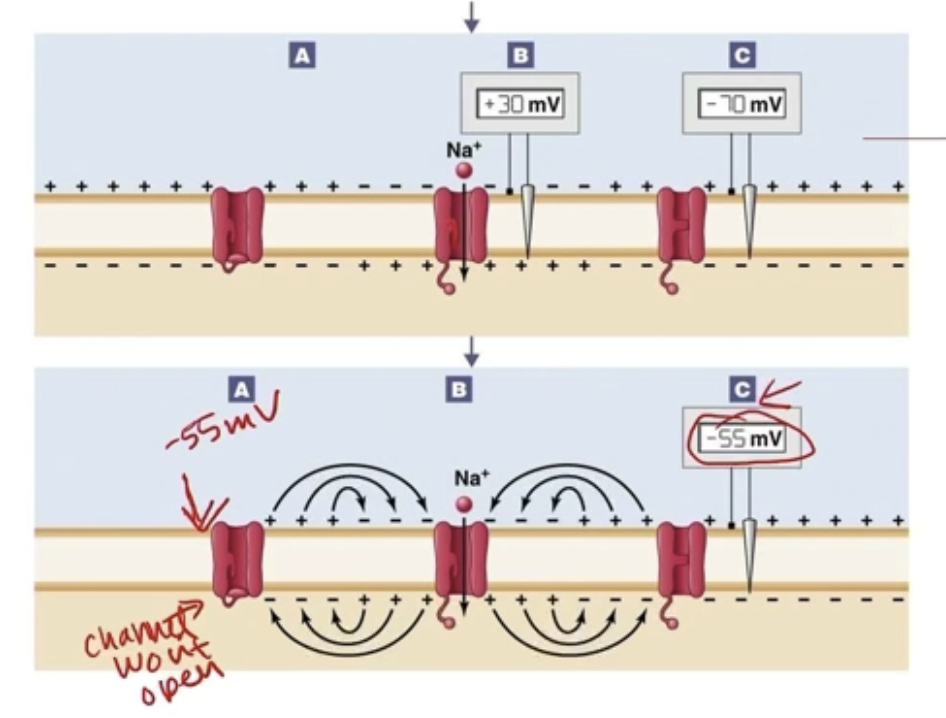
Na+ Channels flow
When Na+ ions flow in, positive current flows passively in both directions
Passive current spread in both directions
Depolarizes the Na+ voltage channels forward and backwards, but Na+ channels backwards can’t open since they’ve been inactivated
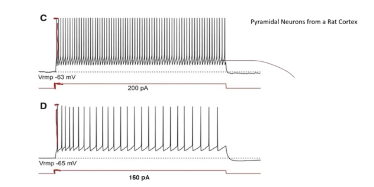
AP potentials stereotypes durations and amplitude - Stimulus strength?
AP all look the same
Same height
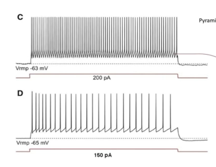
Stimulus strength is indicated by AP frequency
Stronger stimulus = more frequent AP
Weaker stimulus = less frequent AP
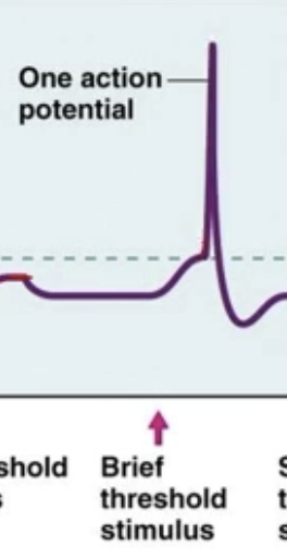
Subthreshold stimulus gives…
0 AP
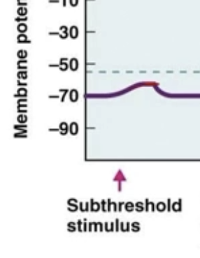
Brief threshold stimulus gives…
1 AP
Sustained stimulus gives…
Multiple AP
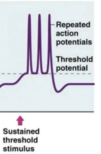
Suprathreshold stimulus
A stimulus strong enough to depolarize the membrane past threshold, even during the relative refractory period
During sustained input → higher firing frequency
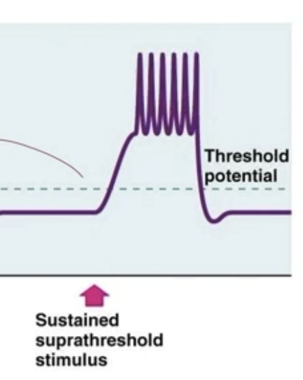
No hyperpolarization between spikes?
Stimulus’s current overpowers the hyperpolarization and keep the AP at depolarization
Spiking immediately after Absolute, enough stimulus to overcome Relative
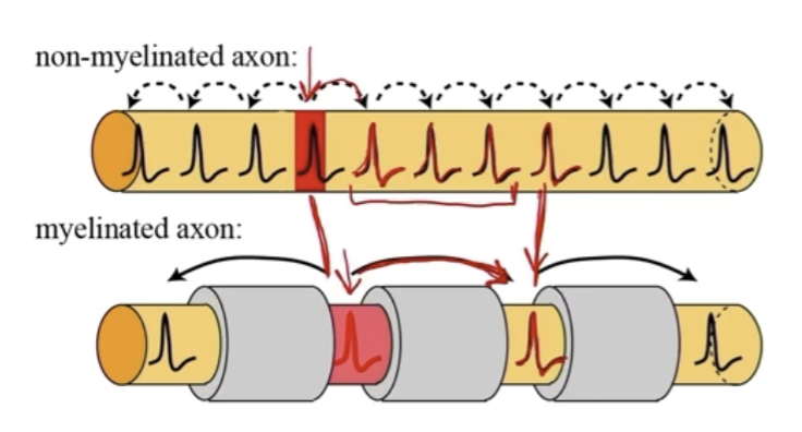
Myelinated Axons
Myelin sheath insulates regions of the axon
Prevents ions from leaving the cytoplasm
Restricts action potential to Nodes of Ranvier
Current passively flows through myelinated areas
Myelination increases length constant
Motor neurons tend to have long myelinated axons
Long, want fast signals
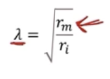
Myelination effect on Length constant
Myelination changes resistance of the membrane, increasing the length constant
Allows signal to travel distance without decaying
𝜆 = length constance
rm = membrane resistance
ri = intracellular resistance
More myelinated and bigger axon diameter = faster
Saltatory conduction
Signal travels faster through internodes than at Nodes of Ranvier
Each AP takes same amount of time
Heavily myelinated: 15-150 m/s
Lightly myelinated: 3-15 m/s
Unmyelinated: 1 m/s
Triggers AP in directly adjacent region
Will have more Voltage-gated ion channels in total
Can’t just myelinate the whole axon, since need AP to “re-up” current at each point
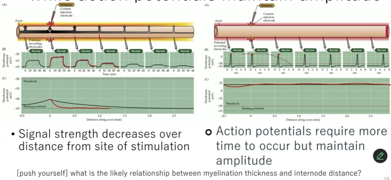
Passive current / Electrotonic spread vs AP
Passive current decreases with distance
Signal strength decreases over distance from site of stimulation
AP maintains amplitude
AP require more time to occur but maintain amplitude
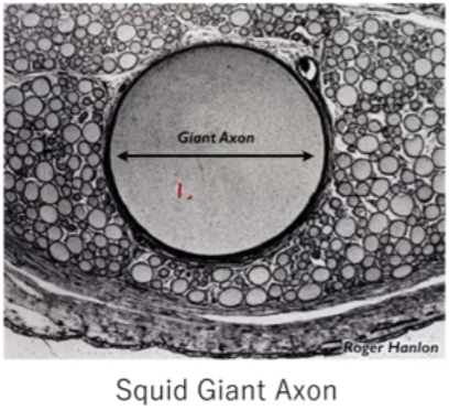
Increase diameter of axon
Larger-diameter axons increase AP conduction speed
Changes intracellular resistance
Increase length constant - allows current to spread farther and decay more slowly
Fastest axon = fat diameter and super myelinated
Diameter size relates inversely to intracellular resistance
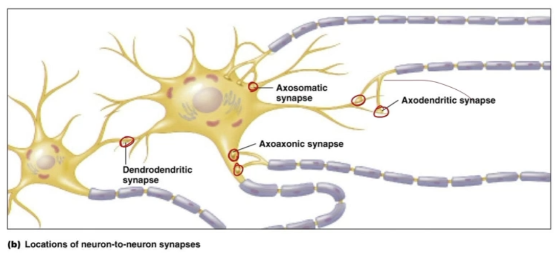
Synapse
Where one neuron passes to another neuron
Can be in a variety of places: soma, axon hillock, but most commonly:
Axodendritric synapse
Presynaptic vs postsynaptic neuron
Pre-synaptic - The sending neuron
Action potential arrives at the axon terminal
Voltage-gated Ca²⁺ channels open
Ca²⁺ enters the terminal
Post-synaptic - the receiving neuron
Neurotransmitter binds to receptors on dendrites or soma
Receptors are usually ligand-gated ion channels
Ion flow produces graded potentials:
EPSPs (excitatory)
IPSPs (inhibitory)
These graded potentials determine whether an action potential will fire
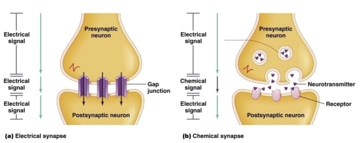
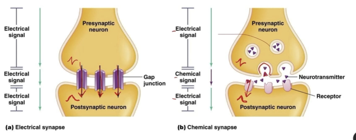
Presynaptic to Postsynaptic neuron
2 strategies:
Electrical synapses
Current passes from pre to post
Ions can move (current flow) through gap junctions
***NO NEUROTRANSMITTERS
Don’t create Graded potentials
Transmit signal, don’t generate anything
Chemical synapses (more common)
Depolarization (AP) in pre triggers release of chemical neurotransmitters (due to Ca2+ influx), which bind to the post’s neurotransmitter receptors
Receptors are typically ligand-gated channels
Ions can go in/out the post, causing depolarization
*ESSENTIALLY: Electrical → chemical → electrical
Create graded potentials (ESPS / ISPS)
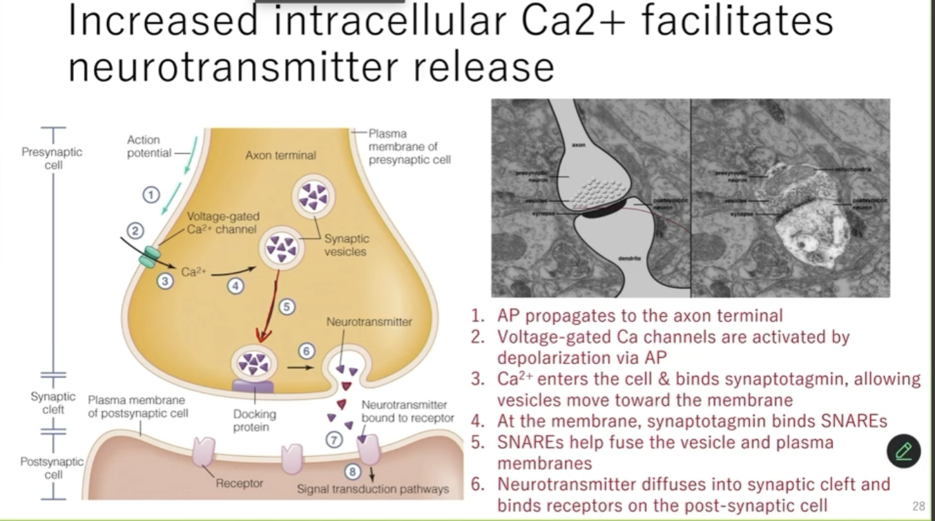
Neurotransmitters exocytosis
Synaptic versicles release neurotransmitter by exocytosis
Neurotransmitter vesicles in pre will fuse with the pre’s membrane, releasing neurotransmitters into the synaptic cleft, which would diffuse towards the post
Exocytosis of Neurotransmitters specific steps
Action potential arrives at presynaptic terminal
Membrane depolarization opens voltage-gated Ca²⁺ channels
Ca²⁺ enters the presynaptic terminal
Ca²⁺ triggers vesicle fusion with the presynaptic membrane
Via binding to Synaptotagmin (movement of vesicles), which binds to SNAREs (helps with fusion of vesicles to presynaptic membrane)
Vesicles release neurotransmitter via exocytosis
Neurotransmitter diffuses across the synaptic cleft
Neurotransmitter binds postsynaptic receptors
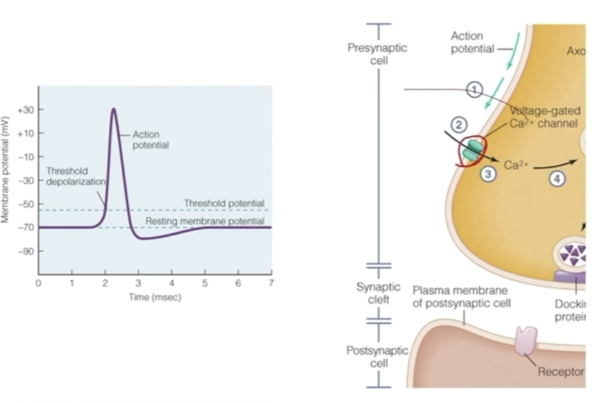
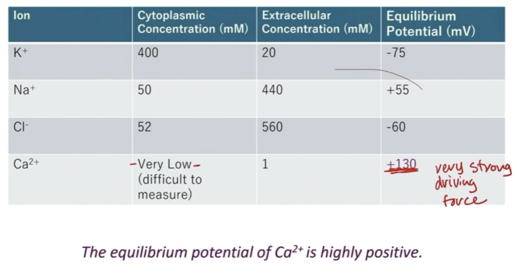
Calcium voltage channels
Depolarization in Axon terminal from AP opens Ca2+ voltage gated ion channels
These channels are only present at the terminal
Low amounts of Ca2+ in cells (higher on outside), very high Eq (+130), so very high driving force
So when Ca2+ channels open, influx of Ca2+ inside the cell
Small amount of huge signaling effect
Calcium rushes in and binds to protein Synaptotagmin, causing a conformational change in synaptotagmin
A protein on vesicle membrane
Causes vesicles of neurotransmitters to move towards the membrane
Synaptotagmin binds to protein SNAREs, helping vesicles and plasma membrane fuse by holding them close together
Vesicle membranes fuse with presynaptic membrane, and releases neurotransmitter into synaptic cleft
Neurotransmitters diffuse across cleft, bind to receptors, …