BIOL 260: Ch. 9b, 10, 11
1/144
There's no tags or description
Looks like no tags are added yet.
Name | Mastery | Learn | Test | Matching | Spaced | Call with Kai |
|---|
No analytics yet
Send a link to your students to track their progress
145 Terms
Muscles of Face Diagram pt.1
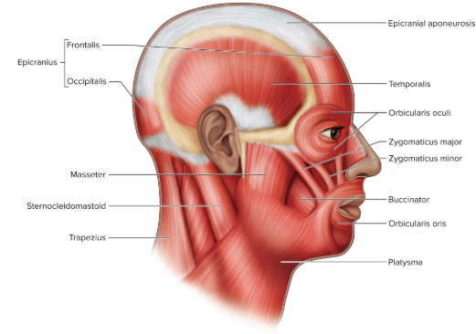
Muscles of Face Diagram pt. 2
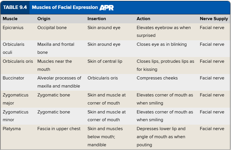
Muscles of Facial Expression Chart
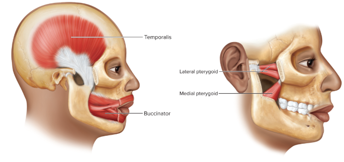
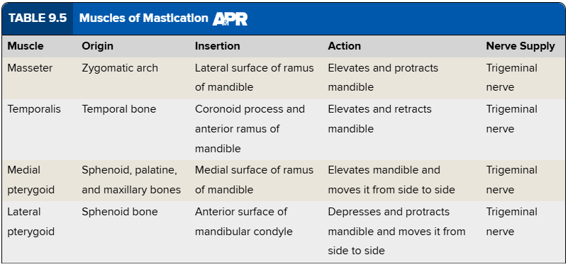
Muscles of Mastication Chart
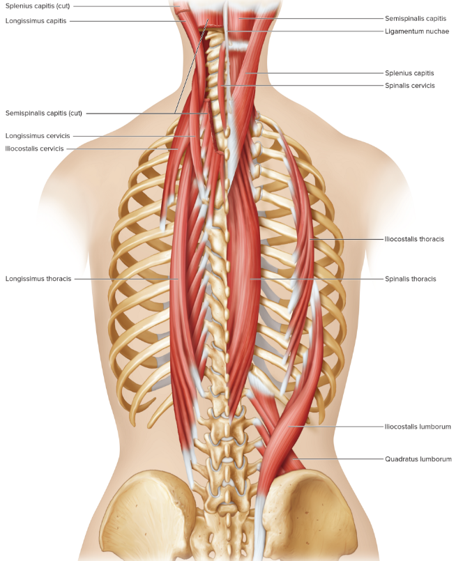
Muscles of Neck and Vertebral Column Diagram
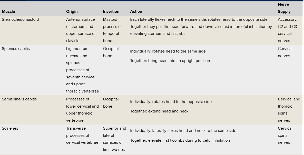
Muscles That Move the Head and Vertebral Column Chart pt.1
Muscles That Move the Head and Vertebral Column Chart pt.2
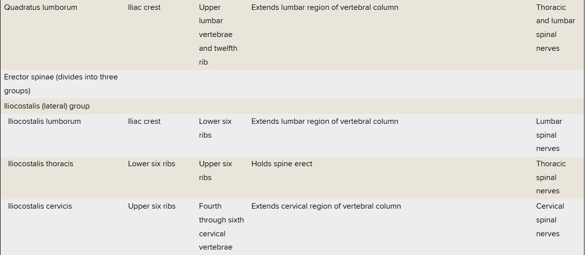
Muscles That Move the Head and Vertebral Column Chart pt.3
Muscles That Move the Head and Vertebral Column Chart pt.4
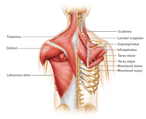
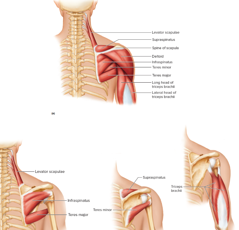
Muscles of Shoulder and Back Diagram
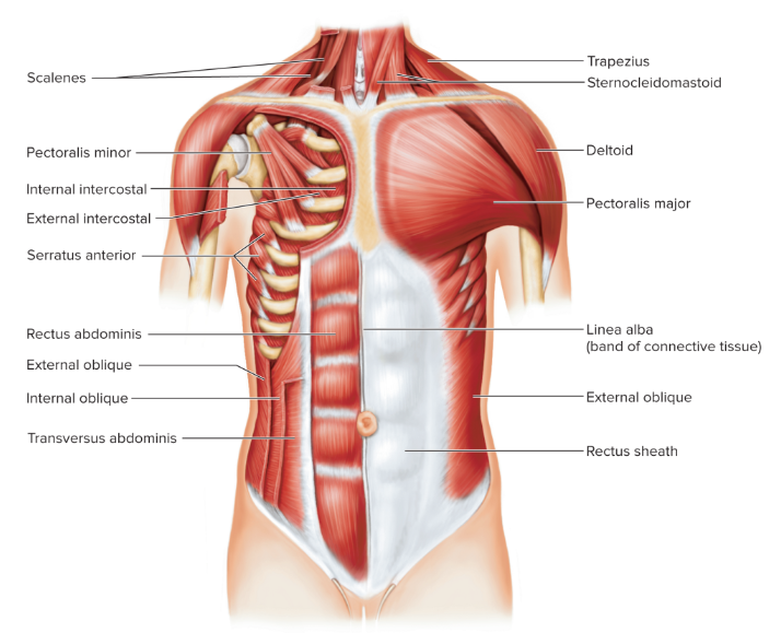
Muscles of Anterior Chest and Abdominal Wall Diagram
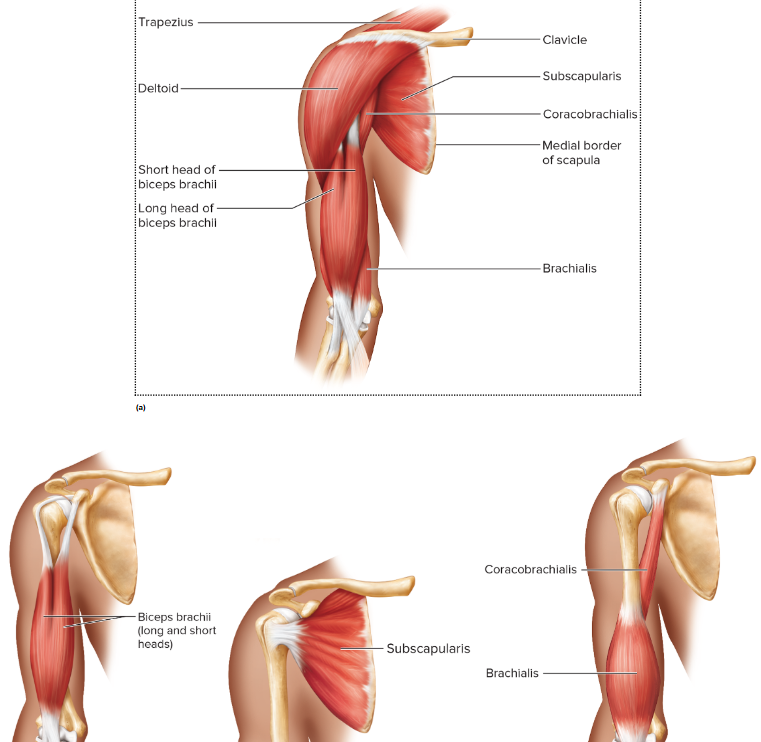
Muscles of Shoulder and Arm Diagram
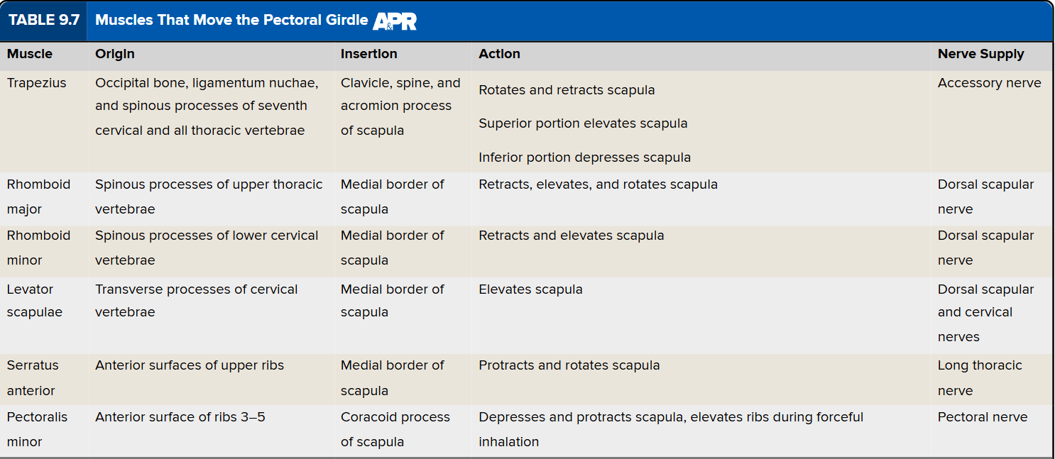
Muscles That Move the Pectoral Girdle Chart
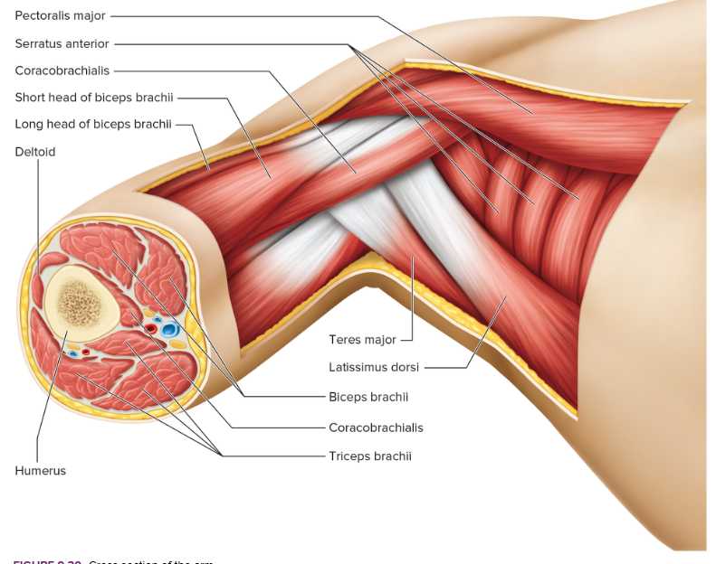
Cross Section of Arm Diagram
Muscles of Shoulder and Arm Diagram pt. 2
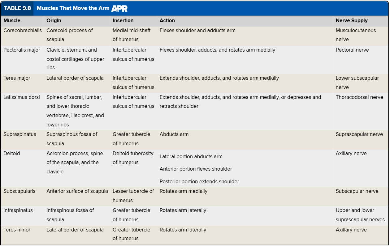
Muscles That Move the Arm Chart
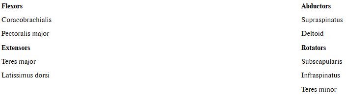
Which muscles that move the arm are flexors, extensors, abductors, and rotators?
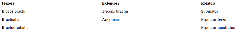
Which muscles that move the forearm are flexors, extensors, and rotators?
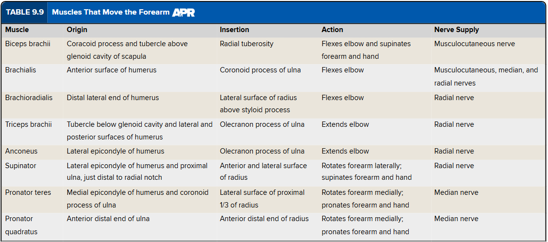
Muscles That Move the Forearm Chart
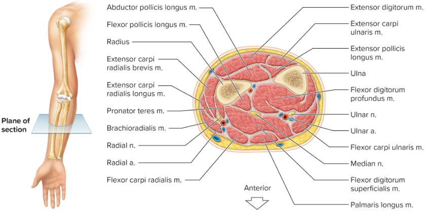
Cross Section of the Forearm
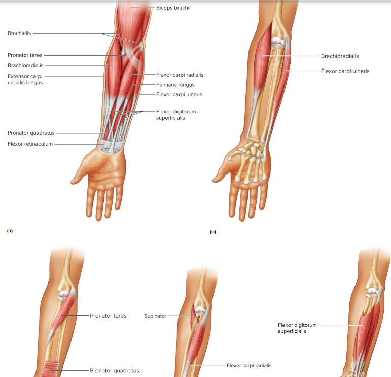
Muscles of Anterior Forearm Diagram
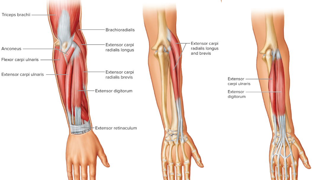
Muscles of Posterior Forearm Diagram
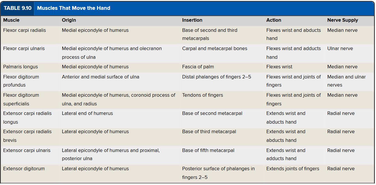
Which muscles that move the hand are flexors and extensors?
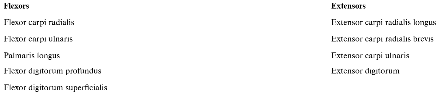
Muscles That Move the Hand Chart
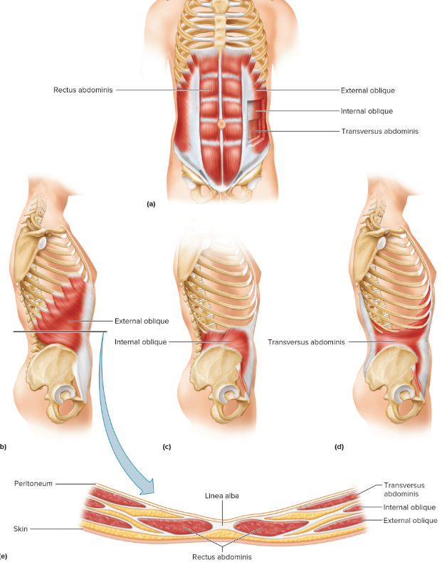
Muscles of the Abdominal Wall Diagram
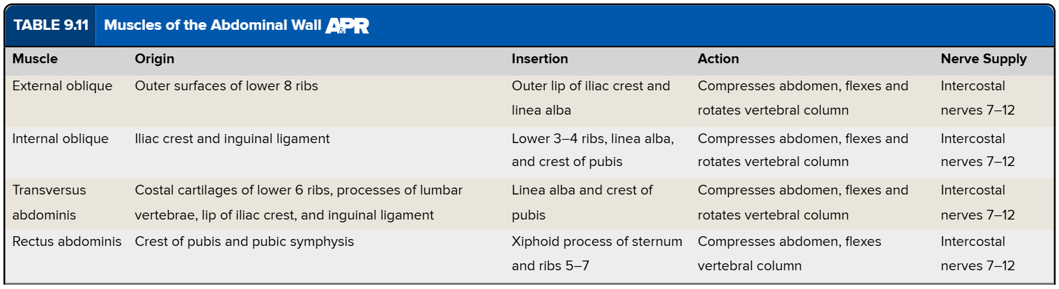
Muscles of the Abdominal Wall Chart
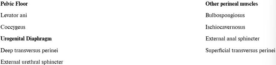
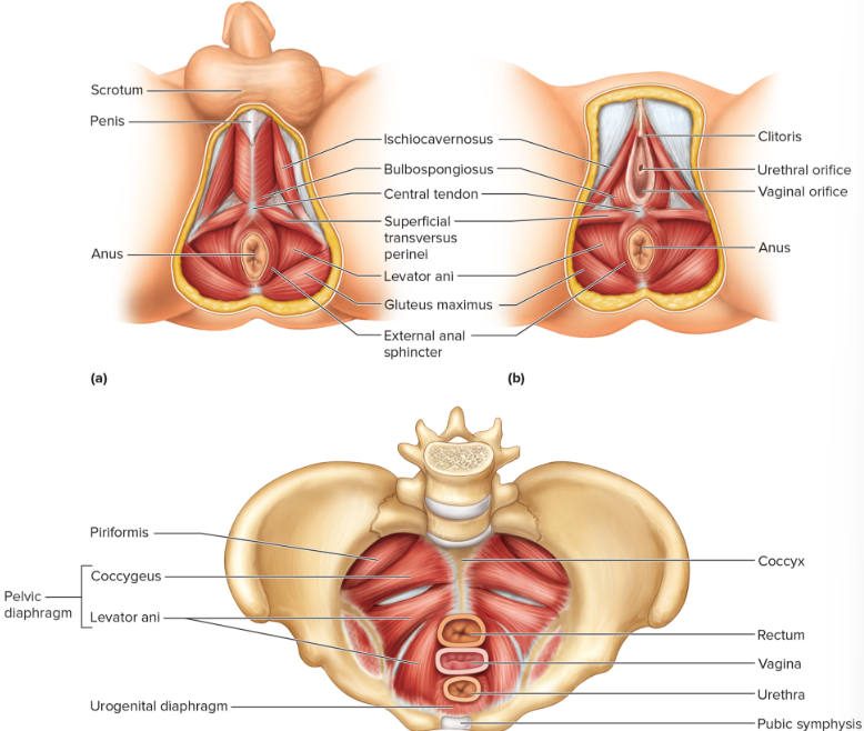
Which muscles are part of the pelvic floor, urogenital diaphragm, or other perineal muscles?
Muscles of Pelvic Floor Diagram
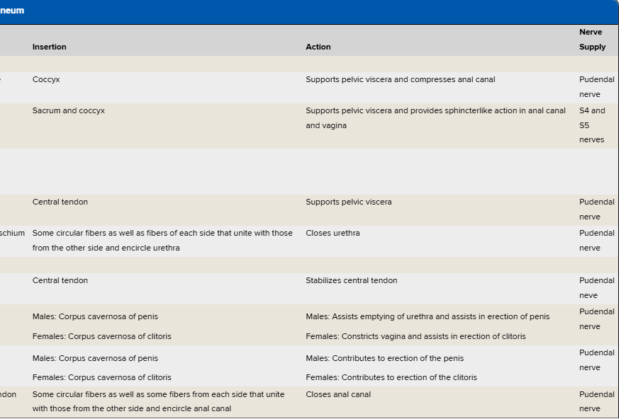
Muscles of the Pelvic Floor and Perineum Chart pt.1
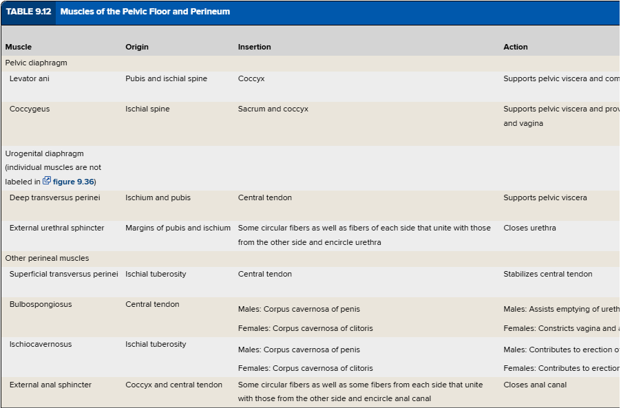
Muscles of the Pelvic Floor and Perineum Chart pt.2
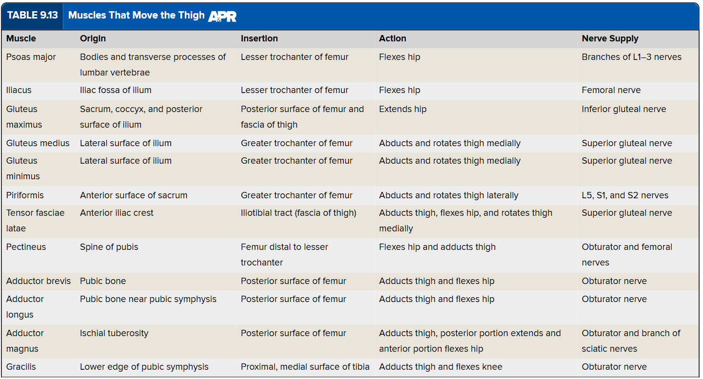
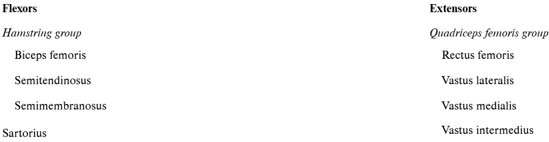
Which muscles that move the thigh are part of the anterior group (flex hip), medial group (adducts thigh), or posterior group (extend hip, abducts or rotates thigh)?
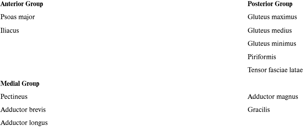
Muscles That Move the Thigh Chart
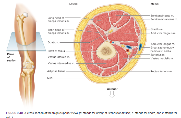
Cross Section of Thigh
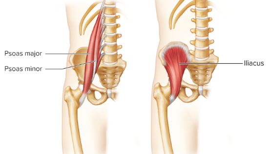
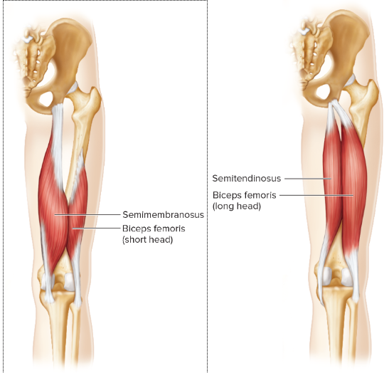
Muscles of the Thigh and Leg Diagram pt.1
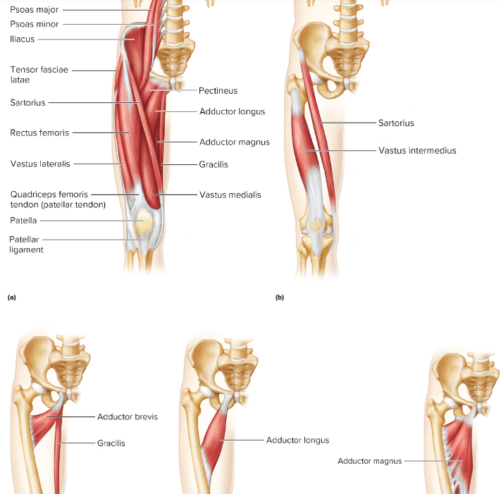
Muscles of the Thigh and Leg Diagram pt.2
Muscles of the Thigh and Leg Diagram pt.3
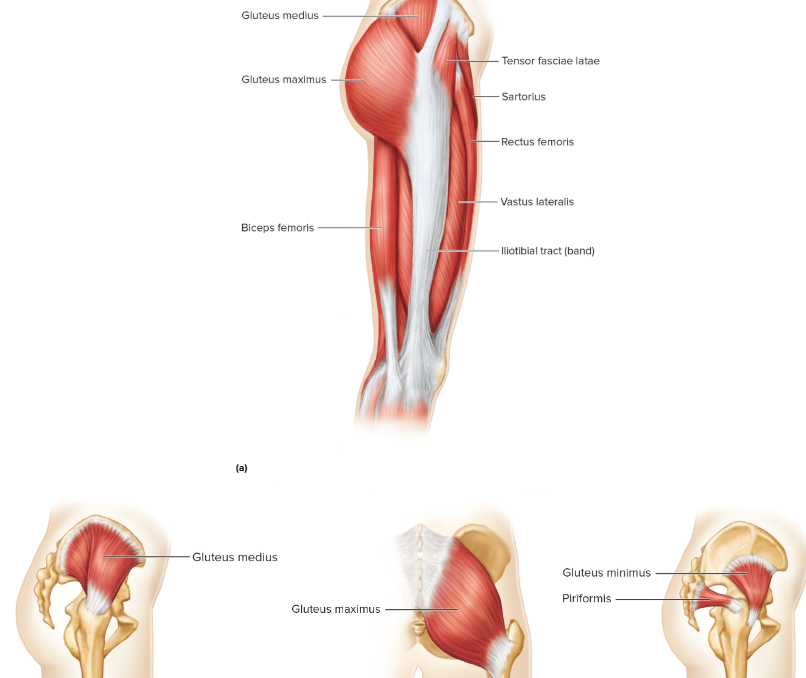
Muscles of the Thigh and Leg Diagram pt.4
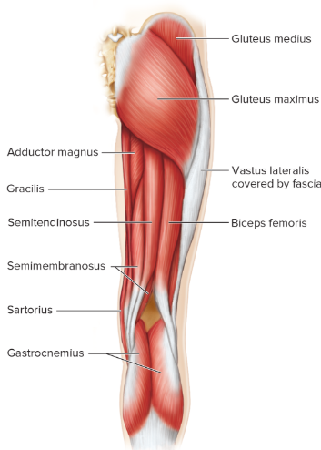
Muscles of the Thigh and Leg Diagram pt.5
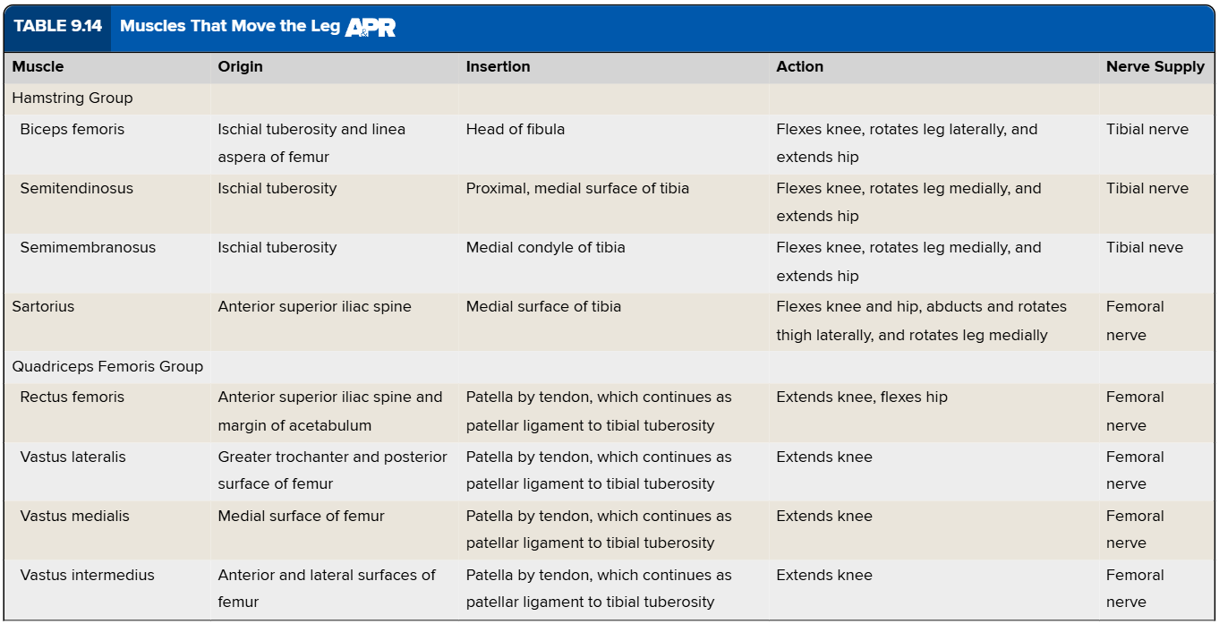
Which muscles that move the leg are flexors and extensors?
Muscles That Move the Leg Chart
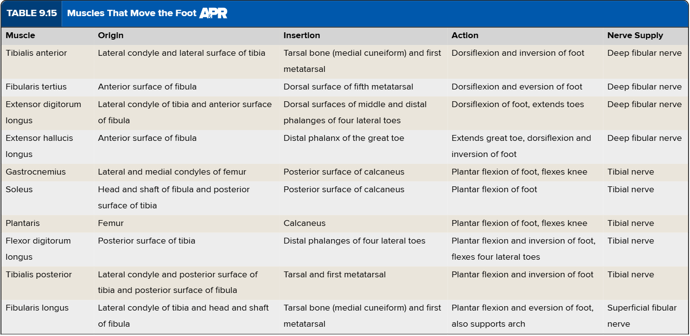
Which muscles that move the foot are dorsiflexors, plantar flexors, invertors, and evertors?
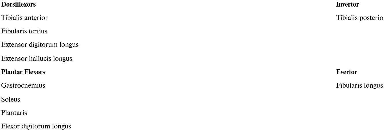
Muscles That Move the Foot Chart
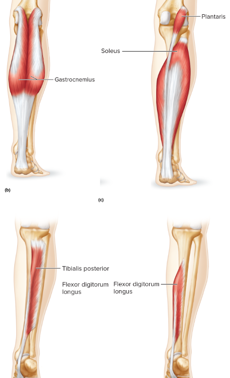
Muscles That Move the Foot Diagram pt.1
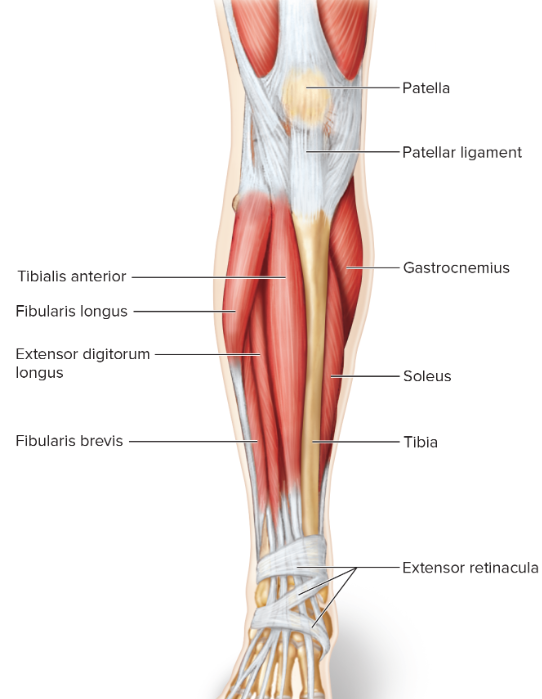
Muscles That Move the Foot Diagram pt.2
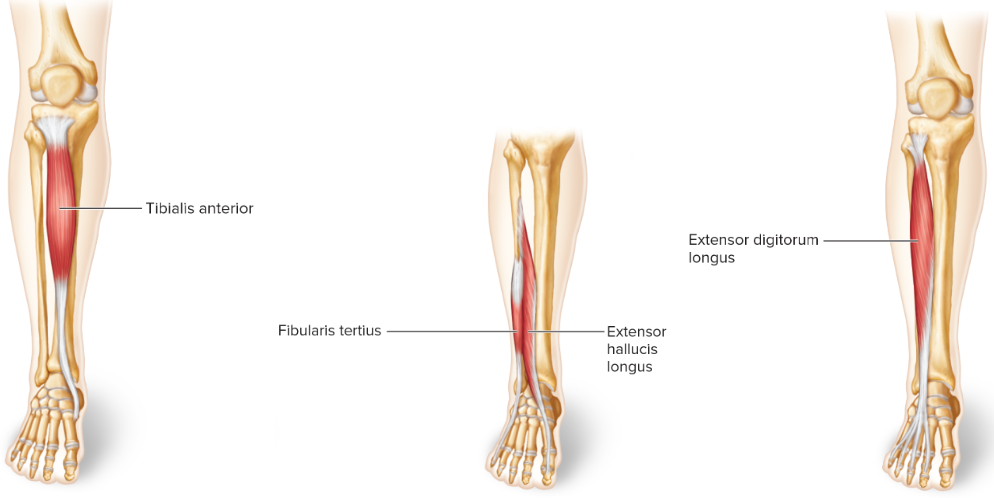
Muscles That Move the Foot Diagram pt.3
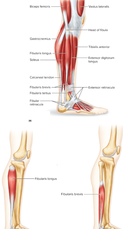
Muscles That Move the Foot Diagram pt.4
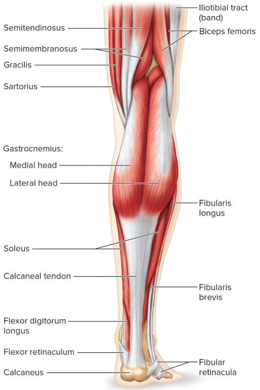
Muscles That Move the Foot Diagram pt.5
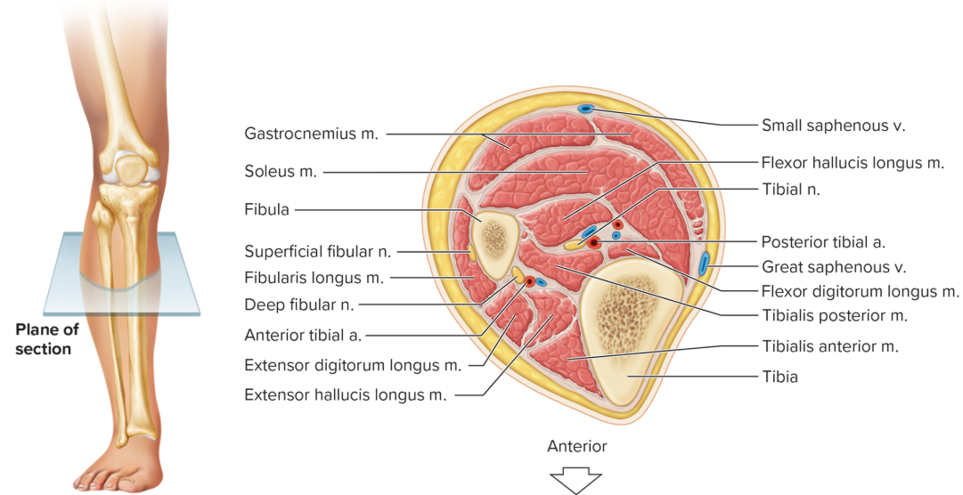
Cross Section of Leg
What changes are associated with an aging muscle?
myoglobin, creatine phosphate, and ATP decline
muscles become smaller and drier
less able to generate force
some muscle tissue is replaced by adipose cells and connective tissue
motor neuron activity declines → reflexes become slower
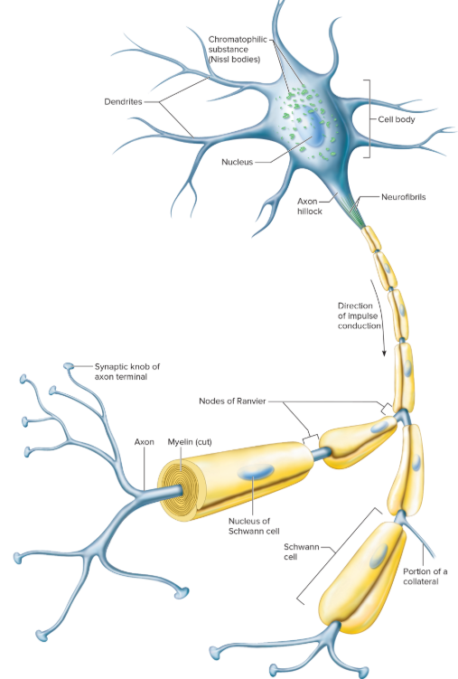
Neuron Diagram
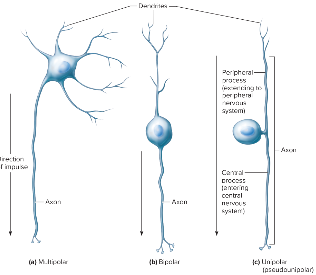
Structural Differences of Neurons Diagram
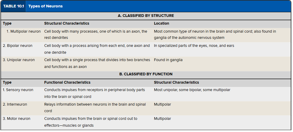
Classification of Neurons Chart
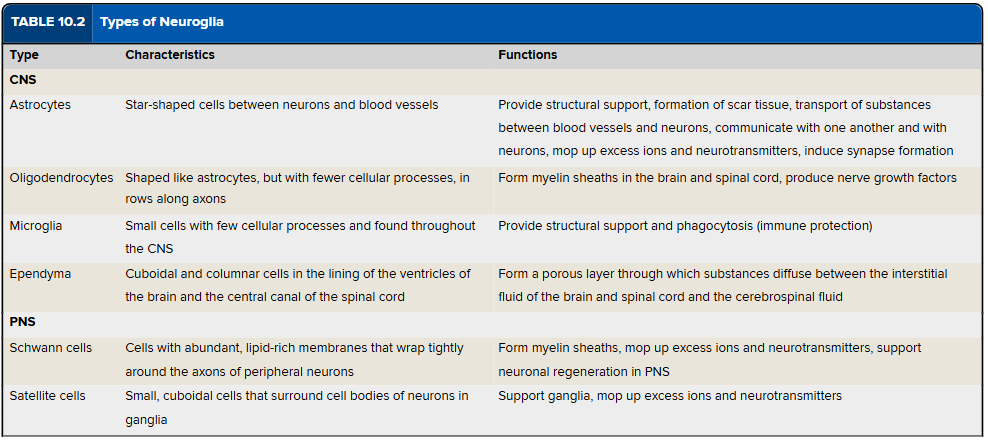
Types of Neuroglia Chart
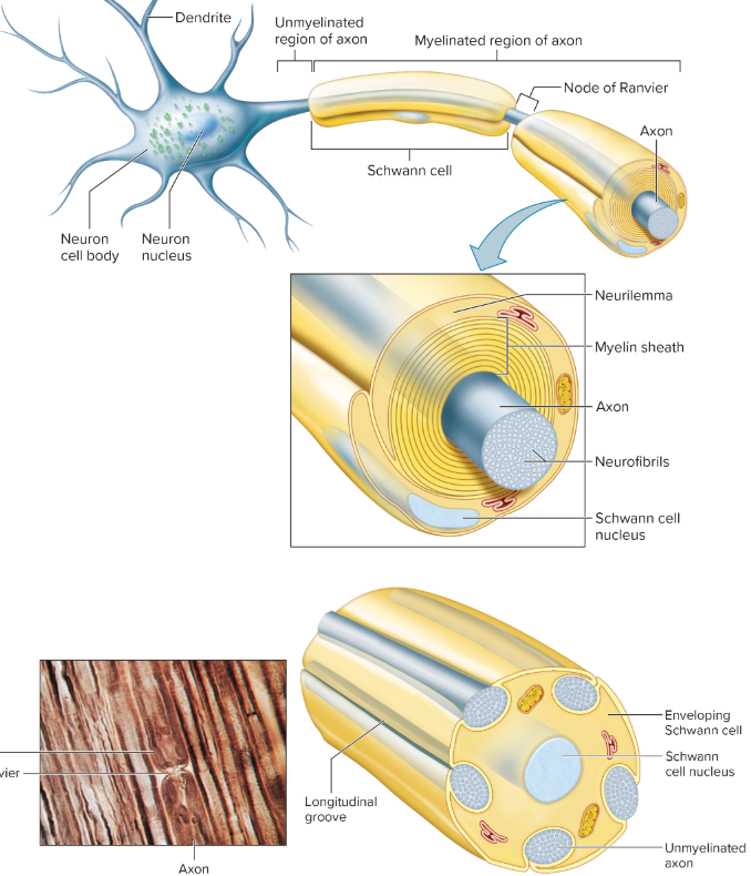
Myelinated Axon Diagram
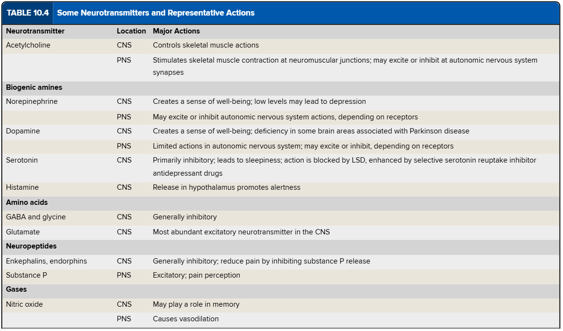
Some Neurotransmitters and Representative Actions Chart
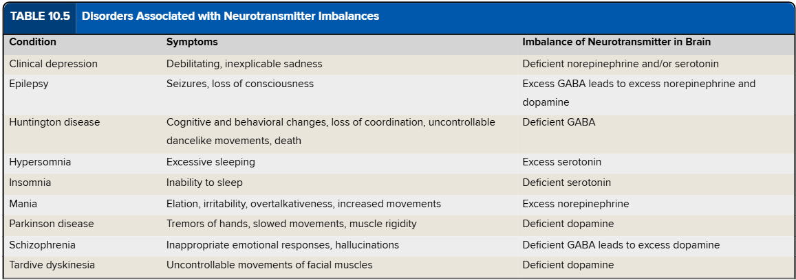
Disorders Associated with Neurotransmitters Chart
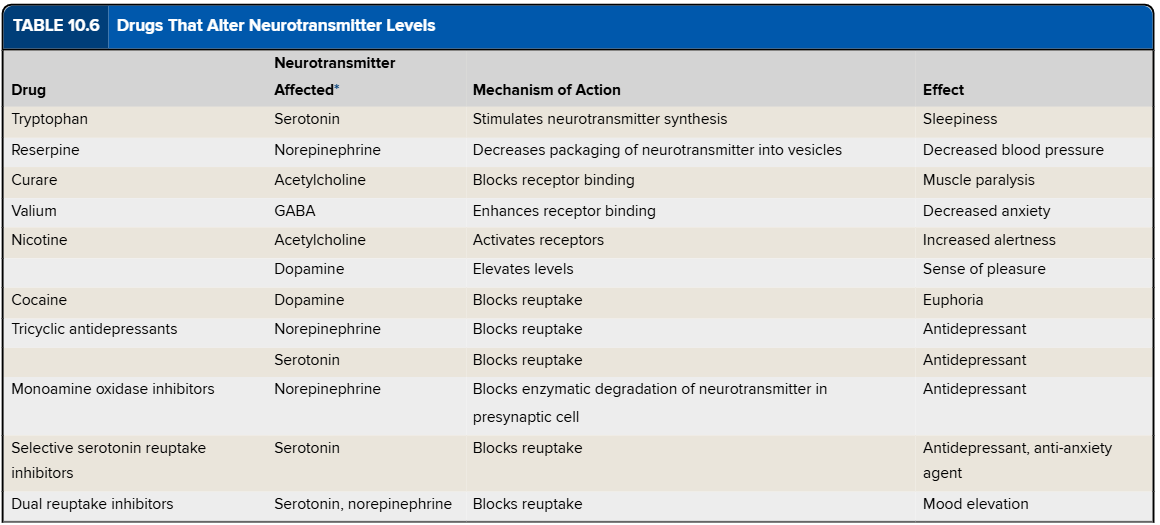
Drugs That Alter Neurotransmitter Levels Chart
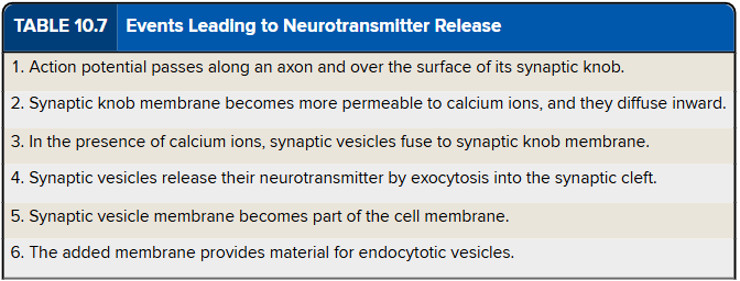
Events Leading to Neurotransmitter Release Chart
Where is grey and white matter found in the brain?
grey - outer layers of cerebral hemispheres; largely the cerebellum
white - deeper in brain
How does the brain connect to the spinal cord?
the brainstem
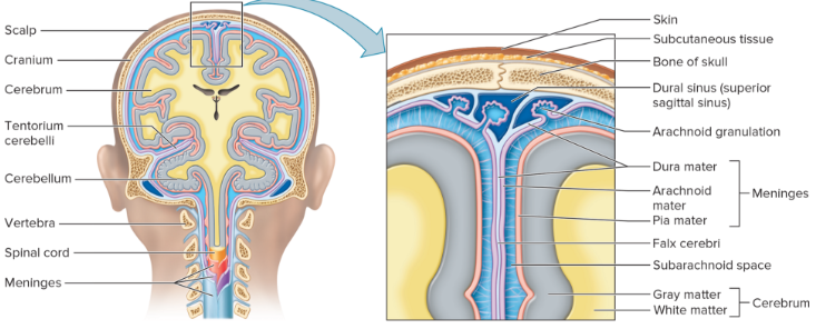
meninges
layers of membrane that cover & protect the brain and spinal cord
What are the three meninges from superficial to deep?
dura mater - tough white dense connective tissue; many blood vessels and nerves; attaches to inside of cranial cavity; internal periosteum of surrounding skull bones
arachnoid mater - thin, weblike membrane; no blood vessels
pia mater - thin w/ many nerves & blood vessels to nourish brain and spinal cord
epidural space
space between dural sheath of spinal cord and bone of the vertebral canal
True or False: The subarachnoid space completely surrounds the brain and spinal cord, allowing them to float in cerebrospinal fluid.
True
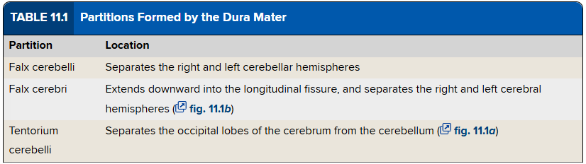
Partitions Formed by Dura Mater Chart
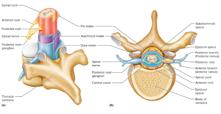
Meninges of Spinal Cord Diagram
Meninges of Brain Diagram
ventricle
cavity in the brain filled with cerebrospinal fluid & continuous with central canal of spinal cord
Ventricles in the Brain Diagram
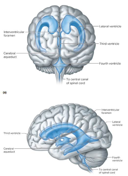
choroid plexus
secrete cerebrospinal fluid; tiny, reddish, cauliflower-like masses of specialized capillaries from the pia mater; covered in layer of specialized ependymal cells
Which ventricles does most CSF form?
lateral & slowly circulates into the 3rd & 4th ventricles
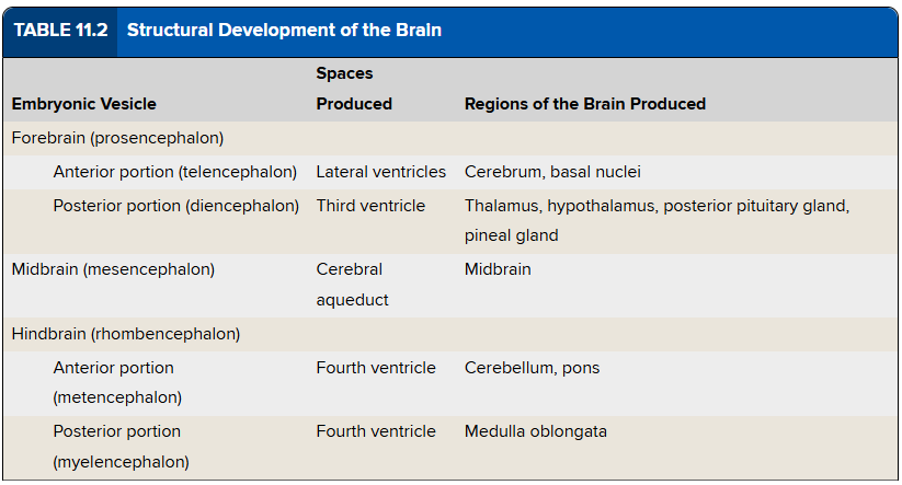
What are the three major cavities/vesicles in the brain development?
forebrain - prosencephalon
midbrain - mesencephalon
hindbrain - rhombencephalon
Which developmental brain cavities divide?
prosencephalon - telencephalon (anterior) & diencephalon (posterior)
rhombencephalon - metencephalon & myelencephalon
Brain Development Diagram
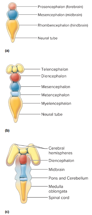
What brain structures does the hindbrain give rise?
cerebellum, pons, medulla oblongata
What gives rise to the cerebrum and basal nuclei?
wall of anterior portion of the forebrain
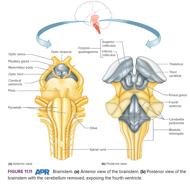
What is the brainstem comprised of?
midbrain, pons, medulla
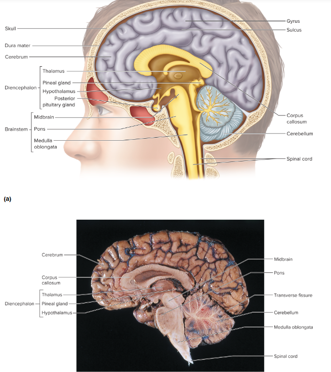
Saggital Section of Brain and Spinal Cord Diagram
Structural Development of the Brain Chart
What is the largest part of the mature brain?
cerebrum
cerebrum
2 cerebral hemispheres
corpus callosum - connects hemispheres
Brain Lobe Diagram
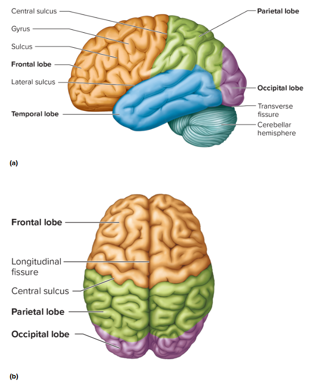
Brain Lobe Diagram 2

What is the difference between gyrus, sulcus, fissure?
gyrus - ridges/convolutions in brain
sulcus - shallow to somewhat deep groove
fissure - very deep groove
Where are fissures and sulci?
longitudinal fissure - separate right and left hemispheres
transverse fissure - separate cerebrum from cerebellum
sulci - divide each hemisphere into lobes
insula (island of Reli)
lobe deep within lateral sulcus of each hemisphere
cerebral cortex
thin layer of gray matter on all brain lobes; outermost portion of cerebrum; 75% of all neuron cell bodies in nervous system
Sensory, Association, and Motor Areas of Left Cerebral Cortex Diagram
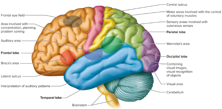
Sensory areas for which senses receive impulses from both sides?
eyes & ears
What are association areas?
not primarily sensory nor motor - analyze and interpret sensory experiences + help provide memory, reasoning, verbalizing, judgement, emotions
Wernicke’s area
left hemisphere; understanding and formulating written and spoken language
Broca’s Area
motor speech area; frontal lobe;
frontal eye field
superior part of frontal lobe - voluntary eye & eyelid movements
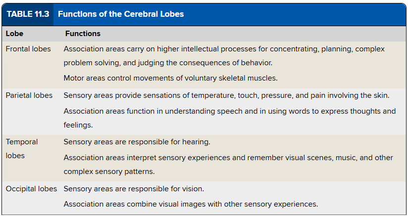
Functions of Cerebral Lobes Chart
Which hemisphere is usually dominant? What for? What is the nondominant specialized in?
left - language related activities (speech, writing, reading); verbal, analytical, computational skills
right - nonverbal functions (motor tasks, interpret musical patters, visual experiences); emotional and intuitive processes
basal nuclei or basal ganglia
masses of gray matters deep within cerebral hemispheres
include caudate nucleus, putamen, globus pallidus
produce neurotransmitter dopamine
interact with motor cortex, thalamus, cerebellum → facilitate voluntary movement
Diencephalon
develops from posterior forebrain
located between cerebral hemispheres & superior to brainstem
surrounds third ventricle
largely grey matter
thalamus
hypothalamus
optic tracts
infundibulum
posterior pituitary gland
mammillary bodies
pineal gland
limbic system
thalamus
selective gateway for sensory impulses ascending to cerebral cortex
What does the hypothalamus regulate?
heart rate and arterial blood pressure
body temp
water and electrolyte balance
control of hunger and body weight
control of movements and glandular secretions of the stomach and intestines
production of neurosecretory substances that stimulate the pituitary gland to release hormones that help regulate growth, control various glands, and influence reproductive physiology
sleep and wakefulness
Brainstem Diagram