4.Macromolecules & Micro-Organisms In Disease
1/23
There's no tags or description
Looks like no tags are added yet.
Name | Mastery | Learn | Test | Matching | Spaced |
|---|
No study sessions yet.
24 Terms
Biomolecules Overview Table
Biomolecule | Description | Examples | Monomer (Building Block) |
|---|---|---|---|
Carbohydrates | Biomolecules consisting of carbon, hydrogen, and oxygen atoms | Sugars, starch, cellulose | Monosaccharides (e.g. glucose) |
Lipids | Diverse group of organic compounds | Fats, waxes, fat-soluble vitamins, monoglycerides, diglycerides, phospholipids | Fatty acids + glycerol |
Proteins | Large biomolecules made of one or more long chains of amino acids | Enzymes, antibodies, hemoglobin | Amino acids |
Nucleic Acids | Large biomolecules in all cells and viruses | DNA, RNA (contain sugar, phosphate, nitrogenous base) | Nucleotides |
What is Monomers?
🔑 Monomer (Building Block) Note
✔ Monomer:
A molecule that reacts with other monomers to form a polymer chain.
Example: Amino acids are monomers that build proteins.
Biomolecule Structures
Carbohydrates
Biomolecule Consisting of Carbon, Hydrogen, and Oxygen Atoms.
-Sugars, Starch, Cellulose
Type | Meaning | Description | Example |
|---|---|---|---|
Monosaccharide | Mono = One | Single sugar unit (monomer of carbohydrates) | Glucose, Fructose |
Disaccharide | Di = Two | Two monosaccharides joined together | Sucrose (glucose + fructose), Lactose (glucose + galactose), Maltose (glucose + glucose) |
Polysaccharide | Poly = Many | Many monosaccharides linked together (polymer) | Starch, Cellulose, Glycogen |
🔹 Carbohydrate Rule
✔ Most carbohydrates end in “-ose.”
Examples from the image:
Glucose
Fructose
Lactose
Maltose
Sucrose
Why are Carbohydrates are important to an organism’s structure and function?
Cell Wall: Plant (Cellulose)
Q: What carbohydrate makes up the plant cell wall?
A: Cellulose
Q: What is the structural role of cellulose in plants?
A: It provides rigidity and structural support to plant cells.
Cell Wall: Fungi (Chitin)
Q: What carbohydrate is found in the cell walls of fungi?
A: Chitin
Q: Is chitin found in plant cell walls?
A: No, chitin is found in fungi, not in plants.
Cellular Respiration
Q: Which carbohydrate is used in cellular respiration to produce ATP?
A: Glucose
Q: Where does the conversion of glucose to ATP occur?
A: In the mitochondria
ATP (Adenosine Triphosphate)
Q: What is ATP?
A: Adenosine Triphosphate, the primary energy currency of the cell
Q: What macromolecule is essential for ATP production?
A: Carbohydrates (specifically glucose)
Carbohydrates | Cell Wall Component | Plant Cell Wall | Made of Cellulose |
Fungi Cell Wall | Made of Chitin |
Carbohydrates | Cellular Respiration | Glucose | Used to make ATP |
ATP | Energy Molecule | ATP (Adenosine Triphosphate) | Produced in mitochondria using glucose |
Plants store energy as starch; animals store it as glycogen
Lipids
Diverse group of organic compounds | Fats, waxes, fat-soluble vitamins, monoglycerides, diglycerides, phospholipids | Fatty acids + glycerol |
Category | Concept | Example / Structure | Details |
|---|---|---|---|
Lipids | Types of Lipids | Triglycerides, Phospholipids, Steroids | All are hydrophobic (repel water) |
Lipid Breakdown | Subunits | Glycerol + Fatty Acids | Not true monomers (no repeating units) |
Lipid Properties | Hydrophobic Nature | Do not dissolve in water | Key in forming cell membranes |
Fun Fact | Monomer Exception | Lipids ≠ true polymers | No repeating monomer subunits like carbs or proteins |
Lipid Functions | Missing from Image | Energy storage, insulation, cushioning, hormone production | Common TEAS test point |
Why are Lipids are important to an organism’s structure and function?
1. Cell Membranes
Q: What role do lipids play in the cell membrane?
A: They form the phospholipid bilayer that surrounds the cell.
Q: Why is the lipid bilayer important?
A: It regulates what enters and leaves the cell.
2. Energy Storage
Q: How do lipids compare to carbohydrates in energy storage?
A: Lipids store more energy per gram and are used for long-term storage.
Q: What type of lipid is primarily used for energy storage?
A: Triglycerides.
3. Insulation
Q: What lipid structure insulates neurons?
A: The myelin sheath.
Q: What is the function of the myelin sheath?
A: It helps conduct electrical impulses along nerves.
Q: How do lipids help regulate body temperature?
A: Fat insulates the body by trapping heat.
4. Hormones
Q: Which type of hormones are made from lipids?
A: Steroid hormones.
Q: Give an example of a steroid hormone.
Function | Structure / Role | Explanation |
|---|---|---|
Cell Membranes | Phospholipid bilayer | Forms the boundary of all cells, controls entry/exit of substances |
Long-Term Energy Storage | Triglycerides | Store more energy per gram than carbs or proteins |
Insulation (Myelin) | Myelin Sheath (nerve cells) | Lipid-rich sheath that insulates neurons and helps transmit impulses |
Insulation (Fat) | Adipose tissue | Helps with temperature regulation and physical protection |
Hormones | Steroid-based (e.g., estrogen, testosterone) | Lipid-derived hormones regulate reproduction, metabolism, etc. |
Extra TEAS Exam Points Not in the Image (But Important!)
Concept | Why It’s Important for the TEAS |
|---|---|
Cholesterol in membranes | Helps maintain fluidity and structure of cell membranes |
Lipids in digestion | Bile salts emulsify fats; lipase enzymes break them down |
Lipid-soluble molecules | Only small nonpolar substances like oxygen, CO₂, and steroids can diffuse across membranes |
Unsaturated vs. Saturated fats | Unsaturated = double bonds, liquid; Saturated = single bonds, solid |
Protein
Why are Proteins are important to an organism’s structure and function?
🔹 Amino Acids
Q: What is the monomer of proteins?
A: Amino acid
Q: How many amino acids are used to build all proteins?
A: 20
🔹 Cell Membrane Proteins
Q: What do protein channels do in the cell membrane?
A: Transport substances across the membrane
Q: What is the function of receptor proteins?
A: Receive chemical signals from outside the cell
🔹 Enzymes, Antibodies, Hormones
Q: What is the function of enzymes?
A: Speed up chemical reactions
Q: What role do antibodies play?
A: Fight infection and recognize foreign invaders
Q: Name a protein hormone.
A: Insulin
Proteins | Large biomolecules made of one or more long chains of amino acids | Enzymes, antibodies, hemoglobin | Amino acids |
Category | Concept | Examples / Details |
|---|---|---|
Monomer | Amino Acid | Building block of protein |
Protein Structure Role | Structural Proteins | Muscle tissue, Hair follicle (keratin), Collagen |
Cell Membranes | Protein Channels & Receptors | Allow transport, act in signal reception |
Functional Proteins | Enzymes | Speed up chemical reactions (e.g., digestive enzymes) |
Antibodies | Immune defense against pathogens | |
Hormones | Chemical messengers (e.g., Insulin) regulating physiological functions | |
Gene |
✅ Extra TEAS Concepts Missing from the Image (But Important!)
Concept | TEAS Relevance |
|---|---|
Protein Structure Levels | Primary, secondary, tertiary, quaternary structures define shape and function |
Denaturation | Proteins lose function if shape changes due to pH or temperature |
Peptide Bond | The covalent bond between amino acids |
Protein Synthesis | Occurs in ribosomes; involves transcription (nucleus) and translation (cytoplasm) |
Examples of Enzymes | Amylase (breaks starch), pepsin (digests protein), DNA polymerase |
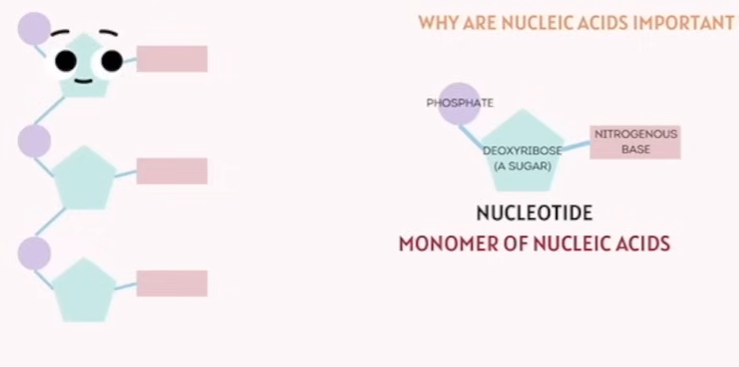
Nucleic Acids
🔹 Nucleotide Structure
Q: What are the three parts of a nucleotide?
A: Sugar, phosphate group, nitrogenous base
Q: What sugar is found in DNA nucleotides?
A: Deoxyribose
Q: What sugar is found in RNA nucleotides?
A: Ribose
🔹 Nitrogenous Bases and Pairing
Q: What base does adenine pair with in DNA?
A: Thymine
Q: What base does adenine pair with in RNA?
A: Uracil
Q: Which bases pair together?
A: Adenine–Thymine (DNA) or Adenine–Uracil (RNA), Cytosine–Guanine
🔹 Function and Importance
Q: What is the main function of DNA?
A: Store genetic information
Q: What is the main role of RNA?
A: Help in protein synthesis
Category | Concept | Details / Examples |
|---|---|---|
Monomer | Nucleotide | Made of sugar (deoxyribose or ribose), phosphate group, nitrogenous base |
Nitrogenous Bases | Base Pairing | A pairs with T (DNA) / U (RNA), C pairs with G |
Types of Nucleic Acids | DNA and RNA | DNA stores genetic info; RNA helps with protein synthesis |
Function | Genetic Information Storage | Carry instructions for making proteins and cellular functions |
Nucleotide is the Monomer of Nucleic Acids
✅ Extra TEAS Exam Points Not in the Image
Concept | Importance for TEAS |
|---|---|
DNA Double Helix | DNA’s structure enables stable storage of genetic info |
Types of RNA | mRNA, tRNA, rRNA each play roles in protein synthesis |
Nucleotide Bond Types | Phosphodiester bonds link nucleotides; hydrogen bonds between bases |
Central Dogma | DNA → RNA → Protein (transcription and translation) |
Micro-Organisms in Biology - Viruses — Key Features
🧠 Flashcard Questions for Viruses (TEAS) 🔹 General Characteristics
Q: Are viruses considered living cells?
A: No, viruses are not cells and are not considered alive.
Q: What kind of parasite is a virus?
A: An intracellular obligate parasite—they need a host cell to reproduce.
Q: What does a virus’s genome consist of?
A: Either RNA or DNA.
Q: What is the outer protein coat of a virus called?
A: Capsid.
Q: What is the typical size range of viruses?
A: 20 to 400 nanometers.
🔹 Diseases Caused by Viruses
Q: Name a viral disease that affects the respiratory tract.
A: Common cold, influenza, or COVID-19.
Q: Which virus causes gastrointestinal illness?
A: Rotavirus.
Q: What viral infection affects the skin?
A: Human papillomavirus (HPV).
Q: Which virus affects the central nervous system?
A: Rabies virus.
Viruses — Key Features
Category | Details |
|---|---|
Size | 20–400 nanometers (nm) |
Structure | Genome (RNA or DNA) inside a protein coat called Capsid; sometimes an Envelope |
Characteristics | - Not a cell |
Disease Examples | - Respiratory: common cold, influenza, COVID-19 |
What is a Virulence
How severe or harmful the disease is
Micro-Organisms in Biology - Bacteria— Key Features
🧠 Flashcard Questions for Bacteria (TEAS) 🔹 General Characteristics
Q: Are bacteria prokaryotes or eukaryotes?
A: Prokaryotes (no nucleus).
Q: What are the two types of Gram stains for bacteria?
A: Gram Positive (thick cell wall) and Gram Negative (thin cell wall).
Q: What are the common shapes of bacteria?
A: Cocci (circle), Bacilli (rod), Staph (clusters), and Strep (chains).
Q: What is the size range of bacteria?
A: 0.2 to 15 microns.
Q: What is the difference between aerobic and anaerobic bacteria?
A: Aerobic bacteria need oxygen, anaerobic bacteria do not.
🔹 Diseases Caused by Bacteria
Q: Which bacterium causes strep throat?
A: Streptococcus.
Q: What bacteria causes skin infections?
A: Staphylococcus aureus.
Q: Name a bacterial cause of community-acquired pneumonia.
A: Mycoplasma.
Q: Which bacteria causes tetanus?
A: Clostridium tetani (implied from "Tetanus").
Q: What is chlamydia caused by?
A: Bacteria called Chlamydia.
Category | Details |
|---|---|
Type | Prokaryote (no nucleus or membrane-bound organelles) |
Size | 0.2 – 15 microns |
Gram Stain | - Gram Positive: thick cell wall- Gram Negative: thin cell wall |
Shapes | - Cocci: spherical (circle) - Bacilli: rod-shaped - Staph: clusters - Strep: chains (line formation) |
Oxygen Use | - Aerobic: needs oxygen - Anaerobic: does not need oxygen |
Characteristics | Cell membrane with cell wall |
Disease Examples | - Chlamydia- Community-acquired pneumonia (Mycoplasma)- Streptococcus (strep throat)- Staphylococcus aureus (skin infections)- Tetanus |
✅ Important TEAS Exam Points (Additional)
Concept | Details |
|---|---|
Prokaryotic Structure | Bacteria have cell membranes and walls but no nucleus. |
Gram Stain Importance | Used to classify bacteria by cell wall thickness, guiding antibiotic use. |
Bacterial Shape | Helps identify bacterial species in lab tests. |
Aerobic vs Anaerobic | Oxygen requirement affects where bacteria can live and how they’re treated. |
What is Gram Positive & Negative in Bacteria.
What type of toxins do they produce and how it works?
Gram Positive vs Gram Negative Bacteria What is Gram Staining?
Gram staining is a laboratory technique used to classify bacteria into two major groups based on their cell wall structure.
This classification helps identify the type of bacteria and guides treatment decisions.
1. Gram Positive Bacteria
Produce
Cell Wall: Thick layer of peptidoglycan (a strong, mesh-like polymer).
Appearance After Staining: They retain the crystal violet stain used in the Gram stain procedure, so they appear purple or blue under a microscope.
Other Features:
No outer membrane.
Generally more susceptible to antibiotics like penicillin.
Examples: Streptococcus, Staphylococcus.
Do not produce endotoxins (since they lack an outer membrane).
Instead, many Gram-positive bacteria produce exotoxins.
Exotoxins are proteins secreted actively by living bacteria.
These exotoxins are often highly potent and can cause specific diseases or symptoms (e.g., the toxins from Clostridium botulinum or Staphylococcus aureus).
2. Gram Negative Bacteria
Produce
Cell Wall: Thin layer of peptidoglycan.
Outer Membrane: Have an additional outer membrane containing lipopolysaccharides (LPS), which can act as toxins.
Appearance After Staining: They do not retain the crystal violet stain but take up the counterstain (usually safranin), so they appear pink or red.
Other Features:
Outer membrane can protect against some antibiotics.
Often more resistant to antibiotics.
Examples: E. coli, Salmonella.
Produce endotoxins.
Endotoxins are part of the outer membrane (specifically the lipopolysaccharide or LPS layer).
These toxins are released when the bacteria die and the cell wall breaks down.
Endotoxins can trigger strong immune responses, causing fever, inflammation, and even septic shock.
Why Does It Matter?
Knowing whether a bacterium is Gram positive or negative helps doctors choose the right antibiotics.
The difference in cell wall structure affects how bacteria respond to drugs and how harmful they can be.
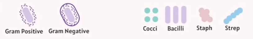
Bacteria Shapes
Cocci?
Bacilli?
Staph?
Strep?
Shapes | - Cocci: spherical (circle) - Bacilli: rod-shaped - Staph: clusters - Strep: chains (line formation) |
Micro-Organisms in Biology - Fungi
What is chitin?
This slide is about Fungi, which are:
Eukaryotes (meaning their cells have a nucleus and other organelles).
Size ranges from 2 to 200 microns.
Characteristics include:
Cell wall made of chitin.
Reproduce both sexually and asexually via spores.
Disease examples caused by fungi:
Mycosis (general fungal infections).
Skin infections like Tinea (Ringworm, Athlete's Foot).
Infections of mucous membranes like Thrush.
Fungal infections in lungs and blood.
Chitin is a long-chain polymer of a sugar called N-acetylglucosamine, which is a derivative of glucose.
It is a structural polysaccharide, meaning it provides strength and protection.
Chitin is the main component of the cell walls of fungi, giving them rigidity and shape.
It is also found in the exoskeletons of arthropods like insects, crustaceans (crabs, lobsters), and some other invertebrates.
Chemically, it’s similar to cellulose (found in plants), but with nitrogen-containing groups.
Mycosis
Superficial mycosis?
Subcutaneous mycosis?
Systemic mycosis?
Mycosis is a general term for fungal infections that affect humans or animals. It happens when fungi invade and grow on or inside the body.
There are different types of mycosis, depending on where the infection occurs:
Superficial mycosis: affects the outer layers of the skin, hair, or nails (like athlete's foot or ringworm).
Subcutaneous mycosis: affects deeper layers of the skin, often through cuts or wounds.
Systemic mycosis: affects internal organs and can be serious, especially in people with weakened immune systems.
Micro-Organisms in Biology - Protozoa 原生动物
Eukaryotes (meaning their cells have a nucleus)
Size: 1 - 50 microns
Characteristics: Unicellular organisms (single-celled)
Disease examples caused by protozoa:
Malaria
Giardiasis
Micro-Organisms in Biology - Animals
Flashcard Questions
Helminths:
Q: What are the three main types of helminths?
A: Roundworms (hookworms), flatworms (tapeworms), and flukes (liver flukes).
Q: What kind of diseases do helminths typically cause?
A: Gastrointestinal diseases.
Q: Are helminths eukaryotic or prokaryotic?
A: Eukaryotic.
Ectoparasites:
Q: Name four common ectoparasites.
A: Mites, fleas, bed bugs, ticks.
Q: What kind of reactions can ectoparasites cause on the skin?
A: Skin inflammation and allergic reactions.
Q: What disease can ticks transmit?
A: Lyme disease.
Q: Are ectoparasites endoparasites or ectoparasites?
A: Ectoparasites (live on the surface of the host).
This slide is about Animals (Helminths and Ectoparasites), which are:
Eukaryotes (cells with a nucleus)
Size: 3 millimeters to 10 meters
Characteristics:
Helminths (Worms):
Round worms, such as hookworms
Flat worms, like tapeworms
Flukes, such as liver flukes
Ectoparasites:
Mites
Fleas
Bed bugs
Ticks
Disease Examples:
Helminths cause gastrointestinal (GI) diseases.
Ectoparasites cause skin inflammation and allergic reactions.
Ticks can transmit Lyme disease.
What is Vector from Infectious Disease
🔹 Active Vector
The vector actively participates in the transmission.
The pathogen develops or multiplies inside the vector.
The vector may inject the pathogen into a new host during feeding (e.g., blood-sucking).
✅ Example:
Mosquito (Anopheles): Transmits malaria by injecting Plasmodium parasites while feeding on blood.
Tick: Transmits Lyme disease after the bacteria multiply inside its body.
🔹 Passive Vector
The vector does not play a biological role in the pathogen’s life cycle.
The pathogen is simply carried on the body or surface of the vector.
Transmission happens by contact, not by injection or feeding.
✅ Example:
Housefly: Can carry bacteria or viruses on its legs from feces to food, causing diseases like typhoid or cholera.
Cockroach: Passively carries pathogens on its body.
🔁 Summary Table
Feature | Active Vector | Passive Vector |
|---|---|---|
Role in pathogen life cycle | Pathogen multiplies or develops inside vector | No development inside vector |
Transmission mode | Biting, injecting | Surface contamination |
Example vector | Mosquito, tick | Housefly, cockroach |
Example disease | Malaria, Lyme disease | Typhoid, food poisoning |
Types of Light Microscopy Explained:
🔬 Key Terms:
Magnification: The process of making something appear bigger.
Resolution: The microscope's ability to distinguish fine details of a specimen.
🔍 Types of Light Microscopy Explained: 1. Brightfield Microscopy
Most common type.
Light passes directly through the specimen.
Image appears darker on a bright background.
Used for stained or naturally pigmented samples.
Setup:
Light → condenser lens → specimen → objective lens → viewer.
2. Darkfield Microscopy
Light is directed from the sides using a light stop.
Only scattered light enters the objective lens.
The specimen appears bright against a dark background.
Ideal for live, unstained specimens (e.g., spirochetes).
Setup:
Light → light stop (blocks central light) → only angled light hits the specimen → scattered light goes to the lens.
🔬 Electron Microscopes Overview
🔬 Electron Microscopes Overview
Use beams of electrons instead of light.
Provide much higher magnification and resolution than light microscopes.
Allow us to view structures at the nanometer scale.
📘 Types of Electron Microscopes 1. Transmission Electron Microscope (TEM)
Views internal structures of cells or microorganisms.
Electrons pass through a very thin sample.
Produces 2D images with very fine detail.
✅ Best for:
Seeing organelles inside cells.
Observing viruses, cell membranes, etc.
2. Scanning Electron Microscope (SEM)
Views surfaces of samples.
Electrons bounce off the surface, creating a 3D-like image.
Shows texture and external structure in great detail.
✅ Best for:
Examining bacteria surfaces, insects, materials, etc.
🔁 Quick Comparison Table:
Feature | TEM | SEM |
|---|---|---|
Image type | 2D | 3D-like |
Focus | Internal structures | Surface structures |
Sample requirement | Must be ultra-thin | Can be thicker |
Resolution | Higher | Slightly lower than TEM |