Cortex
1/95
Earn XP
Description and Tags
complete
Name | Mastery | Learn | Test | Matching | Spaced |
|---|
No study sessions yet.
96 Terms
why does the brain have gyri and sulci?
to increase surface area, which allows for more computing power in a small area.
anterior to the central sulcus is the
primary motor cortex
posterior to the central sulcus is the
primary somatosensory cortex
the primary auditory cortex is located in the ___ fissure
sylvian
what are the gyri/sulci of the temporal lobe?
outer view: superior, middle, and inferior temporal gyri and the superior and inferior temporal sulci
midsagittal view: lingual gyrus, collateral sulcus, occipitotemporal sulcus, occipitotemporal (fusiform gyrus). can see parts of the superior and inferior temporal gyri.
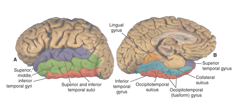
what are the gyri/sulci of the occipital lobe?
surface: lateral occipital gyri
sagittal: cuneus, lingual gyrus, parietooccipital sulcus, calcarine sulcus, occipitotemporal (fusiform) gyrus
the cuneus is defined by what two sulci?
superior edge: parietooccipital sulcus
inferior edge: calcarine
what are the gyri/sulci of the parietal lobe? (11)
outer view: postcentral sulcus and gyrus, superior parietal lobule, intraparietal sulcus, supramarginal gyrus, angular gyrus
sagittal view: precuneus, posterior paracentral lobule, calcarine sulcus, subparietal sulcus, cingulate sulcus (marginal branch)
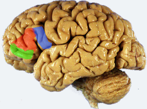
what are the three parts of the inferior frontal gyrus? list from most anterior to posterior
pars orbitalis, pars triangularis, pars opercularis
what lobe is the paracentral lobule associated with?
on the medial surface of the hemisphere, the paracentral lobule includes parts of both frontal and parietal lobes.

the paracentral lobule is the medial continuation of which gyri?
precentral and postcentral
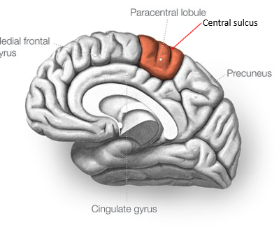
what is the function of the paracentral lobule?
motor and sensory function of the legs
what is the division between the hemispheres, which accommodates the falx cerebri?
the great longitudinal fissure
going directly down the great longitudinal fissure, what is the first white matter tract you hit?
the corpus callosum
what are the Brodmann areas?
they are numbered areas of the cortex based on cytoarchitecture
what are the types of cerebral cortex? How many layers in each?
neocortex - 6 layers
paleocortex - 3 layers
archicortex - 4 layers
which cortex type makes up 95% of the hemisphere surface?
neocortex
the parahippocampal gyrus, cingulate gyrus, and prepyriform cortex are what type of cortex?
paleocortex
the motor cortex and visual cortex are what type of cortex?
neocortex
the hippocampus is made of what type of cortex?
archicortex
in what cortical layers are the axons and dendrites?
layer 1

what cortical layers have corticocortical connections?
2 and 3
(2 has connections within the same hemisphere and 3 has connections to the opposite)

which cortical layer has the most thalamic afferents?
4

which layer has subcortical efferents and some thalamic afferents?
5
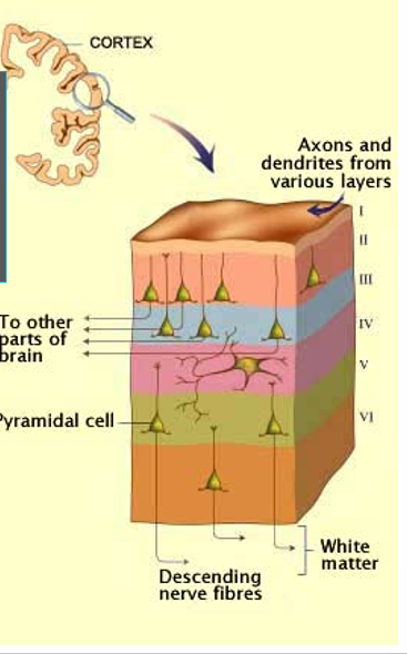
what cortical layer has thalamic efferents?
6

what is the primary computational unit of the cortex?
the column
what are inhibitory neurons between cortical columns for?
to prevent overexcitation of the brain
Betz cells are a type of ___ cell present in the ___
pyramidal cell, motor cortex
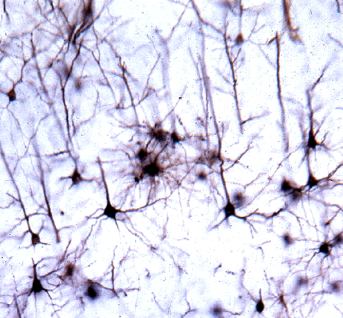
as output cells, ___ make up white matter
pyramidal axons
spiny excitatory (glutamatergic) neurons and projection neurons are all what type of neuron?
pyramidal
granule, stellate, basket cells, and non-spiny inhibitory (GABAergic) interneurons are all what type of cell?
non-pyramidal
white matter bundles that project anterior to posterior are called
association bundles
white matter bundles that project between the hemispheres are called
commissural bundles
white matter bundles that project dorsal to ventral are called
projection bundles
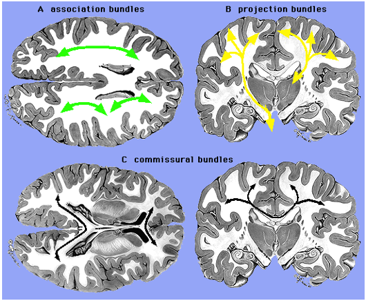
name the three types of white matter tracts and give an example of each
association bundles (front to back) - arcuate fasciculus
projection bundles (dorsal to ventral) - internal capsule
commissural bundles (hemi to hemi) - corpus callosum, anterior commissure
what connects the hemispheres?
major connection: corpus callosum
minor: anterior commissure (thalamus), posterior commissure (recall visual tract)
the corpus callosum connects ___ cortical areas in each hemisphere
homologous (closely related)
how would a split brain patient respond to a picture presented to the left visual field?
It would go to the right brain, but wouldn’t be able to cross to the left for verbal/lingual processing. Patient would not be able to name the object, but would be able to show how to use it with the left hand.
how would a split brain patient respond to a picture presented to the right visual field?
it would go to the left brain already, so would not need to cross for processing. Patient would be able to name the object and can show what it was with the right hand.
___ connect adjacent gyri
U fibers
projection fibers run between the cerebral cortex and various subcortical structures, passing through the ___ and the ___. what are the important fibers in this category?
they pass through the corona radiata and the internal capsule.
Important fibers - corticospinal, corticobulbar, and thalamocortical projections
what is the primary cortex? what is the secondary cortex?
the primary cortex is the first cortical area processing of sensory input or motor output (BA 17, 41, 4, 3, 1, 2)
the secondary cortex is the second stage of processing and makes up the association cortex with the tertiary cortex.
how do the association and primary cortices work together?
sensory info is conveyed to the sensory cortex after synapsing in the thalamus via ascending projection pathways
after processing in the primary sensory cortex, info is passed to sensory association cortices via short association fibers
reciprocal feedback with frontal cortices via longer association pathways allows previous and current sensory input to decide if a response is needed and how to best execute that response
primary motor cortex executes the behavioral response via descending projection fibers
what are the functional divisions of the frontal cortex?
motor cortex, premotor cortex, prefrontal cortex (including the dorsolateral PFC, inferior PFC, and the orbitofrontal cortex)
which division of the prefrontal cortex is associated with cognitive processing? which is associated with affective processing?
cognitive = dorsolateral
Affective = inferior
What neuropsychological testing is used to assess prefrontal cortex functioning?
wisconsin card sorting test
dorsolateral PFC lesions lead to
executive functioning issues, including perseverance, stimulus-bound behavior, echopraxia, echolalia. also eye motor control deficits, working memory impairment, and memory retrieval deficits.
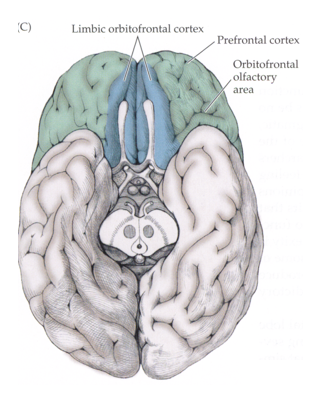
what effects are seen with lesions to the medial (limbic) orbitofrontal cortex?
adynamia and akinetic mutism. patient is self aware but not initiating behavior
what are some symptoms caused by orbitofrontal lesions?
arousal changes, low motivation, reduced insight and impulse control. no deficits seen on card sorting tasks. patients are stimulus bound.
What does it mean for a patient to be stimulus bound?
patients will react to a stimulus for no real reason other than the fact that they encounter them. for instance, the patient may see a glass of water and automatically drink it even if they arent thirsty.
What area contains sound images of words?
Wernicke’s area
What connects Wernicke’s and Broca’s areas?
the arcuate fasciculus
Where are the motor programs used to talk stored?
in Broca’s area
Broca’s vs Wernicke’s aphasia
Broca’s aphasia - patient able to comprehend speech, but has a hard time producing speech.
Wernicke’s aphasia - patient able to produce speech, but has a hard time with comprehension.
Where is Broca’s area?
in the frontal lobe, BA 44 and/or 45 (depends on who you ask lmao)
Where is Wernicke’s area located?
next to auditory area on superior temporal lobe. BA 22
how does the comprehension of a spoken word occur?
spoken word → primary auditory → Wernicke’s → comprehension
Wernicke’s aphasia also affects
nonspeech communication, such as signed language or writing
What is conduction aphasia?
a lesion to the arcuate fasciculus, meaning there is damaged communication between Broca’s and Wernicke’s areas. Patient is typically able to comprehend and produce speech pretty well, but has a hard time repeating after another person.
What are the afferents and efferents of the primary motor cortex?
afferents: primary somatosensory, thalamic VL
efferents: basal ganglia, thalamic VL, superior colliculus, red nuc, pontine nuc, spinal cord
damage to the primary motor cortex results in
dysfunction of contralateral distal muscles
how might damage to the postcentral gyrus affect smiling?
damage to the postcentral gyrus results in paralysis of voluntary movements, so voluntary smiling would be impaired. involuntary smiling would be intact.
how might damage to the limbic cortex affect smiling?
damage to the limbic cortex would affect emotional expression, so involuntary smiling would be impaired. voluntary smiling would be intact.
the postcentral gyrus is the same as
somatosensory cortex, BAs 3, 1, 2
the precentral gyrus is the same as the
primary motor cortex, BA 4
the premotor cortex is ___ to the primary motor cortex
anterior
Brodmann areas 3, 1, 2
somatosensory cortex
Brodmann area 4
primary motor cortex
S1 (primary somatosensory) receives dense input from
VP
how do the receptive fields of the somatosensory Brodmann areas compare?
Areas 3a and 3b have small, simple receptive fields. Areas 1 and 2 have large receptive fields and are direction sensitive
What does BA 1 do?
texture recognition
What does BA 2 do?
detects size and shape of objects
Stereognosis is associated with which brodmann area?
area 5
astereognosis
inability to recognize an object by touch
agraphesthesia
inability to recognize letters or numbers drawn on the person’s hand.
What Brodmann areas have to do with somatosensory empathy?
BAs 1 and 2
this structre lies above the horizontal portion of the intraparietal sulcus, the inferior parietal lobule lies below it.
superior parietal lobule
parietal association cortex function
higher sensory function, language function (particularly in the left hemi, angular gyrus), stereognosis, spatial relations (right hemi)
hemineglect
lesion to parietal association cortex, most common on right side (left neglected). unintentional ignorance of one side of the body, contralateral to the lesion.
vanishing arm
lesions to parietal lobe (nothing really said about it in the powerpoint idk)
Gerstmann syndrome
lesion to left inferior parietal lobule. finger agnosia (inability to recognize fingers), agraphia, right-left co
describe the where pathway
superior longitudinal fasciculus, including terminating axons in posterior parietal cortex, which helps locate the object. Cells in the parietal lobe are tuned to large areas of retina to detect presence/movement of stimulus (
alice in wonderland syndrome
complex visual hallucinations, like objects changing size or distorting shape. associated with occipital association cortex lesion, more common in children.
lesion in temporal association cortex results in
L: verbal memory problems
R: visual discrimination problems
prosopagnosia (inability to identify faces) (left and right)
simultagnosia
inability to attend to more than a limited area of the visual field despite normal vision. can see individual objects, but can’t perceive a scene as a whole
describe the auditory pathway
cochlea → superior olive → inferior colliculus → medial geniculate → auditory cortex
The planum temporale is larger on which side? Why?
larger on the left because of Wernicke’s
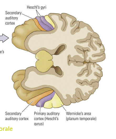
the auditory cortex is organized ___
tonotopically
the insula is covered by the ___
opercula (lips)
the solitary nucleus, central tegmental tract and the insula are all associated with the ___ pathway
gustatory
alien limb syndrome
right temporoparietal lesion. patient describes limb as moving on its own will, as if it were a separate entity.