PSY260 L2 Learning and Plasticity
1/51
There's no tags or description
Looks like no tags are added yet.
Name | Mastery | Learn | Test | Matching | Spaced | Call with Kai |
|---|
No analytics yet
Send a link to your students to track their progress
52 Terms
core nervous system functions
collect sensory information
process information
generate behaviour from given input (motor output)
parts of the nervous system
central nervous system
peripheral nervous system
enteric nervous system
central nervous system
brain and spinal cord
peripheral nervous system
somatic: controls voluntary motor movement and relays sensory information
autonomic: sympathetic (fight or flight) and parasympathetic (rest and digest)
enteric nervous system
“second brain", controls the gastrointestinal system
information processing in the cortex: posterior vs anterior
sensory information processing starts from posterior
basic details, sensory information
information becomes more abstract as it passes to more anterior areas
what do I know about these details? what opinions do i have about this object? how do i relate to this object?
information processing in the cortex: medial vs lateral
processing in medial tends to be internally-focused or self-referential
“me in the centre”
lateral areas tend to be externally-focused or related to external stimuli, task-oriented processing
“what is around me”
what do i sense
what tasks must i accomplish
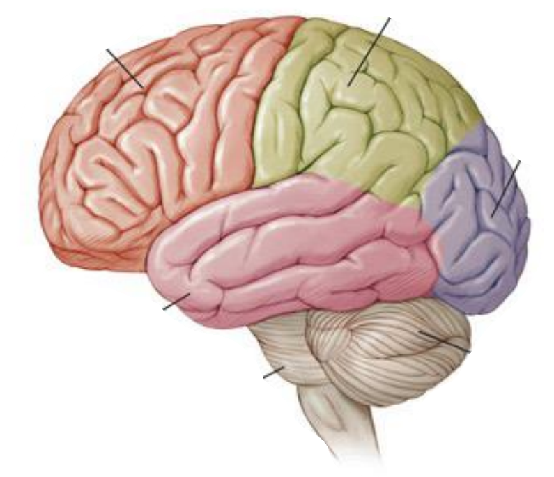
what lobes are shown here?
counter clockwise from the upper left:
frontal
primary motor cortex (the fold between frontal and parietal)
parietal
occipital
cerebellum
brainstem
temporal lobe
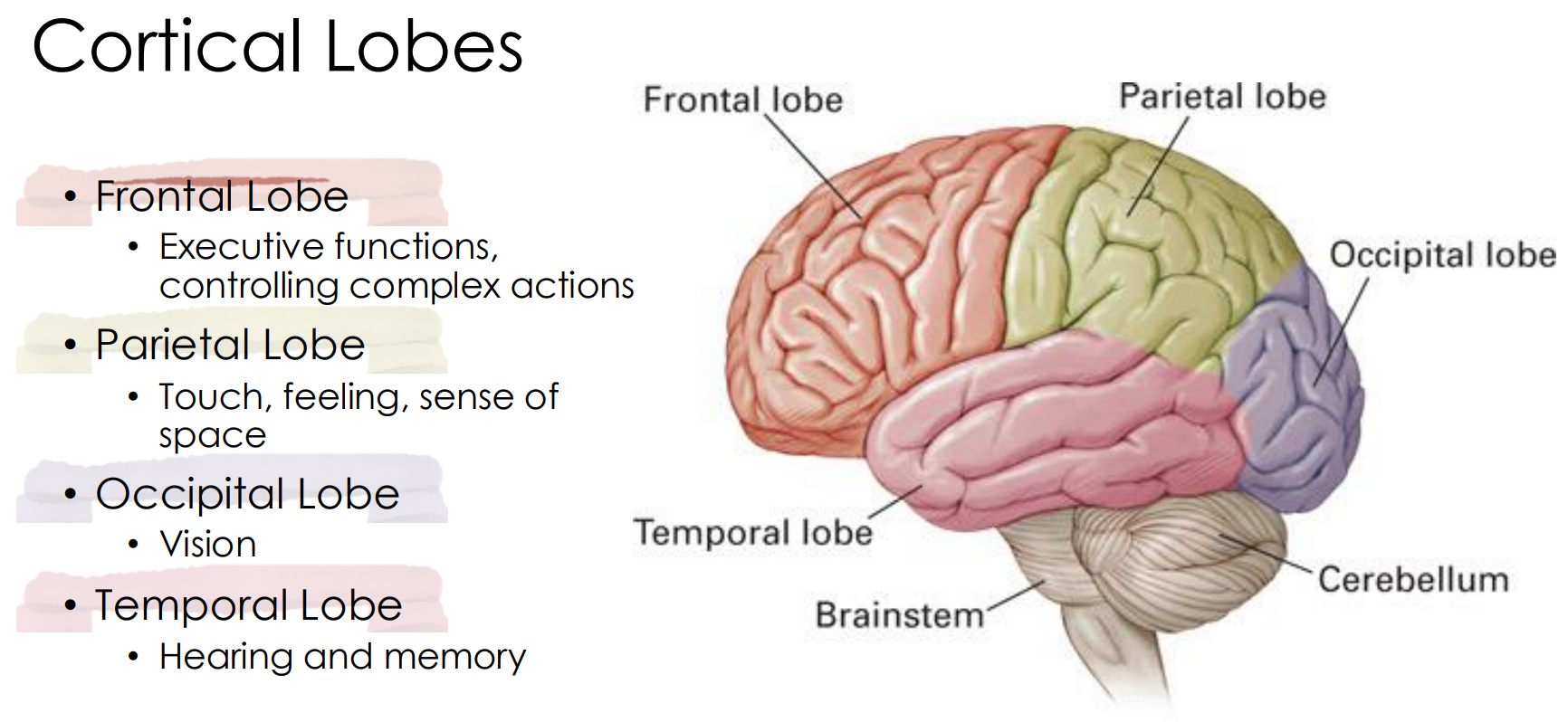
frontal lobe
executive functions, controlling complex actions
decision making, problem solving, planning
more rostral: planning, more caudal: doing (as we move toward the primary motor cortex)
parietal lobe
touch, feeling, sense of space
occipital lobe
vision
temporal lobe
hearing and memory
near a lot of memory-related structures which communicate with each other
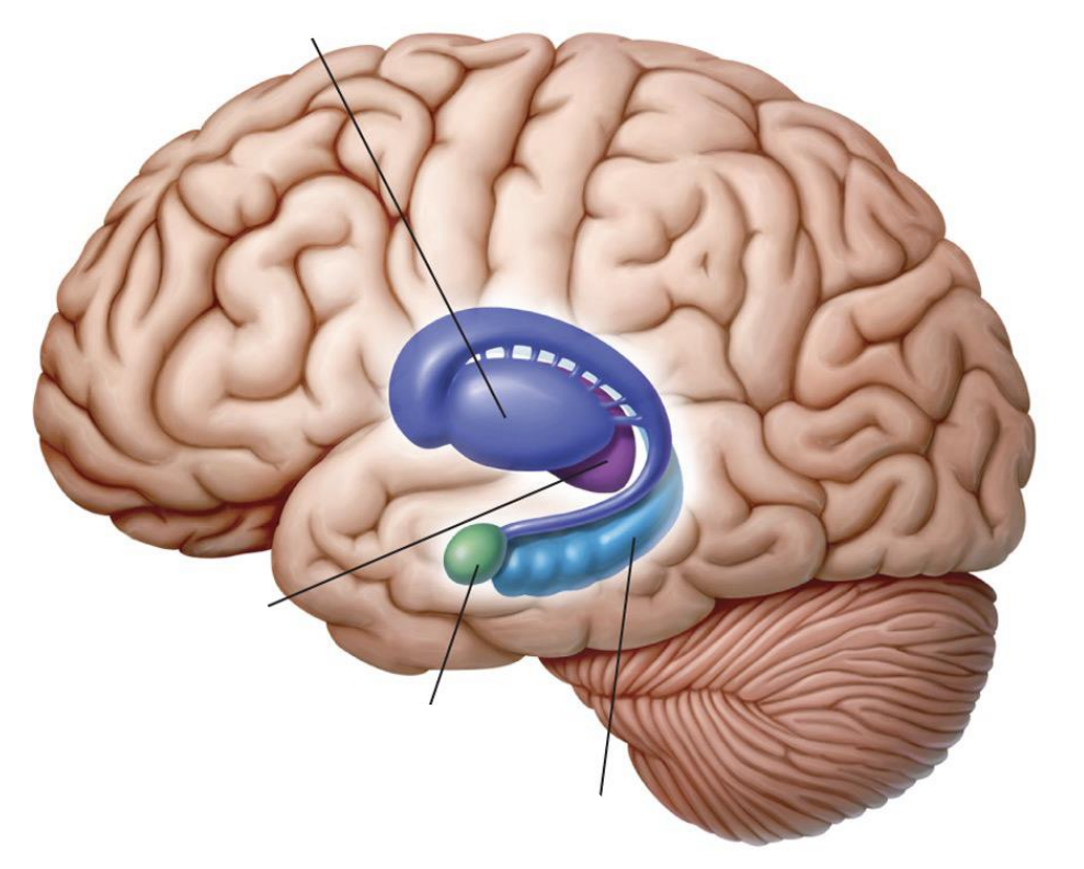
what is shown in this picture?
clockwise from rostral:
basal ganglia
thalamus
amygdala
hippocampus
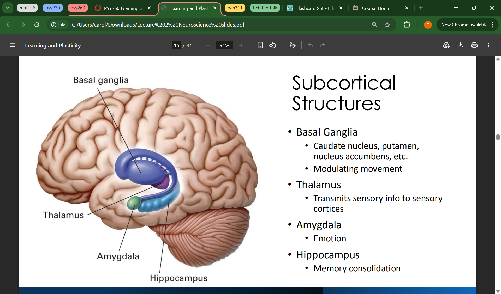
basal ganglia
subcortical structure
reward-based learning, modulating movement
includes caudate nucleus (striatum), putamen, nucleus accumbens, etc
thalamus
subcortical structure
transmits sensory info to sensory cortices
ALL SENSORY INFO GOES HERE FIRST
amygdala
subcortical structure
emotion
important for fear-based learning
hippocampus
subcortical structure
memory consolidation (memory moves from short-term → long term storage)
reflexes
innate responses not done consciously
the simplest expression of the 3 nervous system functions:
stimulus → little processing → response
hardwired, innate, involuntary
processing of reflexes
often handled solely in the spinal cord
interneurons allow for rapid responses that can bypass the brain
immediately activates motor neuron as opposed to first brain processing
common sensory pathway
sensory organ → thalamus → primary sensory cortex
primary sensory cortex function
specialized for initial processing, then relays to other cortical areas
frequency, tone, basic details
understanding/conceptualizing
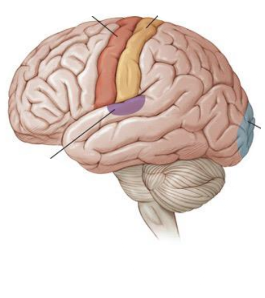
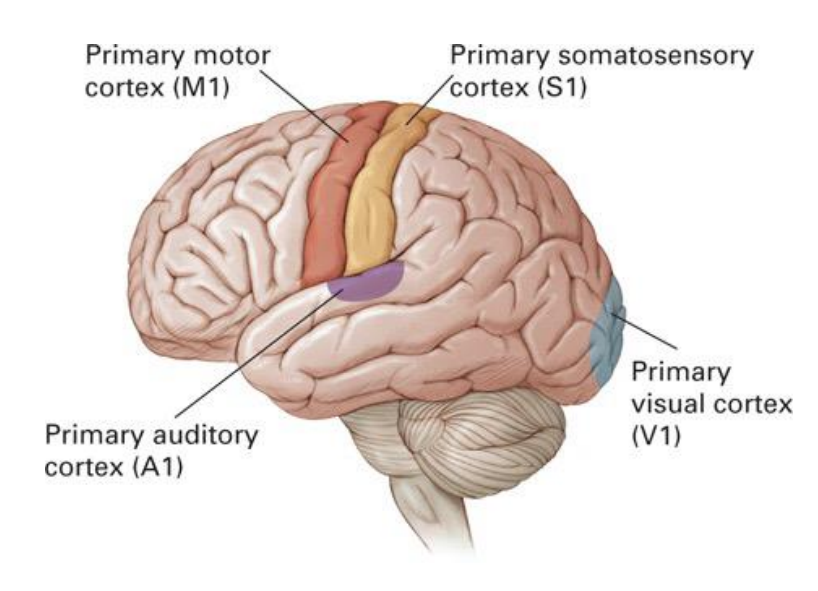
label this image
clockwise from rostral:
primary motor cortex
primary somatosensory cortex
primary visual cortex
primary auditory cortex
voluntary motor output pathway
primary motor cortex (M1) in frontal lobe → down spine
inputs to M1 to form behavioural plans can include:
frontal lobes — planning and logical thinking
cerebellum, basal ganglia — refinements of motor program
comparative neuroanatomy
comparing brains across species gives insight into brain function
bigger isn’t necessarily better — relative proportions and size reflect different specialization for each niche
birds have a disproportionally large cerebellum compared to humans — flying requires motor refinement and balance
humans have a disproportionally large cerebral cortex — higher order thinking, processing
we look at bird and rodent brains in addition to focus of human brain
neocortex: outside lobes recently evolutionarily developed for higher order thinking
lesion analysis approach
very rarely do we intentionally lesion human brain tissue unless there is a medical reason (epilepsy, hippocampus to stop seizures)
compare functions before and after surgery within a single subject
we recruit patients with lesions in similar areas and study what shared symptoms they have
we recruit patients with similar symptoms and study what shared areas of damage they have
no two lesions are the exact same
both methods infer the responsible area
lesion analysis results
data can be messy — no two lesions are exactly the same, no two brains respond the same way to damage
non-human controlled lesions can be done instead to ensure consistency
magnetic resonance imaging (MRI) approach
strong magnetic field aligns subset of atoms in tissue
computer measures signals emitted by different tissues and create photographs
magnetic resonance imaging (MRI) results
extremely detailed structural 3d model of the living brain
safe (no radiation)
has revolutionized medical, scientific research into the brain
good spatial resolution, no temporal (just a photo)
functional magnetic resonance imaging (MRI) approach
detect changes in blood flow to different parts of the brain Blood Oxygen Level Dependent response
indirect measure of neural activity (relying on the assumption that more blood flow = more activity),
estimates timeline on a broad scale — takes time for O2 to replenish
functional magnetic resonance imaging (MRI) results
allows for functional 3d modelling of the brain over time during tasks or at rest
has revolutionized medical and scientific research into the brain
good spatial, less good temporal
electroencephalogram (EEG) approach
electrodes on the scalp with gel conductor detects electrical charges from neural activity
electroencephalogram (EEG) results
relatively portable, inexpensive, easy to use
good temporal (better than MRI), low spatial
measures from the surface — unknown how deep signals come from
scalp can distort
magnetoencephalogram (MEG) approach
electrodes on the scalp record magnetic field produced by electrical currents in the brain
smaller than MRI but still needs dedicated space
magnetoencephalogram (MEG) results
expensive, difficult to use, not portable
better spatial resolution than EEG (magnetic fields don’t get as distorted by the scalp)
more accurate measures closer to the scalp
good temporal resolution
excitatory neurons neurotransmitter
glutamate
makes it more likely for cells around to fire
inhibitory neurons neurotransmitter
gamma-aminobutyric acid (GABA)
makes it less likely for cells around to fire
glial cells
provide support to nerve cells
structurally, functionally
form myelin sheath
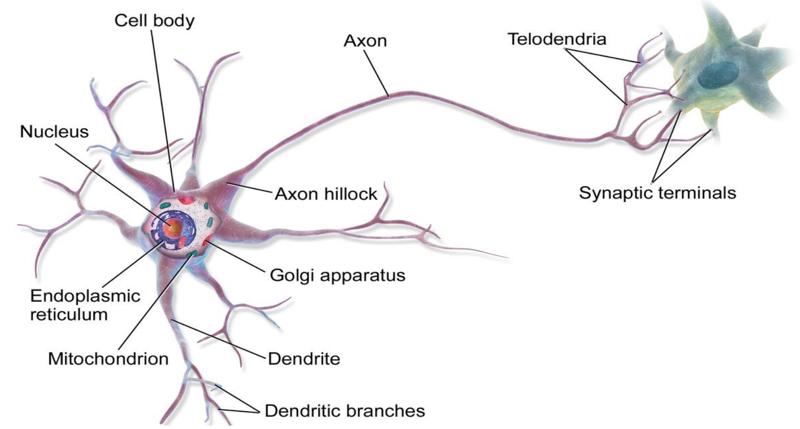
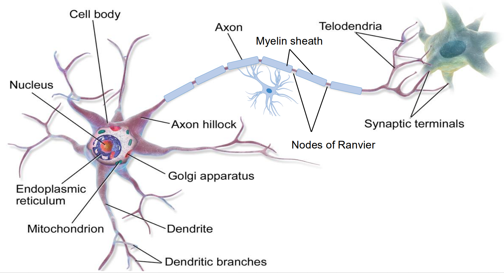
label this image


label this image
axon conducts action potentials away from the cell body
myelin sheath cover the axon and help signal travel faster
nodes of ranvier allow signal to jump across rapidly
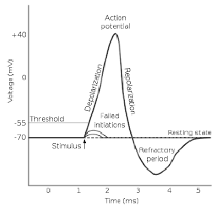
action potential
resting potential is negative
if stimulus threshold is reached, sodium channels open, and Na+ ions enter the cell
at around 40 mV, K+ ions exit the cell
neuron becomes hyperpolarized (refractory period)
very unlikely to meet stimulus threshold during this phase
back propagation
echo of electricity rippling out to dendrites — telling dendrites it has fired
EPSPs
excitatory post-synaptic potential caused by neurotransmitters
a temporary depolarization of the postsynaptic membrane caused by inflow of ions
makes the neuron more likely to fire an action potential
neural communication: chemical
NTs are contained in synaptic vesicles in the terminal of the presynaptic neuron
action potential causes them to fuse with the cell membrane and release NTs into the synaptic cleft
NTs bind to receptors on the post-synaptic neuron
examples of neurotransmitters
modulatory (controlling other NTs)
acetylcholine
dopamine
norepinephrine
GABA (inhibitory)
glutamate (excitatory)
hebbian plasticity
Hebb’s theory on how neurons change over time
proposed that synaptic connections are determined competitively like natural selection
cells that fire together wire together
cells out of sync lose their link
suggests an experience-based “natural selection” for the most useful synaptic connections — the most useful synaptic connections survive
repeated experience with a stimulus may help neurons tuned to that stimulus “win” and expand their synaptic connections
tuned especially (not exclusively) to particular stimuli
cells that fire together…
“wire together”
two neurons that fire at the same time are more likely to connect and trigger each other
cells out of sync
“lose their link”
neurons not firing are more likely to die
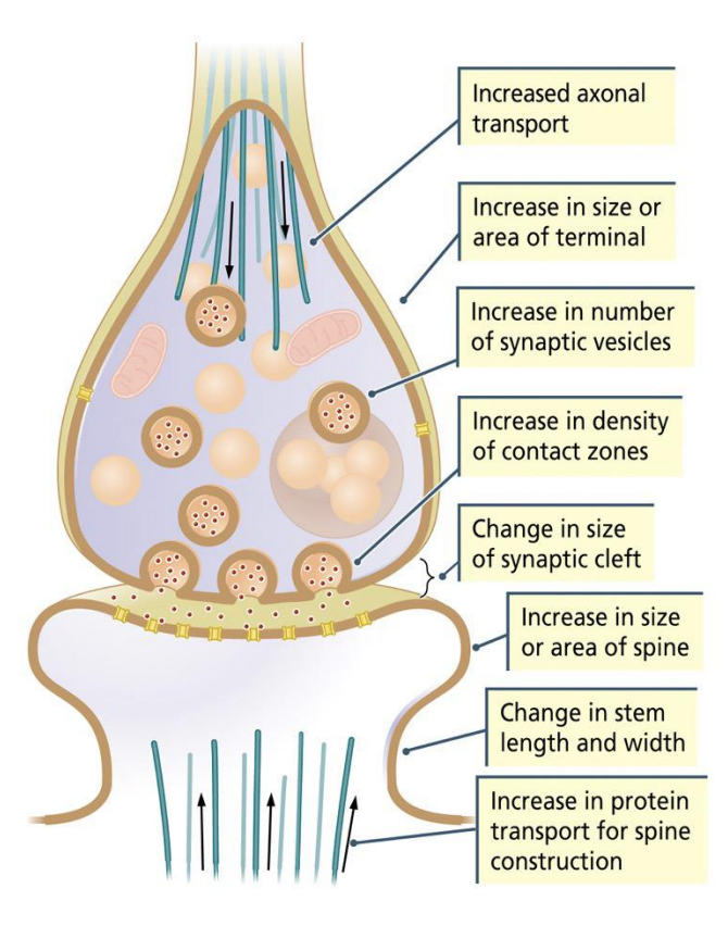
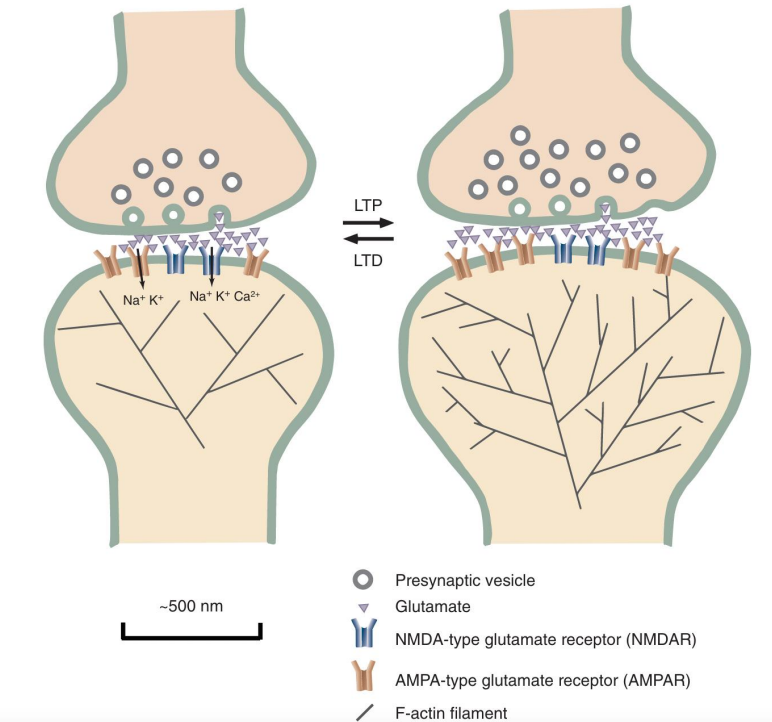
long-term potentiation
when a neuron connection is used repeatedly, synapses between them get stronger
sprouting of new synaptic contacts between co-activated neurons
new synaptic bulbs can be formed
more vesicles formed, more AMDA receptors, more glutamate emitted and received, more F-actin to provide more structure to cell (BIGGER)

long-term depression
when a neuron connection is used very little, or they fire at different times, synapses between them get weaker
to limit unnecessary connections, retracting/dismantling of synaptic contacts between non-cooperating neurons
fewer vesicles formed, less AMDA receptors, less glutamate emitted/received, less F-actin (SMALLER)
pruning unnecessary associations
examples of unnecessary associations
phobias
unneeded memories, emotions
examples of neural plasticity in effect
make more/less transmitter
have more/fewer receptors
make synapses bigger/smaller
eliminate synapses/make new synapses