cell bio exam 3 practice questions
1/30
There's no tags or description
Looks like no tags are added yet.
Name | Mastery | Learn | Test | Matching | Spaced | Call with Kai |
|---|
No analytics yet
Send a link to your students to track their progress
31 Terms
Which of the following morphological changes is NOT typically seen in a cell that is undergoing apoptosis?
A. The cell rounds up.
B. The nuclear envelope disassembles.
C. The cell swells.
D. Large cells break up into membrane-enclosed fragments.
E. The nuclear chromatin breaks into fragments.
C
While cells undergoing necrosis normally swell and burst, apoptotic cells shrink and condense.
T or F: Apoptosis is the final fate of almost all cells in an adult animal.
False.
Even though the number of apoptotic cell deaths in developing and adult animal tissues is astonishing, not all cells die in this way.
T or F: Even perfectly healthy cells may undergo apoptosis.
True
It can be triggered in perfectly healthy cells that are no longer needed
T or F: DNA damage that cannot be repaired inhibits apoptotic pathways.
False
Cells with extensive DNA damage can kill themselves by undergoing apoptosis.
T or F: Apoptosis is the main form of programmed cell death in plant cells as well as in animal cells.
False
Apoptosis is found primarily in animals.
Initiator and executioner caspases share all of the following features EXCEPT that:
A. they are cysteine proteases (they have a cysteine residue at their active site).
B. their inactive form is a monomer.
C. they undergo cleavage during activation.
D. their active form is a dimer.
E. they are inhibited by IAPs.
B
Caspases are cysteine proteases that are normally activated in apoptosis through proteolytic cleavage by other caspase molecules and formation of active dimers. Both initiator and executioner caspases can be inhibited by inhibitors of apoptosis (IAPs). While inactive initiator caspases are monomers, inactive executioner caspases are dimers.
v-FLIPs are viral proteins that were first identified as modulators of apoptosis; they contain two death effector domains, which are also found in some initiator caspases such as procaspase-8. These v-FLIP proteins can be recruited to the DISC through the binding of the death effector domain to similar domains in the adaptor proteins but are otherwise catalytically inactive. What do you think is the effect of v-FLIP expression in the host cell?
A. It promotes apoptosis mainly via the extrinsic pathway.
B. It inhibits the extrinsic pathway of apoptosis.
C. It activates only the mitochondrial pathway of apoptosis.
D. It inhibits the intrinsic pathway of apoptosis.
E. It enhances the caspase cascades in both the intrinsic and extrinsic pathways.
B
By binding to the adaptor proteins, these viral proteins interfere with caspase activation at the death-inducing signaling complex (DISC), and therefore make the cells resistant to Fas-mediated apoptosis, which is normally triggered by cytotoxic lymphocytes and proceeds via the extrinsic pathway.
Fill in the blank: In the intrinsic pathway of apoptosis, a protein called ______ is released from the mitochondria into the cytosol and binds to the adaptor protein Apaf1, causing it to oligomerize into a wheel-like assembly called an apoptosome, which then recruits initiator caspase-9 proteins
cytochrome c
In the mitochondrial pathway of apoptosis, caspase activation occurs after cytochrome c is released into the cytosol and induces the formation of apoptosomes.
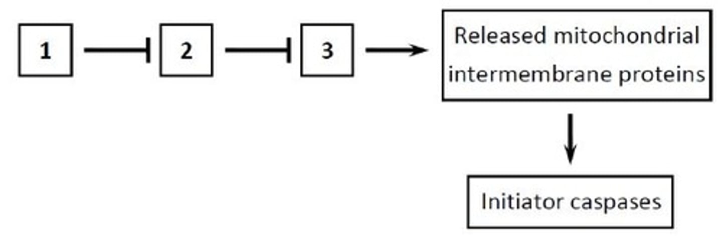
The Bcl2 family is comprised of anti-apoptotic (A), BH3-only (B), and effector (E) proteins. In the following diagram representing the regulation of the intrinsic pathway of apoptosis, what class of activated Bcl2 family proteins (A, B, or E) corresponds to boxes 1 to 3, respectively? Your answer would be a three-letter string composed of letters A, B, and E only, e.g. ABE. #3
BAE
The pro-apoptotic effector Bcl2 family proteins such as Bax and Bak induce the release of cytochrome c and other mitochondrial intermembrane proteins into the cytosol. Anti-apoptotic Bcl2 family proteins, such as Bcl2 and BclXL, inhibit these effector proteins and are in turn inhibited by the pro-apoptotic BH3-only proteins such as Bid and Bim. Some BH3-only proteins may also directly activate Bax and Bak (not shown).
Which of the following proteins activates the mitochondrial pathway of apoptosis?
A. The tumor suppressor protein p53, when activated in response to extensive DNA damage.
B. The BH3-only protein Bid, when cleaved by the initiator caspase-8 (from the extrinsic pathway).
C. The anti-IAP protein Omi, when activated by dephosphorylation.
D. The BH3-only protein Bad, when activated by dephosphorylation.
E. All of the above.
E
Activated BH3-only proteins or anti-IAP proteins can result in the activation of the intrinsic pathway of apoptosis. In response to DNA damage that cannot be repaired, the tumor suppressor protein p53 activates the expression of genes encoding BH3-only proteins (such as Puma).
Would this mutation promote or inhibit apoptosis? Mutations in the pro-apoptotic effector Bcl2 family proteins Bax and Bak that prevent their association with the outer mitochondrial membrane.
Inhibit
Association of Bax and Bak with the mitochondrial membrane is necessary (although not sufficient) for the activation of the intrinsic pathway of apoptosis.
Would this mutation promote or inhibit apoptosis? A mutation in the BIR domain of the IAP protein DIAP1 that prevents binding to either caspases or anti-IAP proteins.
Promote
Interfering with the binding of IAPs to the caspases promotes apoptosis.
Would this mutation promote or inhibit apoptosis? A mutation in the anti-IAP protein Reaper that prevents its binding to the IAP proteins.
Inhibit
Interfering with the binding of IAPs to anti-IAPs represses apoptosis.
Would this mutation promote or inhibit apoptosis? A mutation in the CARD domain of caspase-9 that prevents its binding to Apaf1.
Inhibit
The association of procaspase-9 with Apaf1 in apoptosomes is necessary (although not sufficient) for the activation of the intrinsic pathway of apoptosis.
Soon after the discovery of nerve growth factor (NGF), researchers injected newborn mice with rabbit antiserum (i.e. serum that contains antibodies) against NGF. They observed massive nerve cell death compared to appropriate control injections. Up to 99% of the neurons in some parts of the developing peripheral nervous system died after about a week of daily injections. These results suggest that:
A. NGF signaling sensitizes developing nerve cells to apoptotic signals.
B. developing neurons undergo apoptosis in the presence of rabbit proteins.
C. developing neurons require NGF for apoptosis.
D. developing neurons undergo cell death in the absence of NGF.
E. NGF signaling is sufficient for survival of the developing neurons.
D
The antiserum injection results in the perturbation of NGF function, which is similar to knocking-down NGF using genetic techniques. The increase in cell death after injection suggests that NGF is important for nerve cell survival. We now know that the cells undergo apoptosis in the absence of the survival factor.
Apoptotic cells are efficiently phagocytosed by neighboring cells or macrophages. Which of the following DOES NOT normally happen in this process?
A. The apoptotic cell releases some of its cytoplasmic content to induce a local inflammatory response.
B. The apoptotic cell exposes phosphatidylserine at its surface, which interacts with receptor proteins on the surface of phagocytes via "bridging" proteins.
C. The apoptotic cell loses or inactivates "don't eat me" signals.
D. The apoptotic cell rounds up and detaches from its neighbors, which facilitates phagocytosis.
A
T or F: Either excessive or insufficient apoptosis can contribute to disease.
True
Contribution to disease can come from either excessive apoptosis (e.g. in strokes) or insufficient apoptosis (e.g. in some autoimmune disease and cancers).
T or F: Excessive apoptosis, in many cases, leads to autoimmune disease and cancer.
False
Lack of apoptosis leads to autoimmune disease and cancers.
T or F: In about half of human cancers, the tumor suppressor protein p53 is mutated.
True
The p53 protein normally arrests the cell cycle or initiates apoptosis in response to DNA damage, which explains why the loss of p53 function is of benefit to, and is widely observed in, cancer cells.
T or F: Drugs that interfere with the function of Bcl2 family proteins such as Bax and Bak may treat cancers by stimulating apoptosis.
False.
Bax and Bak promote apoptosis, so inhibiting them would not treat cancers (which already lack appropriate apoptosis).
Is this cleavage carried out by an initiator or executioner caspase? The initiator caspase-2
Initiator
Initiator caspases are cleaved and activated when brought into proximity.
Is this cleavage carried out by an initiator or executioner caspase? The executioner caspase-3
Initiator
Initiators cleave and activate their downstream executioner caspases and initiate caspase cascades.
Is this cleavage carried out by an initiator or executioner caspase? The BH3-only protein Bid
Initiator
While most caspase substrates are cleaved by executioners, Bid is one of few exceptions.
Is this cleavage carried out by an initiator or executioner caspase? The endonuclease inhibitor iCAD
Executioner
The great majority of over a thousand identified caspase substrates are thought to be cleaved by executioner caspases to orchestrate apoptosis.
Is this cleavage carried out by an initiator or executioner caspase? The nuclear protein Lamin A
Executioner
The great majority of over a thousand identified caspase substrates are thought to be cleaved by executioner caspases to orchestrate apoptosis.
T or F: Microtubules become greatly stabilized in mitosis compared to interphase.
False
Mitosis is accompanied by a dramatic reorganization of the microtubule cytoskeleton, with a significant increase in dynamic instability.
T or F: