Lab - Arteries and veins
1/100
There's no tags or description
Looks like no tags are added yet.
Name | Mastery | Learn | Test | Matching | Spaced | Call with Kai |
|---|
No analytics yet
Send a link to your students to track their progress
101 Terms
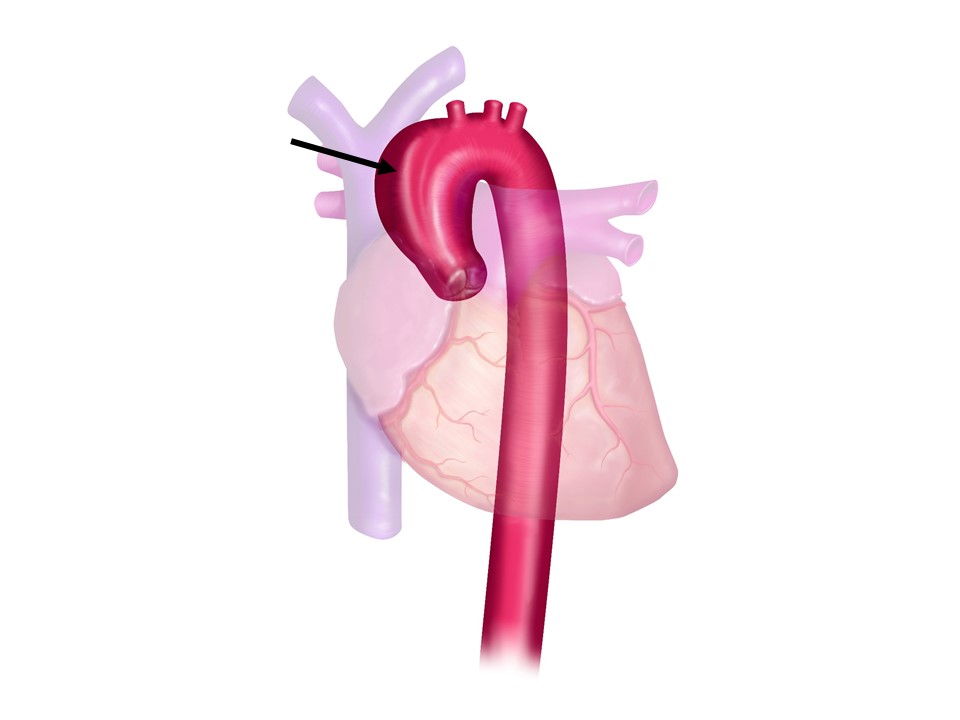
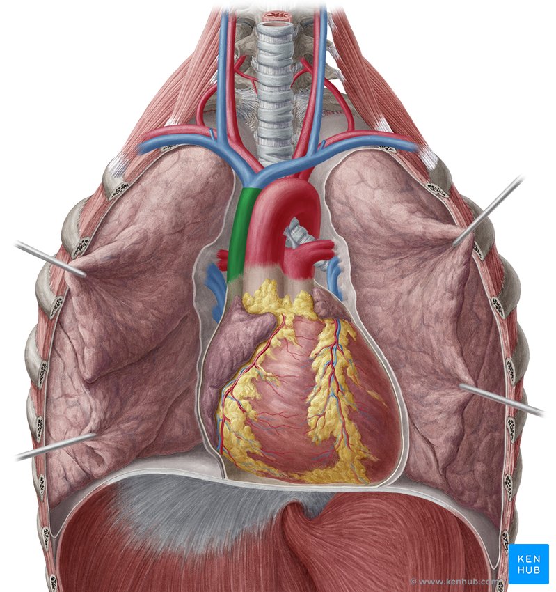
Ascending aorta
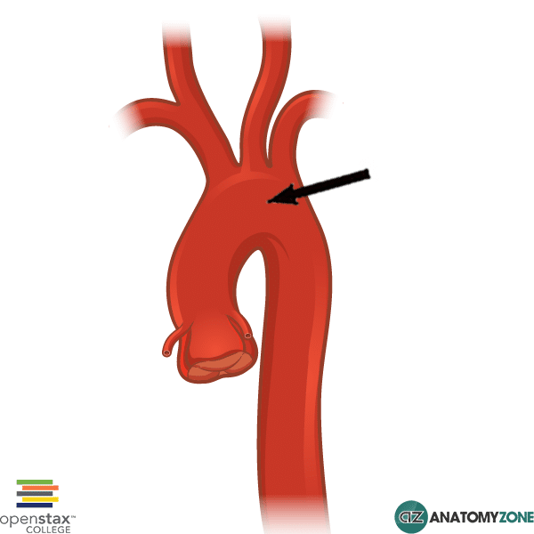
aortic arch
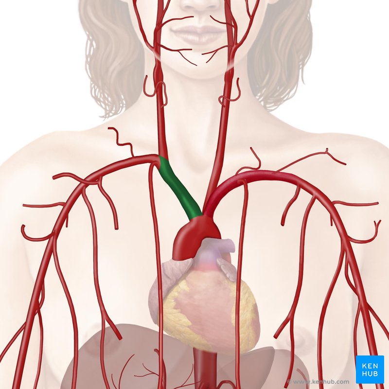
brachiocephalic trunk
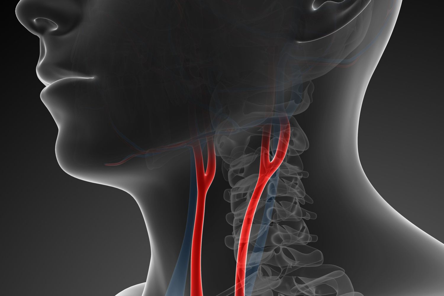
common carotid arteries
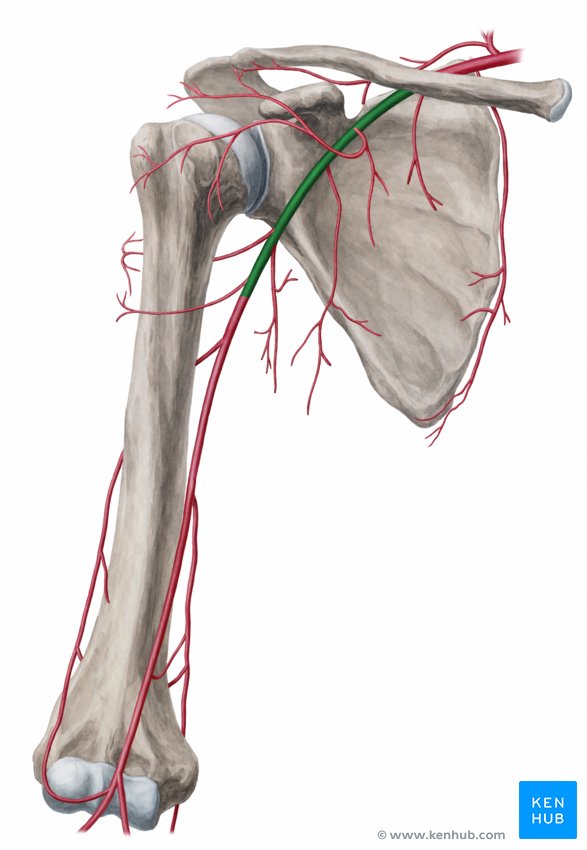
axillary artery
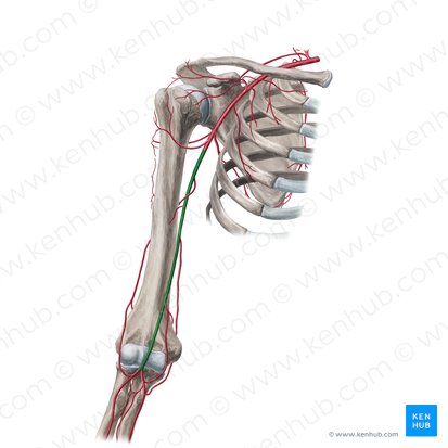
brachial artery
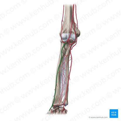
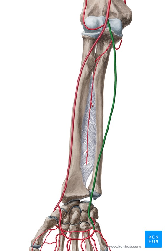
Radial artery
ulnar artery
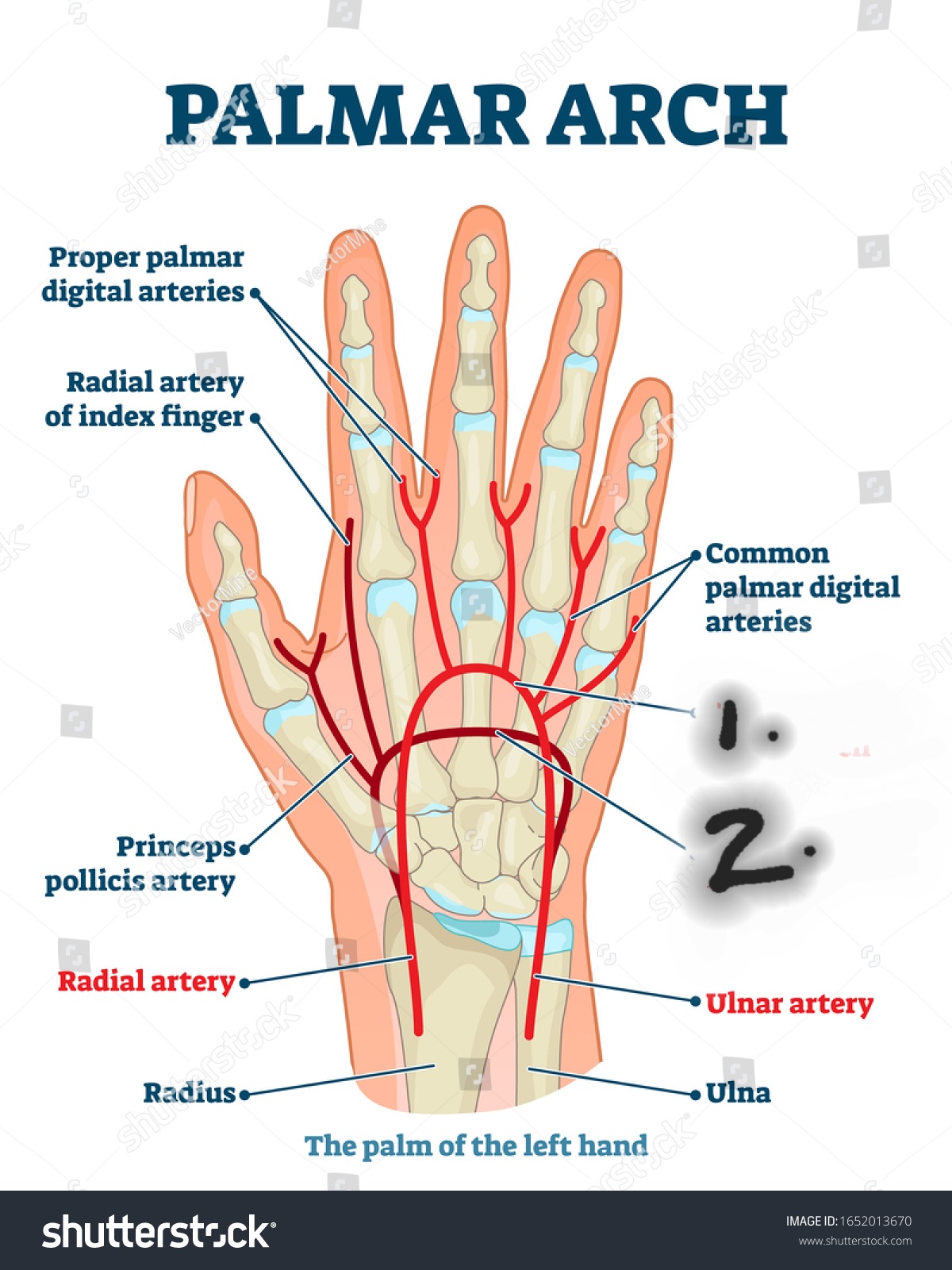
Name one and two
Palmar arches (one superficial palmar arch and two deep palmar arch)
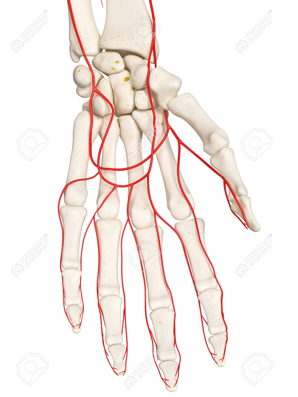
digital arteries
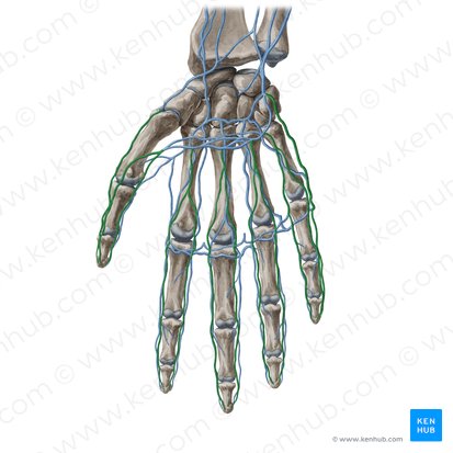
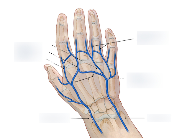
digital veins
IMAGINE IT IN BLUE!!!
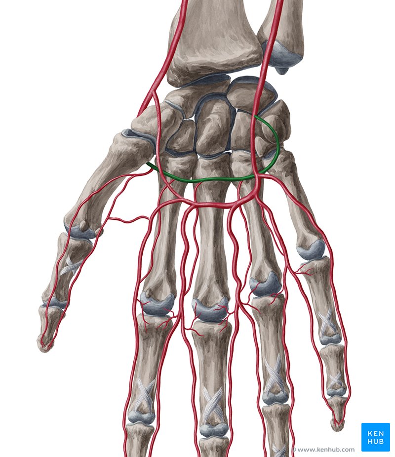
palmar venous arches (superficial and deep)
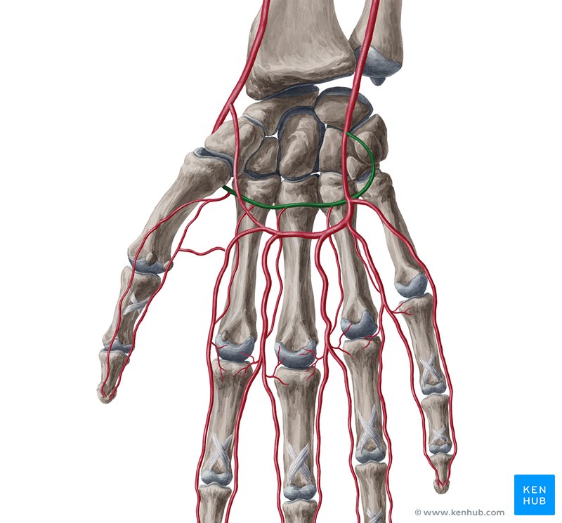
deep palmar arch

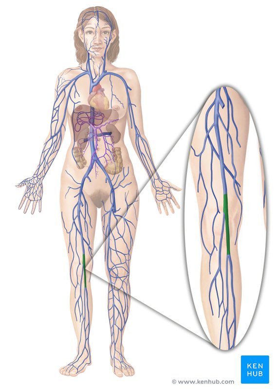
What is the blue one called?
Median cubital vein
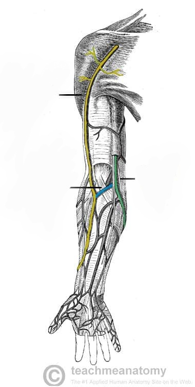
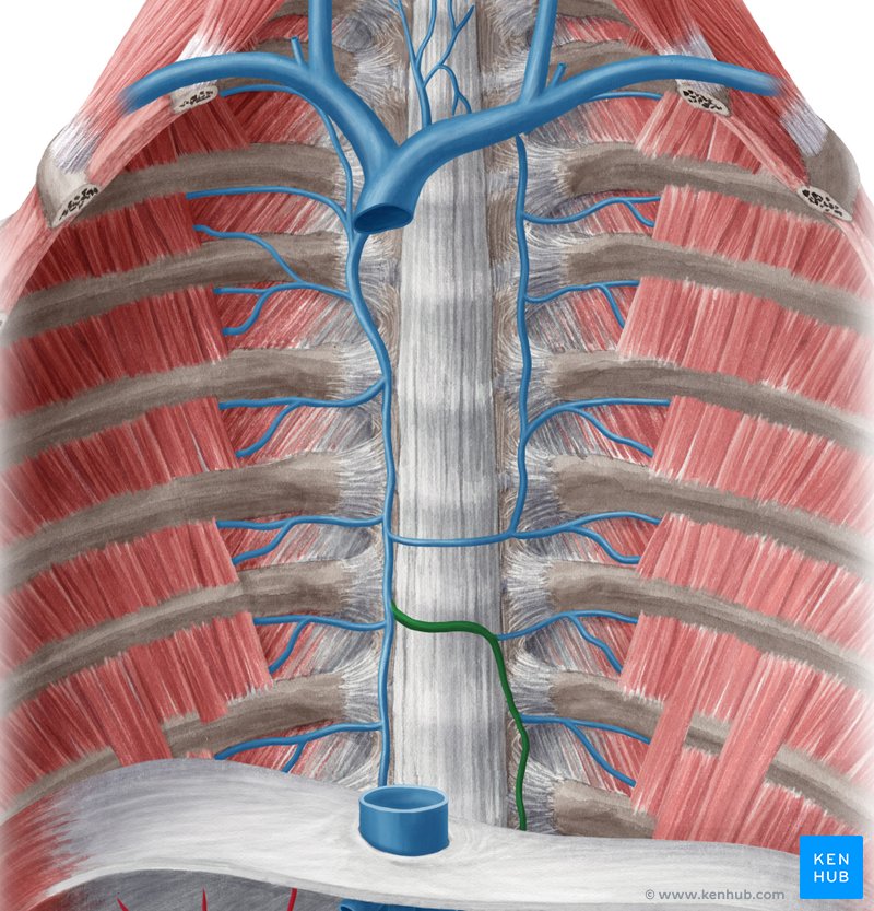
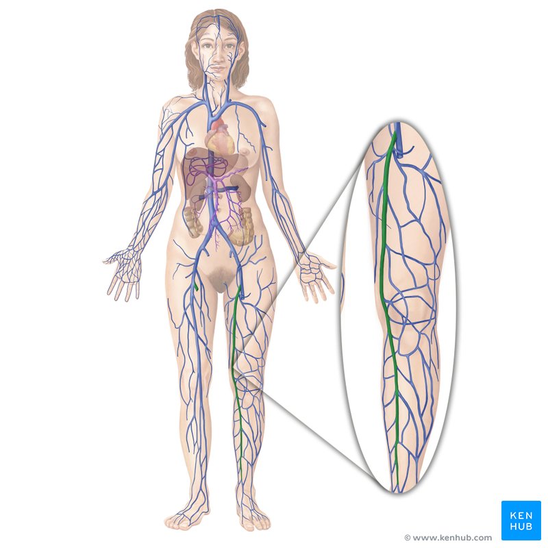
What is the green vein called?
Basilic vein
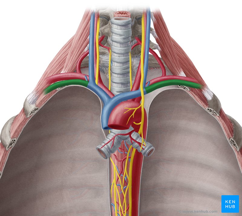
subclavian vein
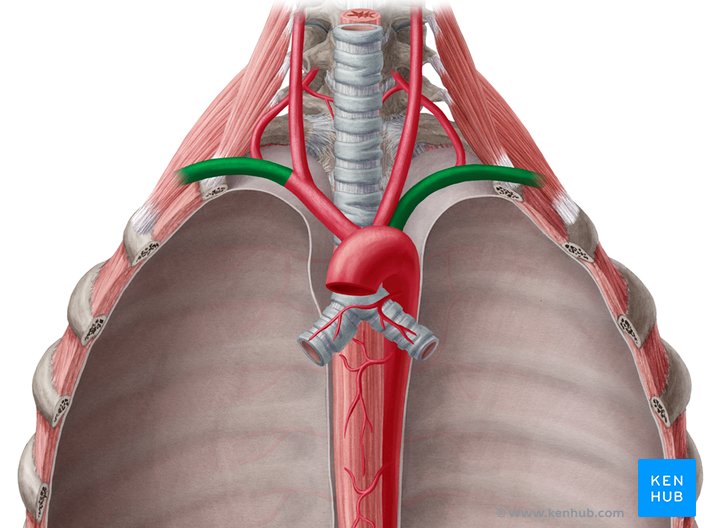
Subclavian arteries
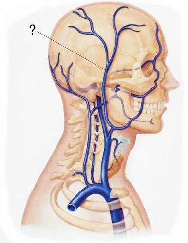
external jugular vein
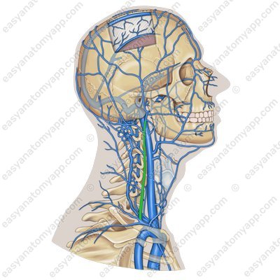
vertebral Jugular vein
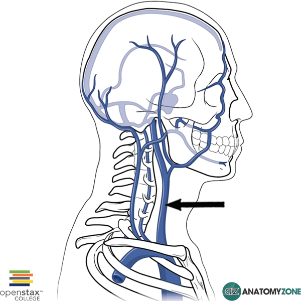
internal jugular vein
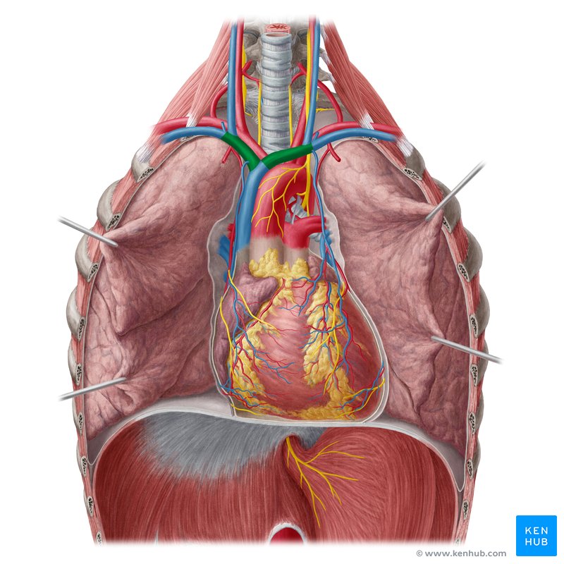
brachiocephalic vein
superior vena cava
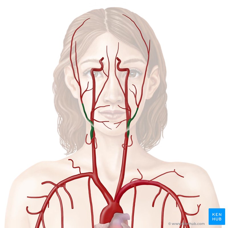
external carotid artery
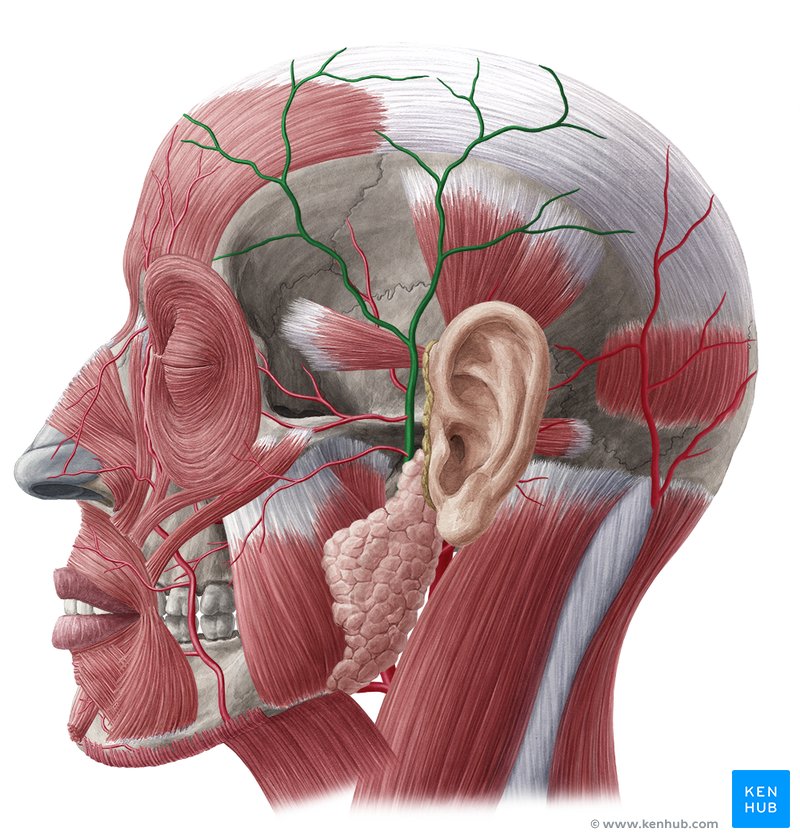
(superficial) temporal artery
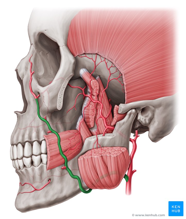
facial artery
internal carotid artery
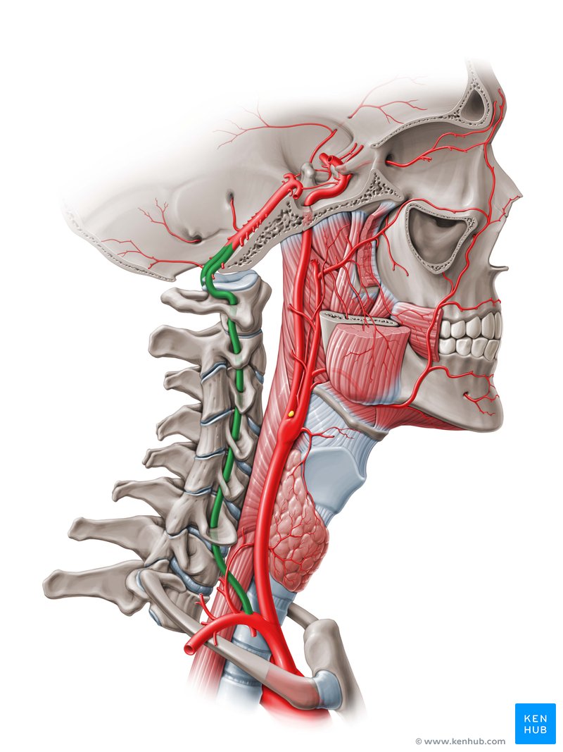
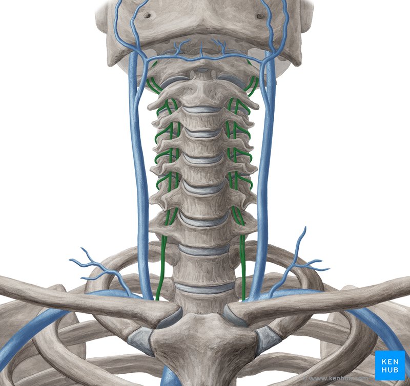
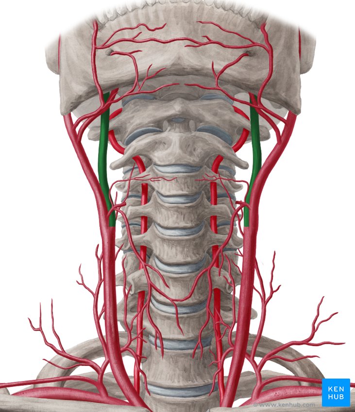
vertebral artery
(superficial) temporal vein
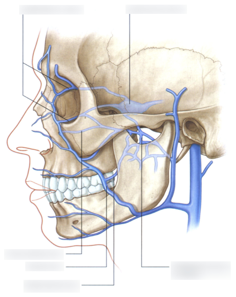
facial vein
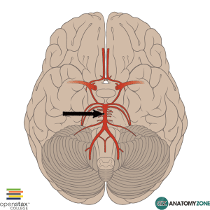
basilar artery
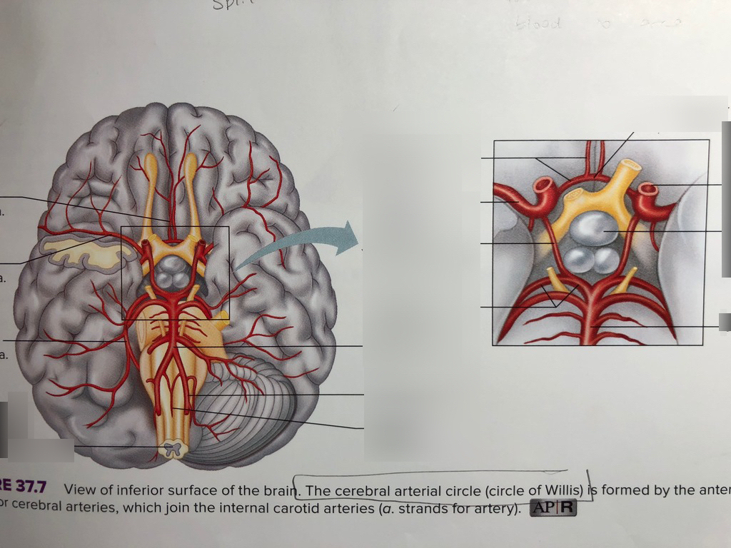
Cerebral Arterial Circle
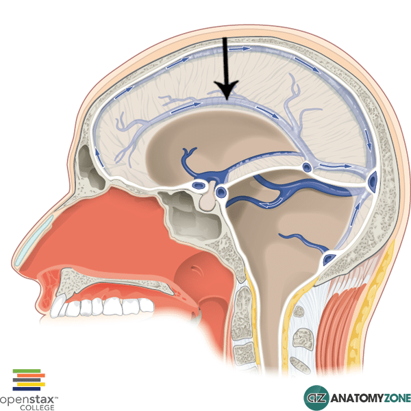
superior sagittal sinus
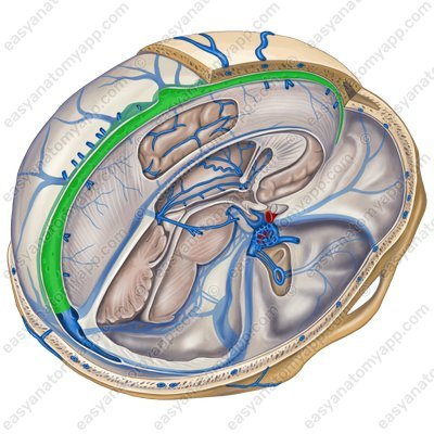
inferior sagittal sinus
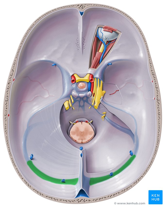
transverse sinus
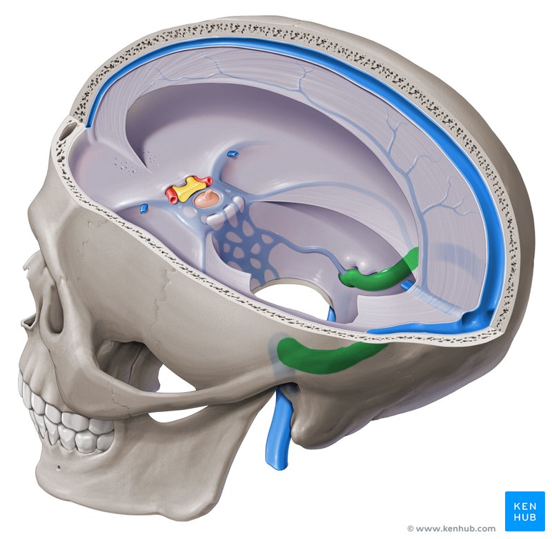
sigmoid sinus
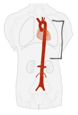
thoracic aorta
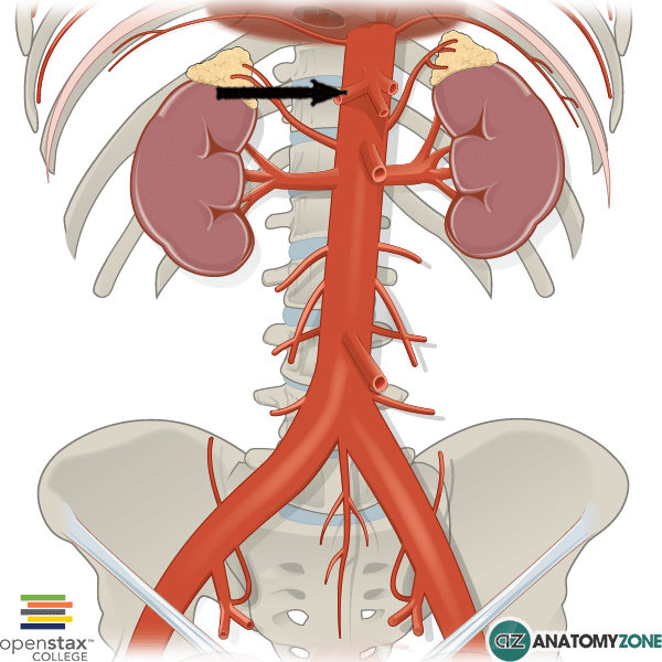
abdominal aorta
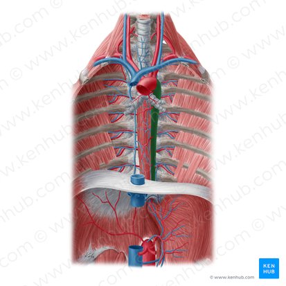
superior phrenic arteries
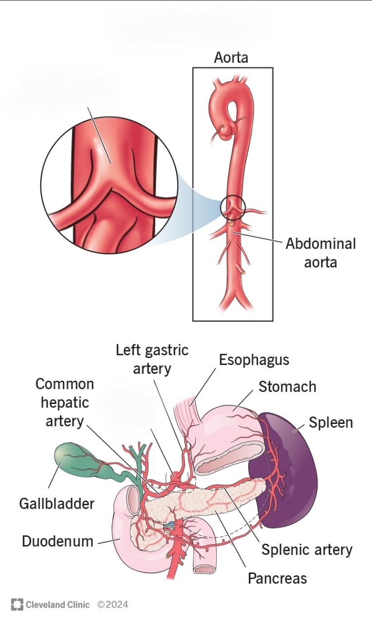
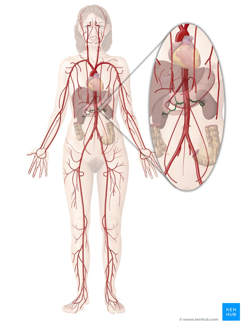
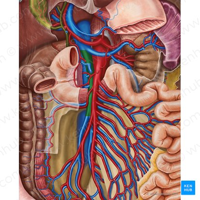
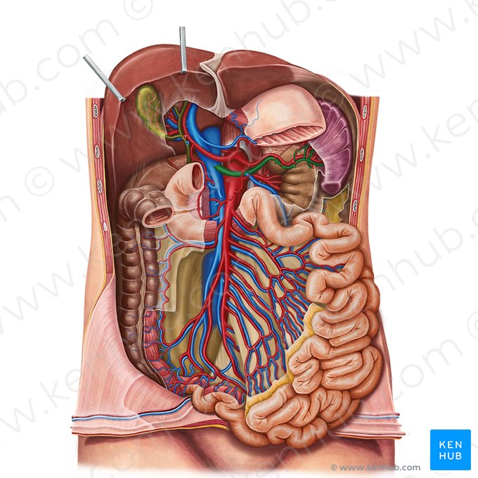
celiac trunk
renal arteries
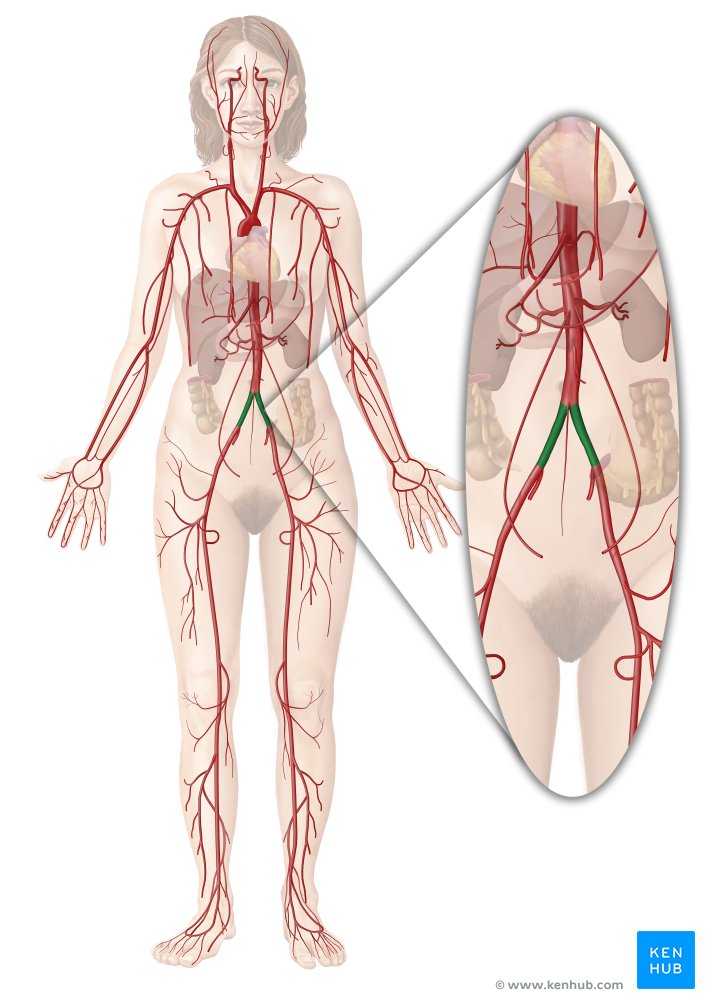
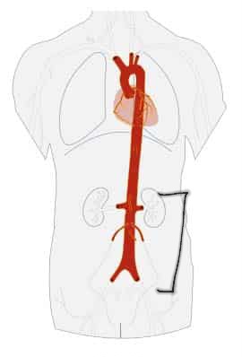
common iliac arteries
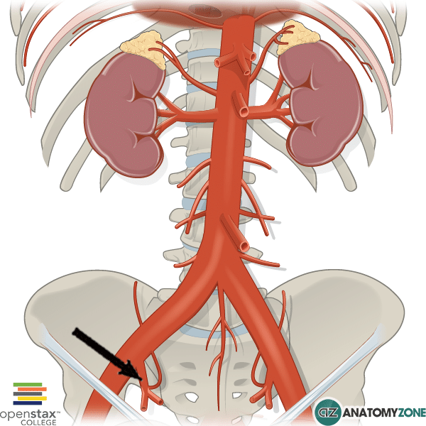
internal iliac arteries
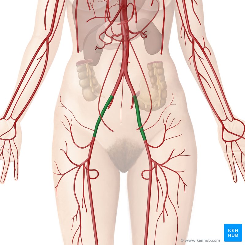
external iliac arteries
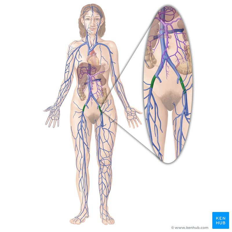
external iliac veins
internal iliac veins
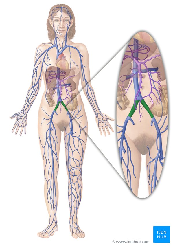
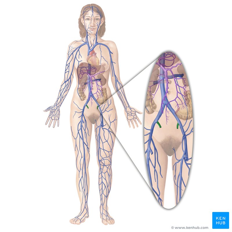
common iliac veins
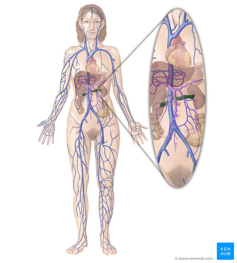
Renal veins
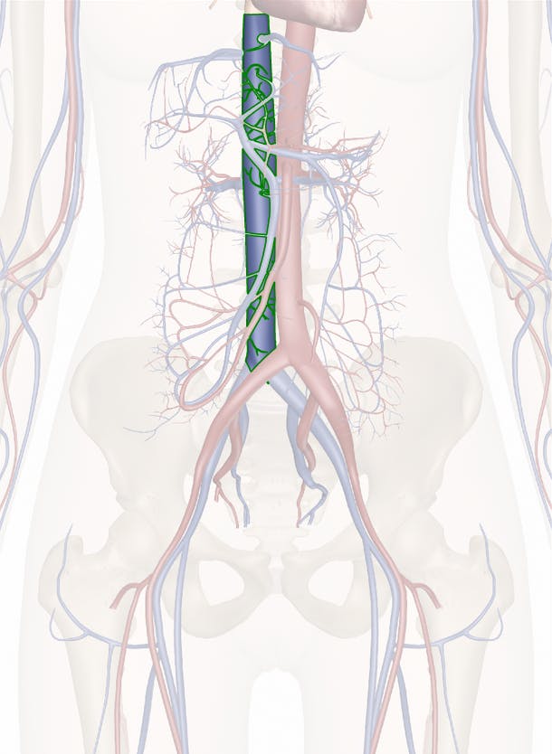
inferior vena cava
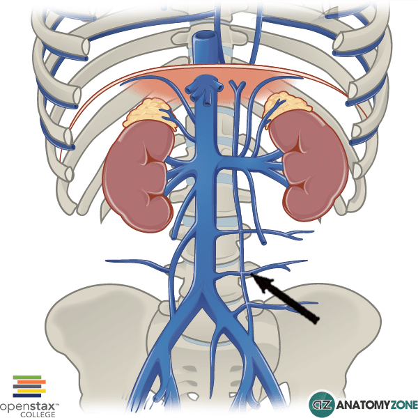
Lumbar veins
hemiazygos vein
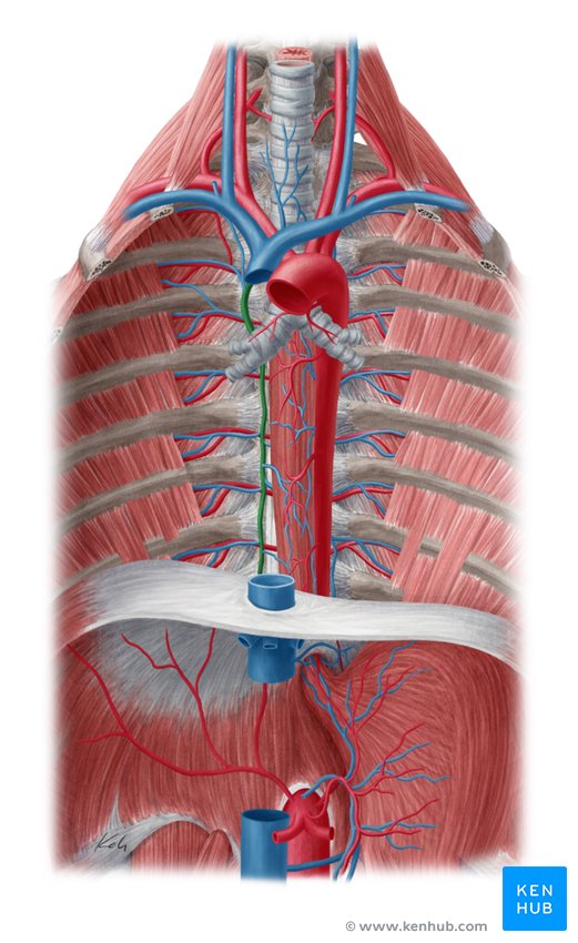
Azygos vein

superior vena cava
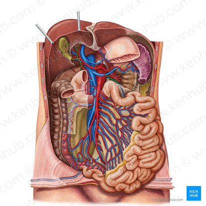
Common hepatic artery
Superior mesenteric artery
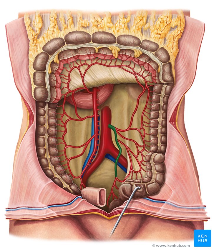
Inferior mesenteric artery
inferior mesenteric vein
superior mesenteric vein
hepatic portal vein
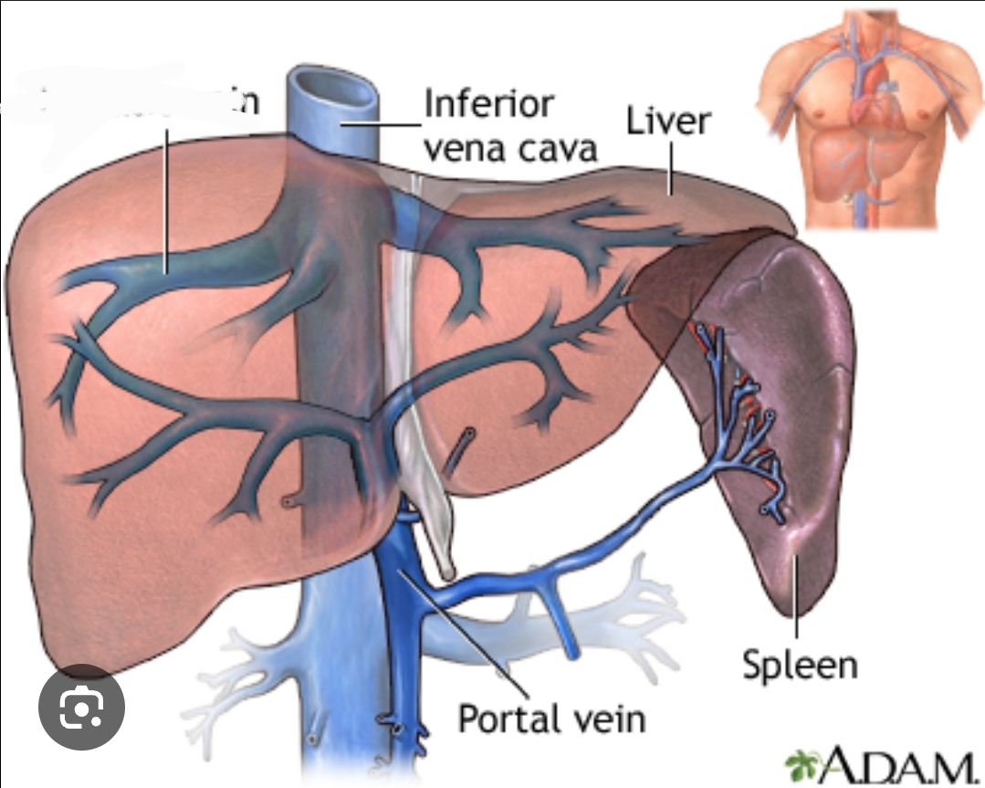
hepatic veins

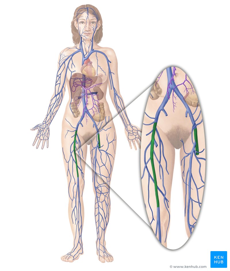
femoral vein

IMAGINE IT RED
popliteal artery
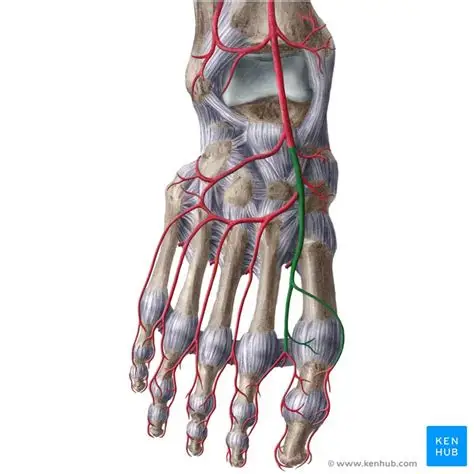
dorsalis pedis artery
What is the indented region called?
superficial dorsal venous arch
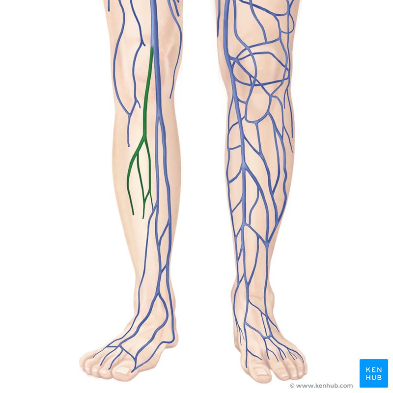
small saphenous vein
great saphenous vein
popliteal vein
femoral vein
external iliac vein
common iliac veins
Artery
Vein
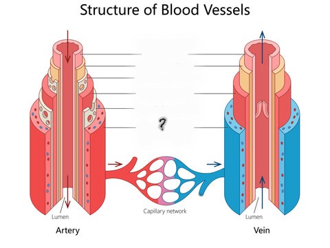
Tunica externa
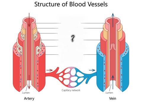
Tunica media
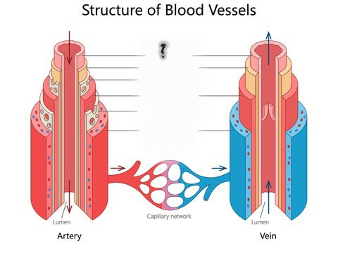
Tunica intima
Internal elastic membrane
External elastic membrane
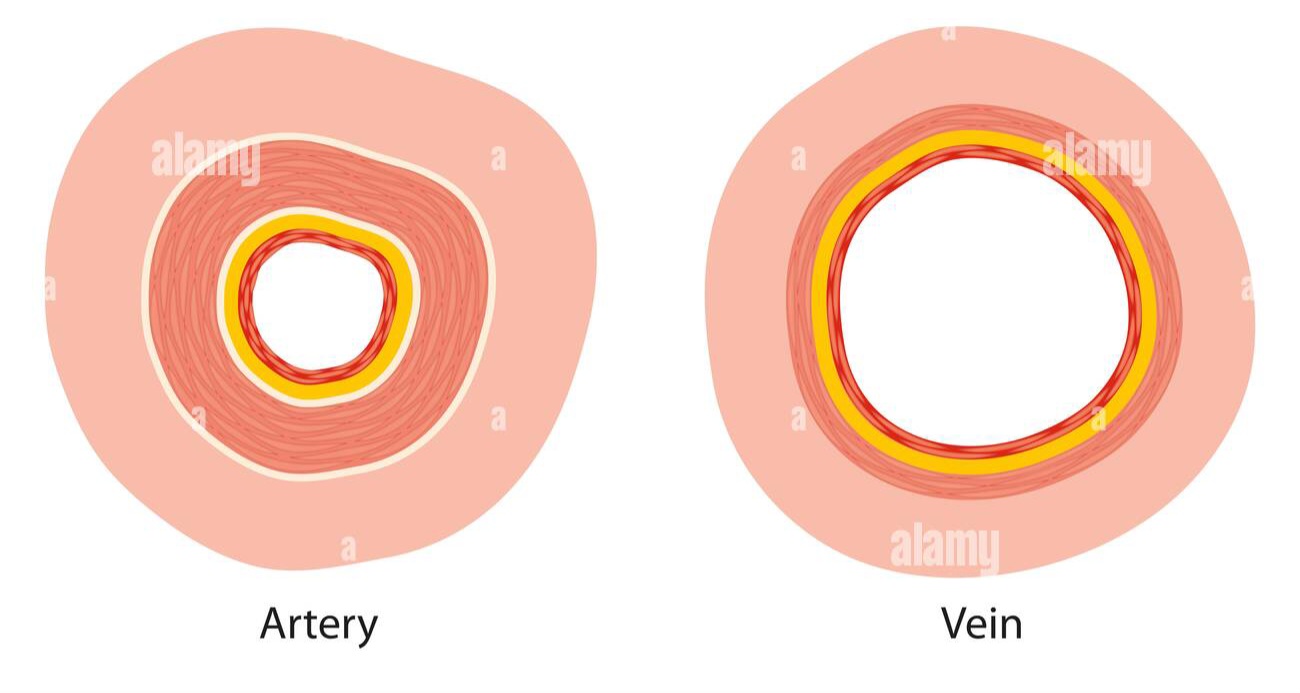
What is the space called?
Lumen
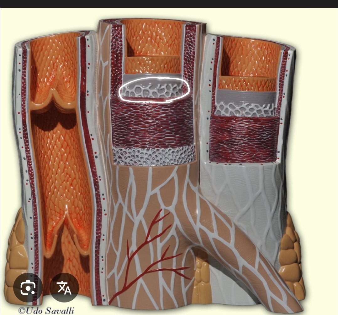
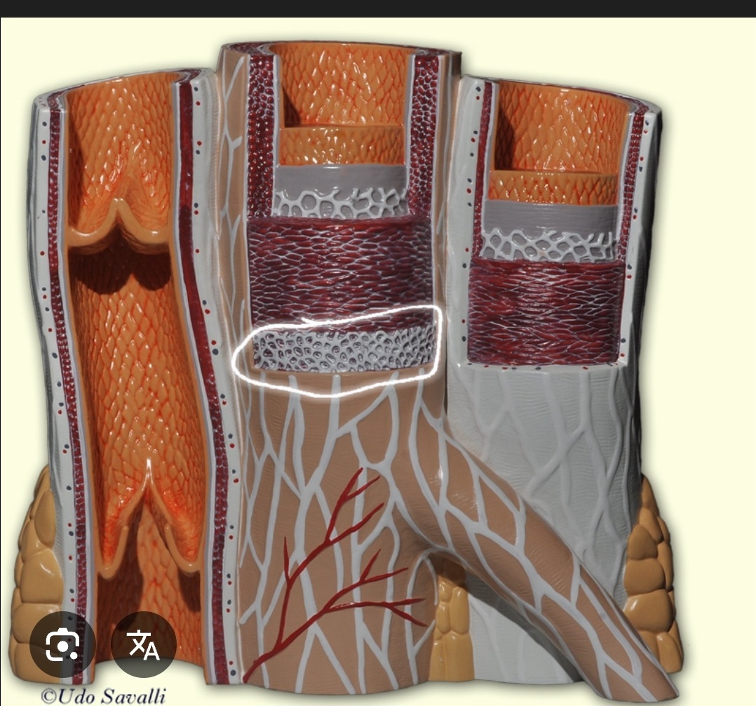
What is the fat tissue found in blood vessels?
Adipose tissue
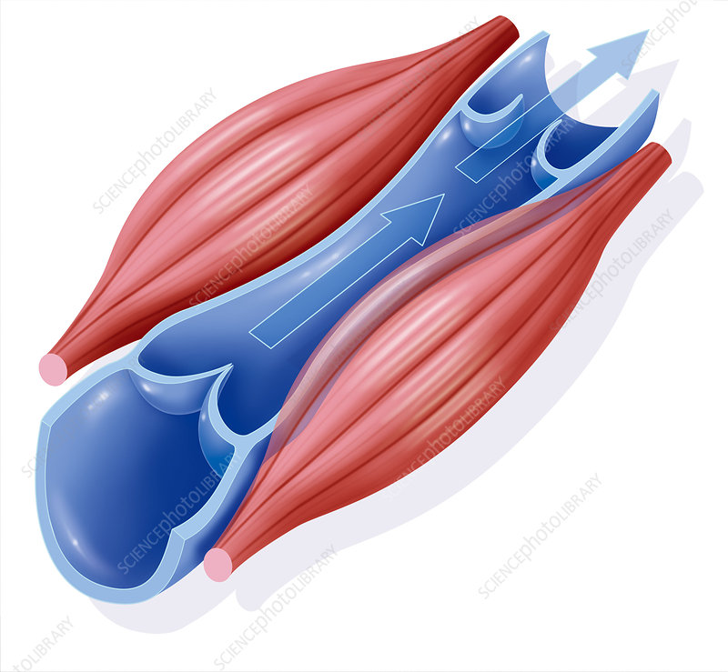
Venous valve
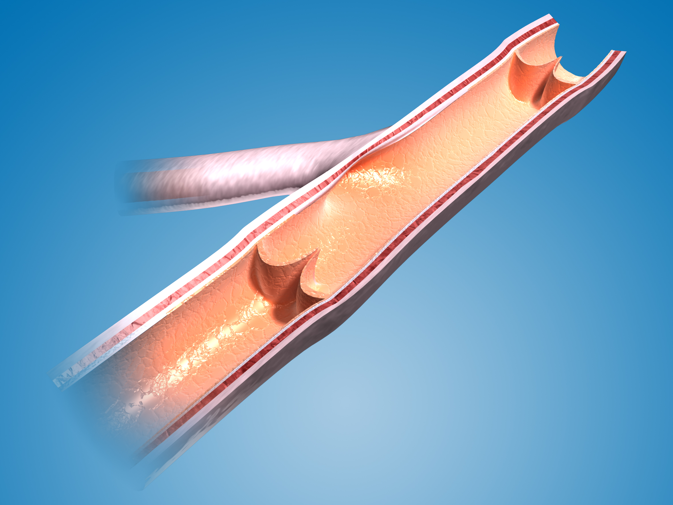
Examine the model of the Progression of Atherosclerosis. Describe the difference in the location of the adipose tissue on this model versus the Vein and Arteries Model.
Atherosclerosis is a chronic, progressive disease affecting the arteries. It involves the buildup of fatty deposits, known as plaques, within the inner lining of arteries. This process leads to hardened and narrowed arteries, which eventually restrict blood flow.
Examine the preserved specimens of human arteries. Describe the difference between the normal artery and the artery with arteriosclerosis. What component(s) of peripheral resistance is/are decreased or increased by the disease condition?
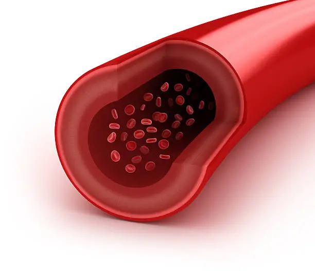
Normal Artery: Highly elastic and resilient, capable of distension and recoil to accommodate pulsatile blood flow. Walls are soft but firm; texture is pliable and smooth.
Preserved Specimen: Stiffer and less elastic due to cross-linking of proteins by fixatives. The vessel may feel brittle or tough, depending on hydration level, and can break or crack if handled aggressively.
Normal Artery: The lumen is open, flexible, and maintains circular or elliptical shape under normal pressure. Endothelial lining is intact and exhibits fine smooth layers.
Preserved Specimen: Lumen can be collapsed or partially deformed, especially if surrounding fixation prevents maintenance of physiological pressure. The inner surface may show slight wrinkling or folding.
Where is the thymus located?
On top of the heart
Spleen
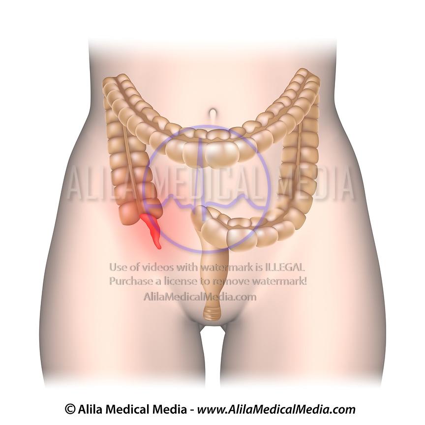
Appendix

What is number 2
Palatine tonsil
Lymph nodes, where might they be found?
They are located around the neck, arms, chest, groin, and abdomen
Lymphatic vessels, where might they be found?
Throughout the entire body
Peyer’s patches (not shown b/c they are microscopic, but where would they be found?)
Located in the small intestine especially the ileum
wrong
.
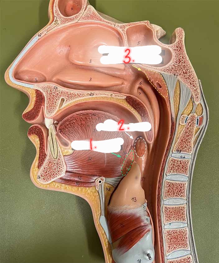
what is number 1?
Lingual tonsil
hilum
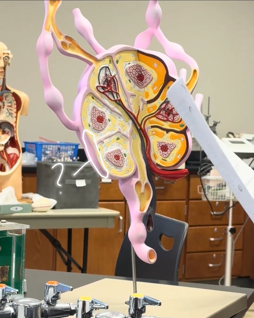
capsule
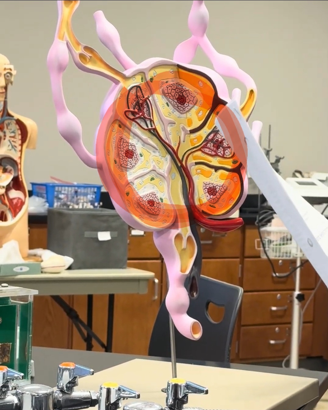
cortex
medulla
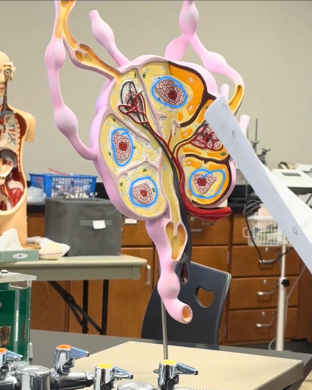
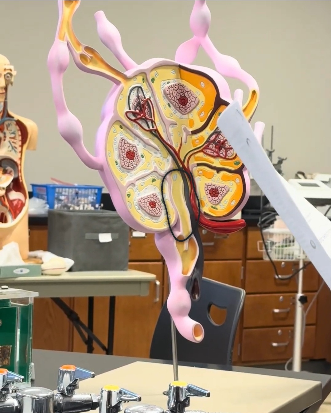
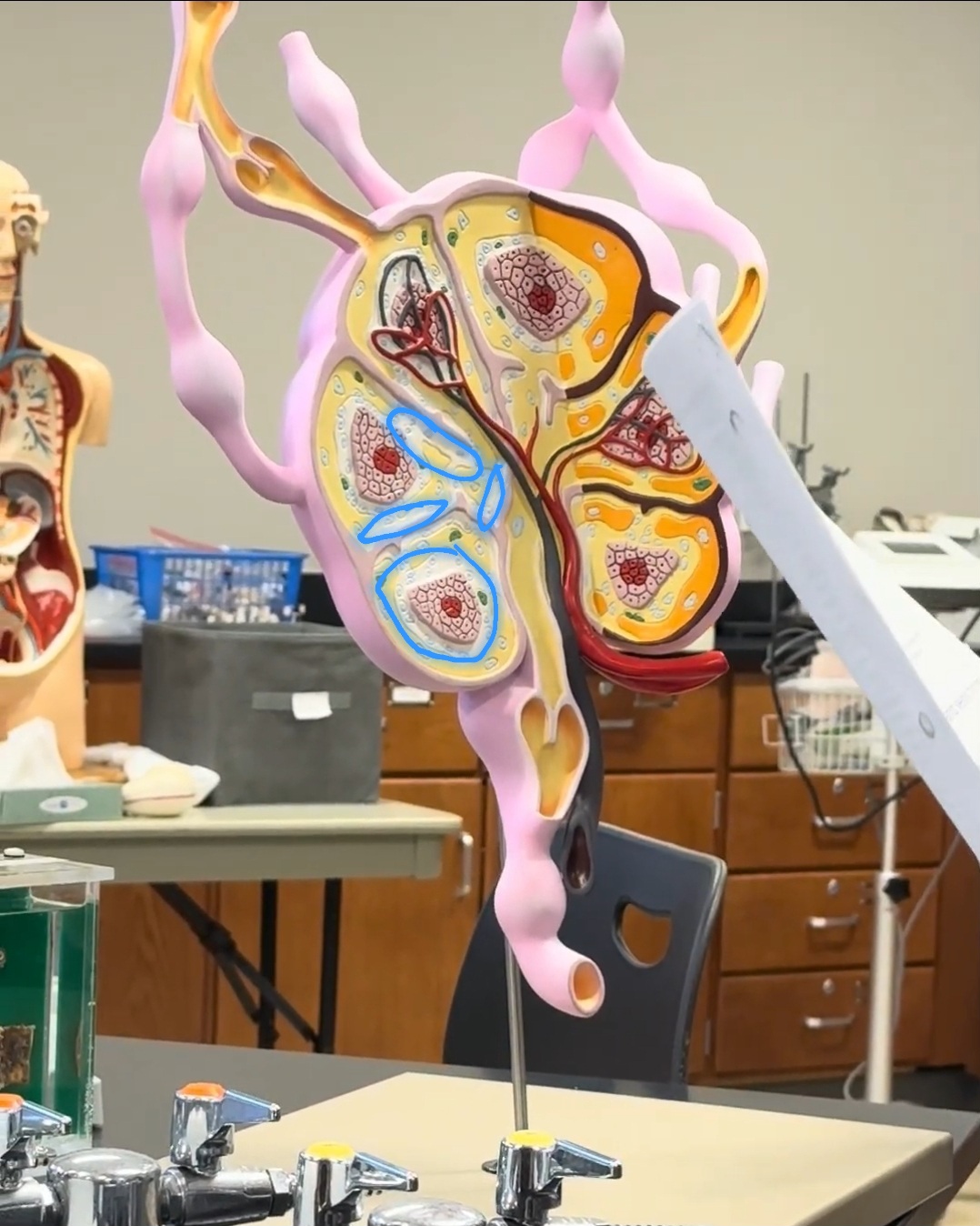
What is the blue circled area called?
lymphoid follicle
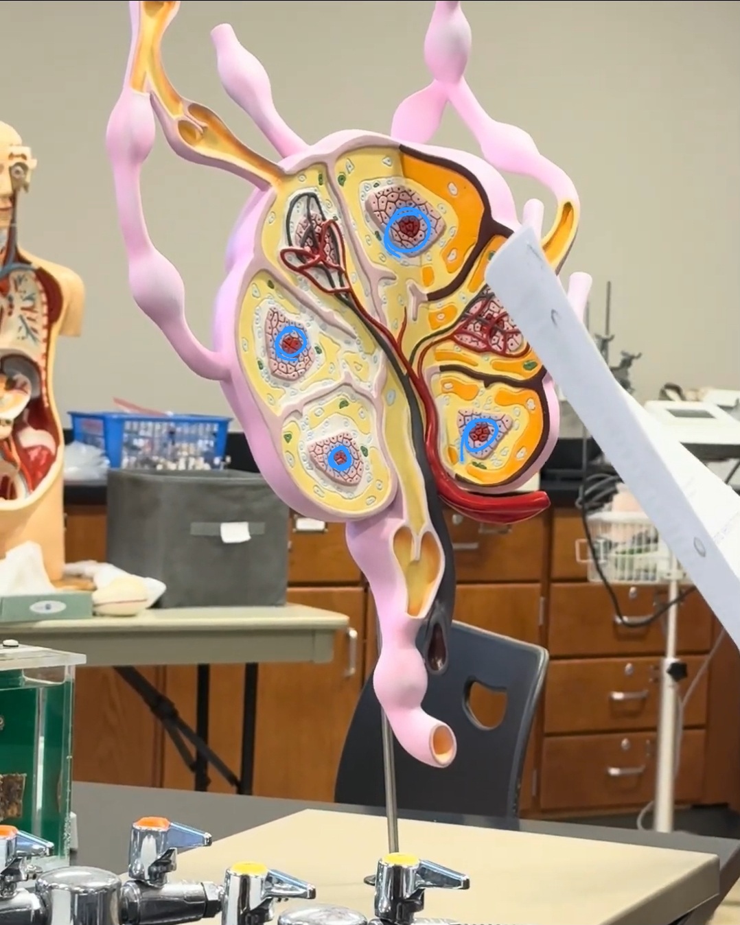
germinal center of follicle
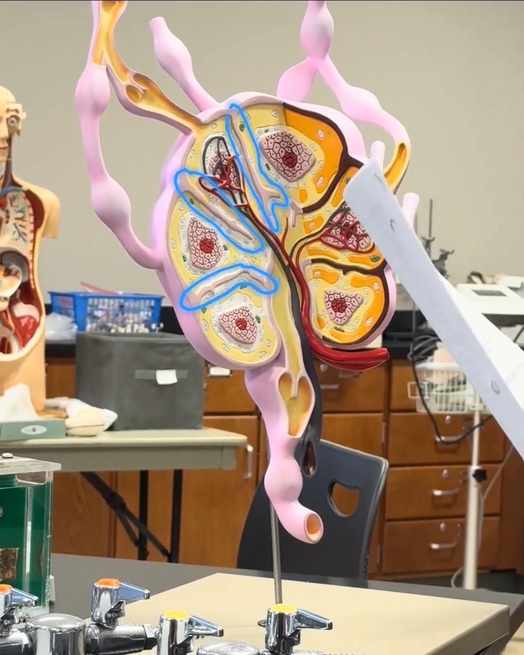
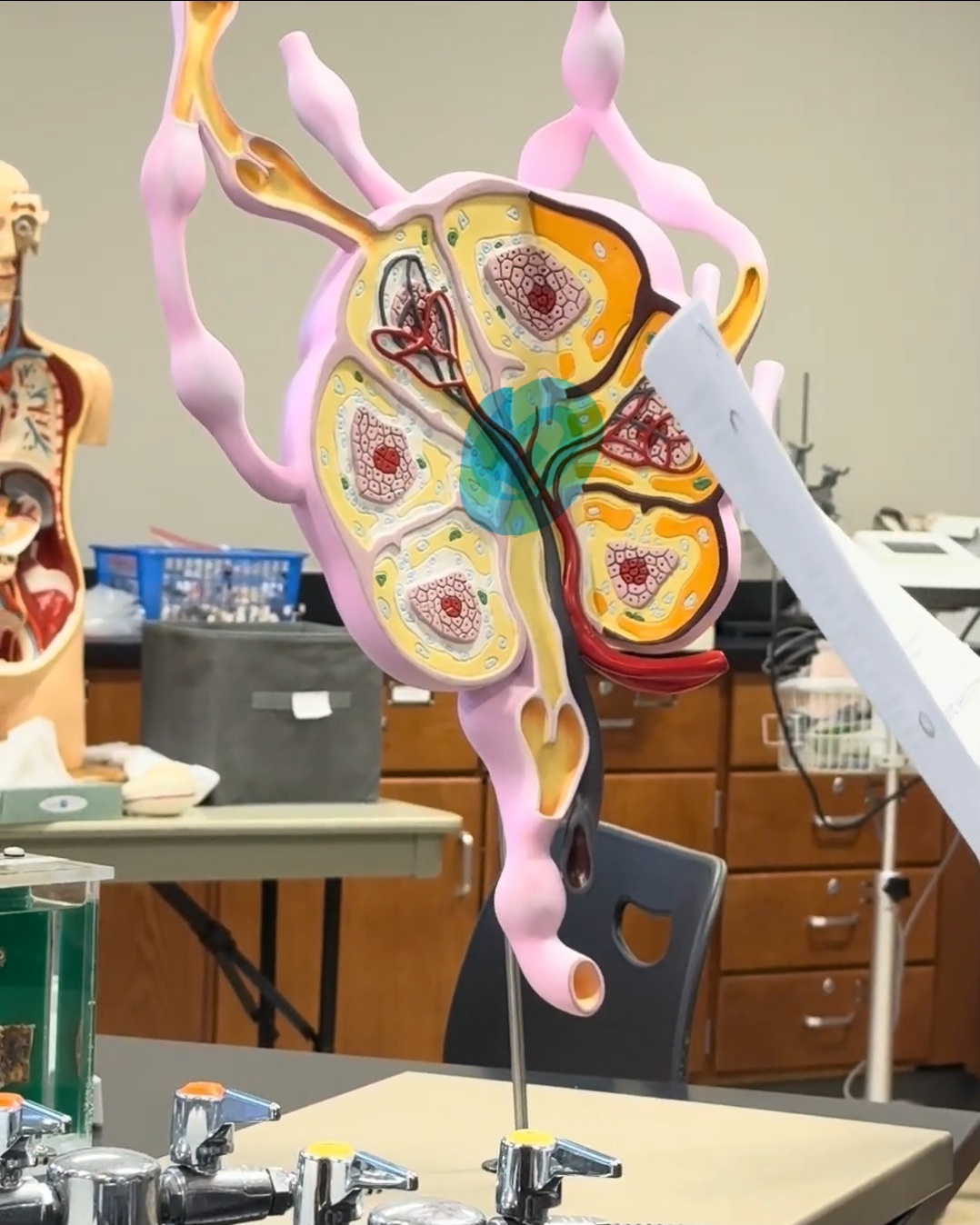
trabeculae
What are the white channels called?
medullary sinus
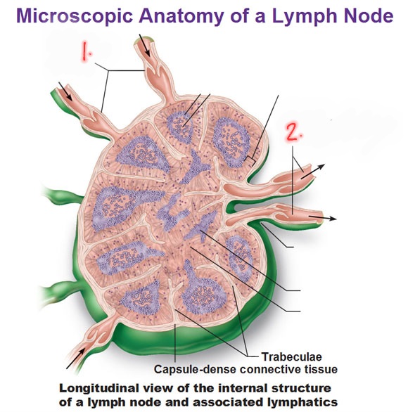
What is number 1?
Afferent vessels (top)