SLHS 539: 1/20 “Gross Anatomy of the Brain”
1/27
There's no tags or description
Looks like no tags are added yet.
Name | Mastery | Learn | Test | Matching | Spaced | Call with Kai |
|---|
No study sessions yet.
28 Terms
Cerebrum
The largest division of the brain, and is divided into four hemispheres, each of which is divided into four lobes.
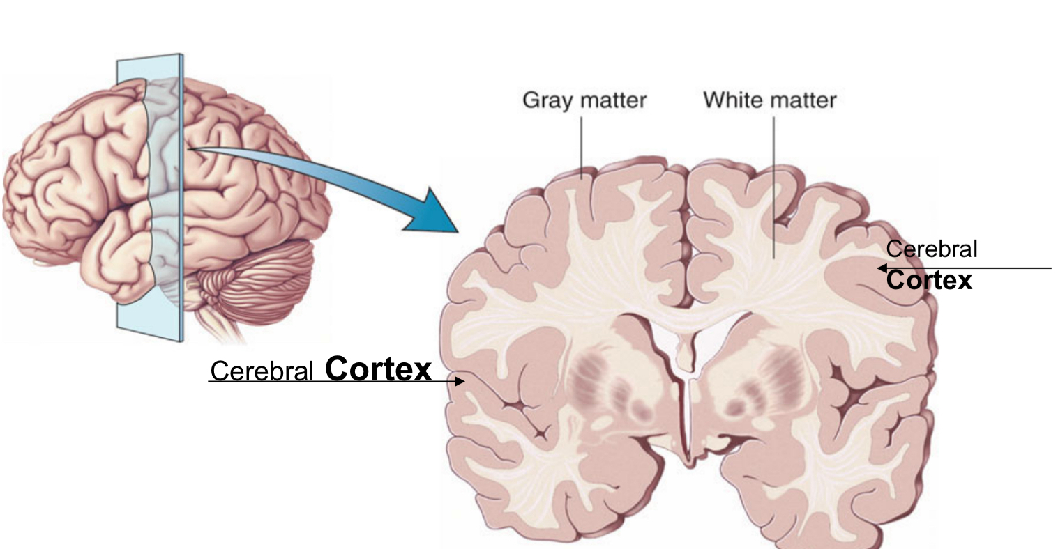
Cerebral Cortex
The outermost layer of gray matter making up the superficial aspect of the cerebrum.
“The bark of the brain”
CNS
Central Nervous System
Organization of the CNS
The Brain
Functional Organization
Neuronal Circuits Organized into
Gray matter and White matter
What does gray matter consist of and what are it’s functions?
It consists of non-myelinated axon terminals, dendrites, and cell bodies.
It functions in signal processing (integration), neurocrine/neurohormone secretion.
What is white matter consist of and what are it’s functions?
It consists of myelinated axons.
It functions in formation of tracts, ascending or descending, commissural, association, projection.
Why is the brain so wrinkled?
The folds in the brain allow it to hold billions of neurons.
Why does the brain have convolutions?
Convolutions=Folds
They allow the brain to have more surface area.
What are the three major types of cells in the cortex?
Pyramidal cells (with descending projections)
Interneurons (local circuits to facilitate or inhibit info) Ex. stellate cells, basket cells
Glial cells: Astrocytes, Oligodendrocytes, Microglia (glial cells make up half the cells in our brain)
What are the six layers in the the cerebral cortex responsible for?
They are responsible for higher level function.
Six Layers of the Cerebral Cortex
I. Molecular Layer
II. External Granular Layer
III. External Pyramidal Layer
IV. Internal Granular Later
V. Internal Pyramidal Layer
VI. Multiform Layer
—> The size and content of these layers vary by region. (important because of cytoarchitectonics — different size/content = different functions: Brodmann Layers)
Depending on the role of the brain region, there will be more or less…
Afferent vs. Efferent Projections
Korbinian Brodmann
A German Anatomist who segmented the cerebral cortex into 52 distinct regions based on histological characteristics (cytoarchitectonics) of the cerebral cortex.
Are the Brodmann Areas based on cytoarchitectonics or function?
Cytoarchitectonics (—which secondarily lead to different functions)
Brodmann Areas
52 different areas
Cerebral Cortex = __ millimeter layer
1-3 millimeter layer.
The convolutions (folds) of cerebral cortex comprise of what three elements?
Sulci (singular, sulcus) = small grooves
Fissures = large grooves
Gyri (singular, gyrus) = bulges between adjacent sulci or fissures
Image **
Specific Sulci/Fissures
Central Sulcus: Separates the primary motor from sensory cortex
Sylvian/Lateral Fissure: Separation of frontal from temporal lobe
Transverse Fissure: Separates cerebellum from cerebrum
Longitudinal Fissure: *
Brian Image **
What are the four main lobes of the cerebral hemispheres and their functions?
Frontal Lobe: Reasoning, planning, speech and movement (motor cortex), emotions, and problem solving
Occipital Lobe: Processing, integration, interpretation, etc. of vision and visual stimuli
Parietal Lobe: Spatial awareness and perception (touch, pressure, temperature and pain)
Temporal Lobe: Perception and recognition of auditory stimuli (hearing), olfaction and memory (hippocampus)
What happens when there is damage to the prefrontal cortex?
Difficulty with planning, problem-solving, thinking, reasoning, and performing executive functions
What happens when there is damage to the orbital prefrontal cortex?
Personality disorder, emotional disregulation and impulsive social behaviors.
What is the medical prefrontal cortex responsible for? When it is damaged, what deficits occur?
Responsible for attention, motivation and responsiveness.
When it is damaged, there are initiation deficits.
Phineas Gage
Tamping iron went through his eye, and he suffered from personality disorder, emotional disregulation and impulsive social behaviors.
Sensory and Motor Homunculi
The sensory cortex, like the motor cortex, follows the organization of organs and parts of our body.
Superior Parietal Lobule vs. Inferior Parietal Lobule
Superior Parietal Lobule: Sensory integration, visual-spatial and construction tasks.
Inferior Parietal Lobule: Language (dominant), body schema, and spatial orientation (non-dominant)
Primary Visual Cortex
Located in the occipital lobe, and is the primary area responsible for sight, recognition of size, color, light, motion, dimensions, etc.