Looks like no one added any tags here yet for you.
all cells must have…
plasma membrane, nucleus, and cytoplasm
all cells must…
1. maintain its own shape
2. eat nutrients and make new chemical structures
3. dispose wastes
integral proteins
embedded in phospholipid bilayer
peripheral proteins
not embedded into the bilayer, can be attached loosely or anchored
what are the six major roles of membrane proteins?
1. transport
2. cell surface receptors
3. identity markers
4. enzymes
5. cell-adhesion proteins
what molecules can pass in simple diffusion?
urea, respiratory gases, small fatty acids, small nonpolar molecules
what molecules can pass in facilitated diffusion?
larger polar molecules or charged ions
what are the two types of facilitated diffusion?
1. channel-mediated: channels embedded into the bilayer!! (leak and gated channels)
2. carrier-mediated: carrier protein! it needs a key to be opened! (key being molecule passing)
isotonic
cytosol = solutes, no net movement of water
hypotonic
solutes > cytosol, water rushes into cell (lysis)
hypertonic
solutes < cytosol, water rushes out (crenation)
primary active transport
uses ATP to get solute against gradient
secondary active transport
catches a ride with other molecule going against the gradient
symport: same direction (hitchhiking)
antiport: opposite direction
resting membrane potential
potential energy at which the cell is resting
what are the two conditions of maintaining a membrane potential?
1. unequal distribution of ions
2. unequal amounts of positive and negative charges
3 OUT NA
2 K IN
K+ (potassium): creates a negative charge outside of the membrane
Na+: makes outside of the cell more positive
what are the four tissue types?
epithelium: covers
connective: connects
muscle: contracts
nervous: conducts
intercellular junctions
tight junctions: like a nail! prevents subtances from moving between cells
desmosomes: a screw, binds neighboring cells together
gap junctions: a pipe, allows direct passageway!
basement membrane
a membrane that a layer epithelium tissue attaches to, separates epithelia and connective tissue
basal surface
the bottom deep epithelium tissue that attaches to the basement membrane
apical surface
the superficial layer of epithelium cells on top
lumen
the empty space in the body within tubes, tracts, cravities, etc.
lateral surface
surface that faces away from the body
simple
one layer of epithelial cells
stratified
2 or more layers
pseudostratified
appears stratified, but all cells are still connected to the basement membrane
squamous
FLAT!! cells, looks like fried eggs
cuboidal
as tall as they are wide, cubed shape
columnar
taller than they are wide, columns!!
transitional
special type of epithelium that stretches and shrinks based on pressure
simple squamous ET
allows rapid movement of molecules
-alveoli, vessel walls, serous membranes
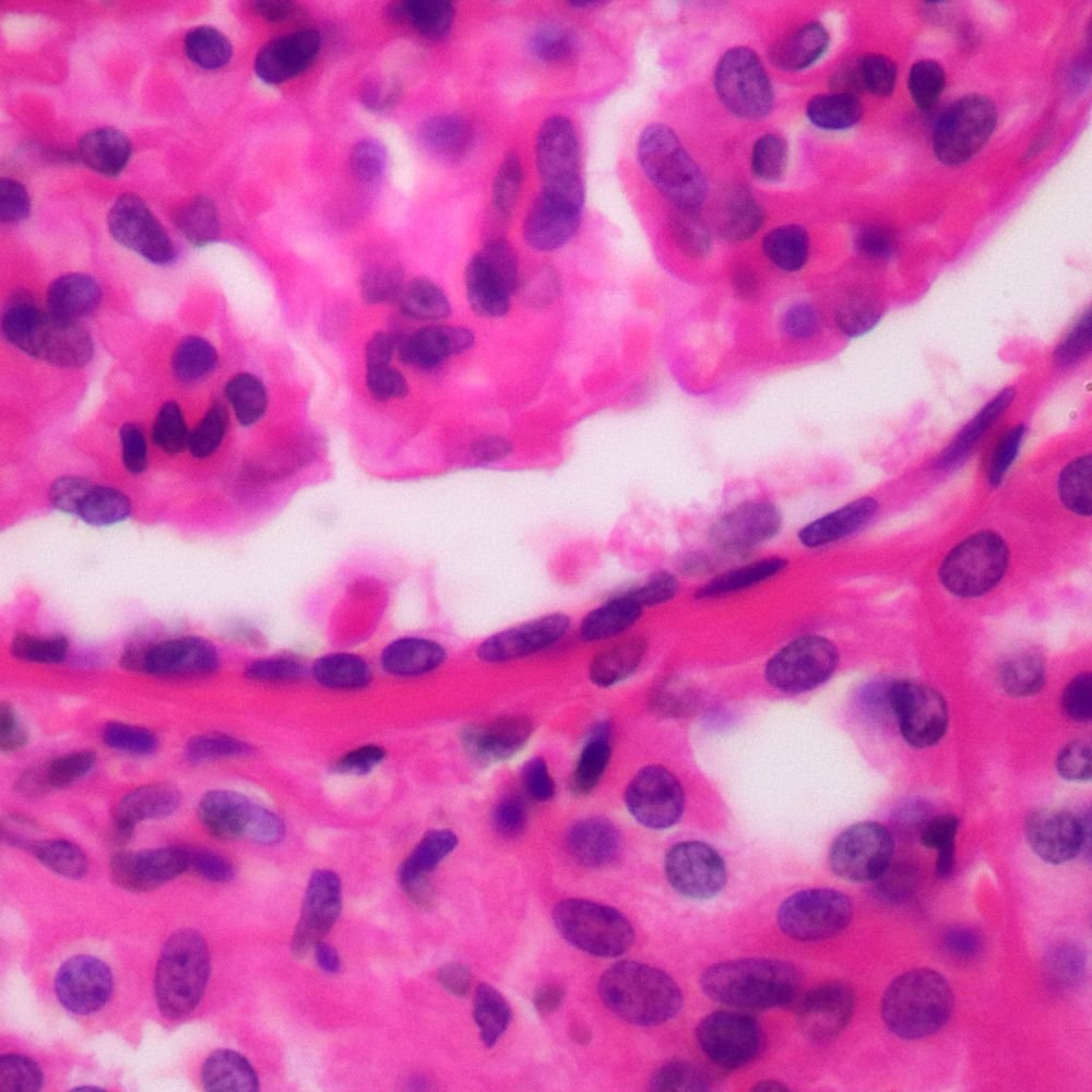
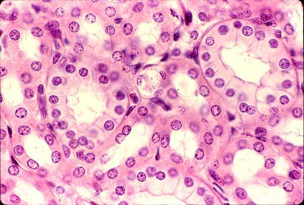
simple cuboidal ET
absorption and secretion, components for glands!
-kidney tubules, thyroid glands, exocrine glands
nonciliated simple columnar ET
secretory and absorption
-contains microvilli, may have spaces in between them for goblet cells! lives in digestive tract, stomach, anal canal
-goblet cells form mucin, which will make mucus!!
ciliated simple columnar ET
moves mucus along!! goblet cells may be spread out
-bronchioles, uterine tubes (help with passing oocyte like crowd surfing)
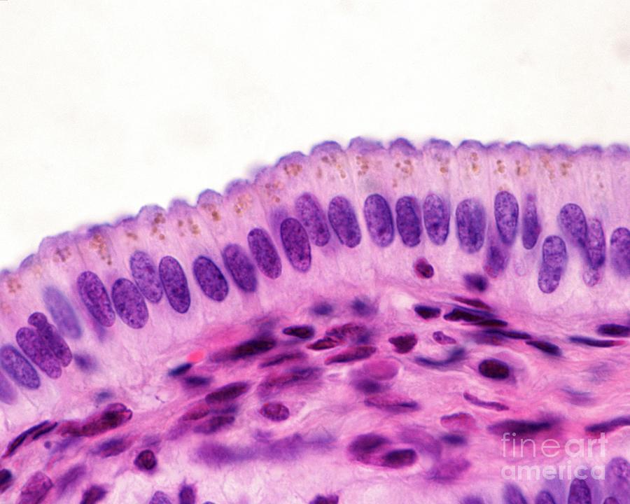
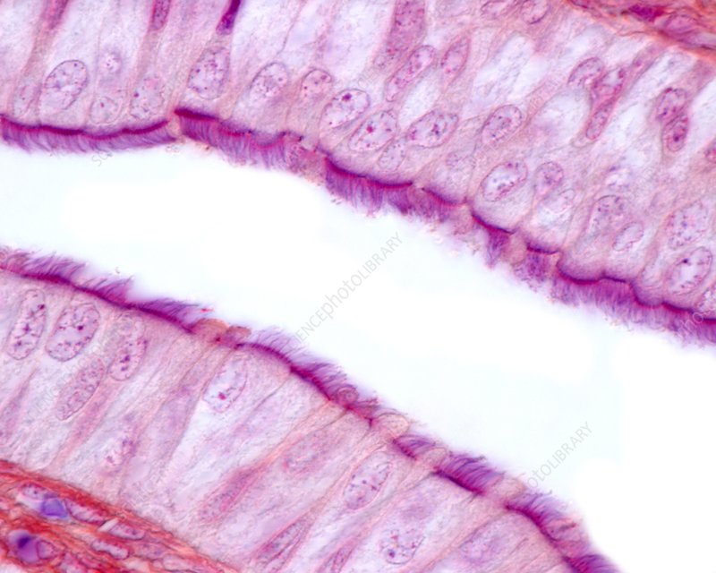
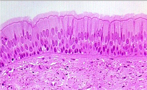
pseudostratified ciliated columnar ET
traps foreign particles moved by cilia!
-large passageways of respiratory system
pseudostratified ciliated columnar ET
very rare, only present in the make urethra and epididymis!! only for protection
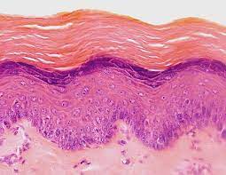
stratified squamous keratinized ET
layers of dead skin cells filled with keratin, basal region divide and eventually make it to apical region
-protect tissue from abrasion
-epidermis!
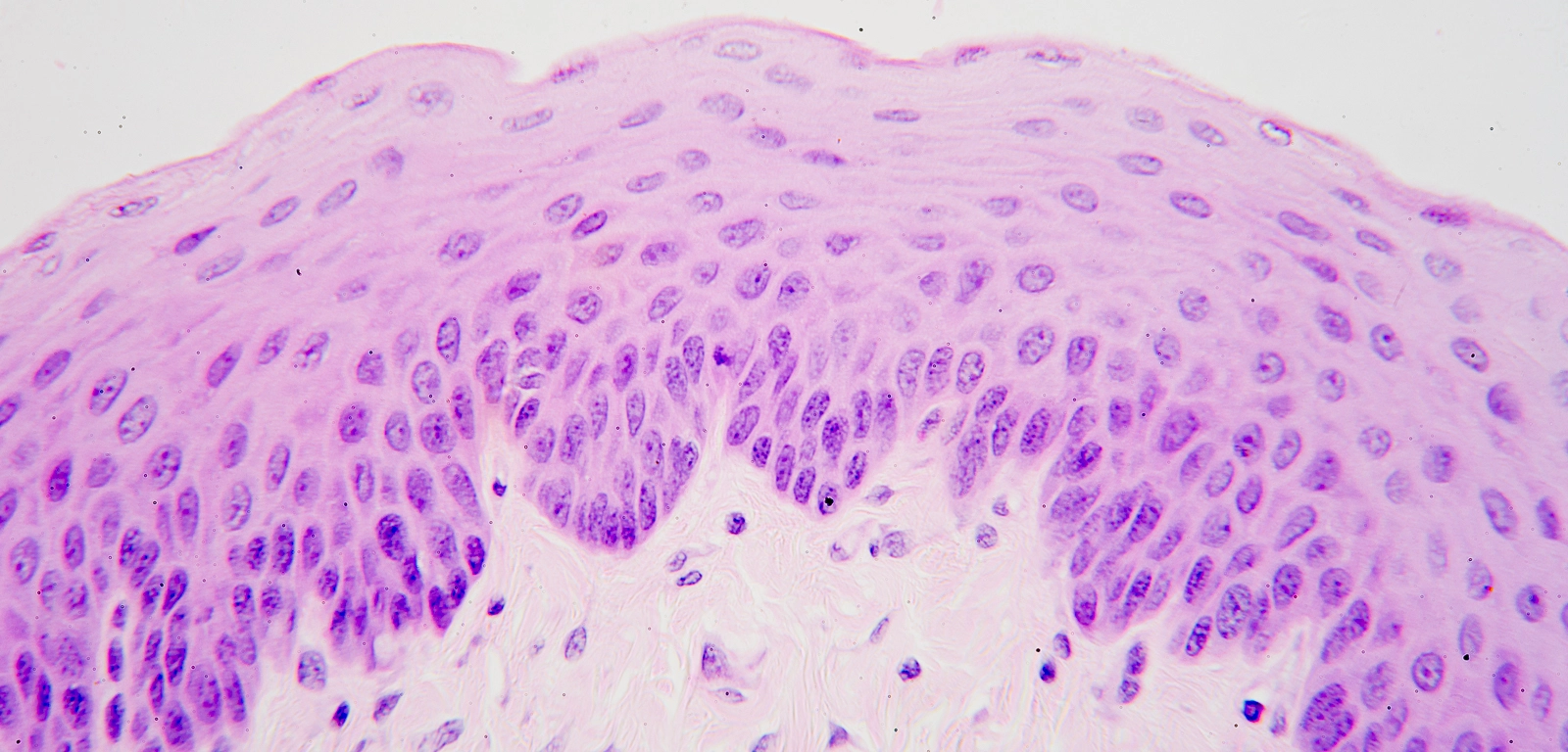
stratified squamous nonkeratinized ET
all cells mostly alive! kept moist so that fluid can go through
-oral cavity, pharynx, vagina
stratified cuboidal ET
protection and secretion
-exocrine glands, sweat glands, parts of male urethra
stratified columnar ET
rare! protection and secretion
-large ducts of salivary glands, parts of male urethra
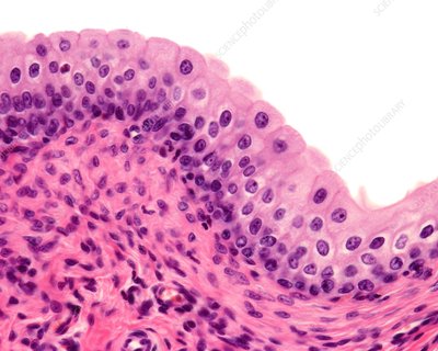
transitional epithelium
binucleated cells, stretches when bladder fills!!
-limited to the urinary tract
endocrine glands…
secrete hormones to the blood
exocrine glands…
is connected via a duct to the outside!
unicellular exocrine glands
no duct, close to surface, common type goblet cell
multicellular exocrine glands
numerous cells!
acini: cells that make the secretions, ducts transport to the surface
simple glands
single-no branched duct
compound glands
branched ducts
tubular glands
secretory gland is the same size as the duct
acinar glands
secretory side makes an expanded sac
tubuloacinar gland
tubular + acinar gland
merocrine glands
secretions into a vesicle, like a backpack!! released by exocytosis!!
apocrine glands
membrane pinches off and becomes secretion
holocrine glands
ruptured cell becomes secretion
extracellular matrix
a large network of proteins and ground substance (basically a space between cells in connective tissue)
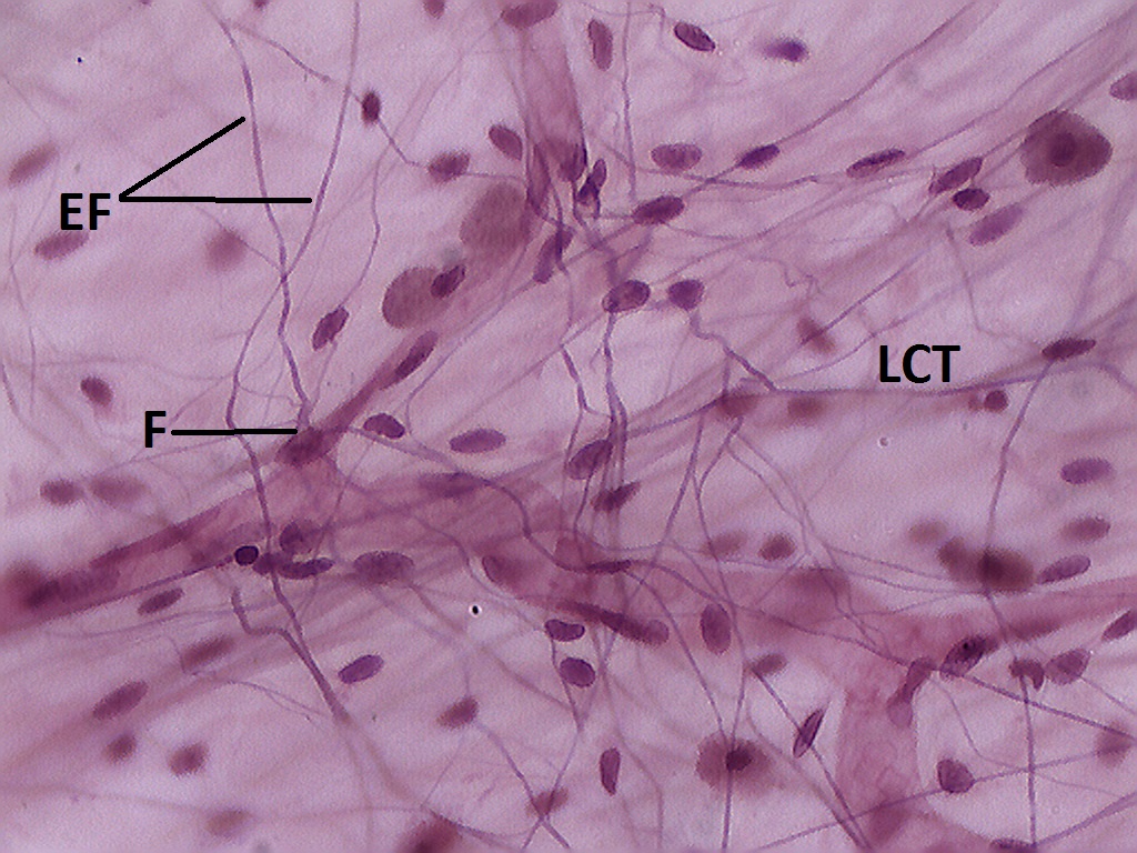
collagen fiber
strong cable like fibers but unbranched
reticular fiber
thinner, used for framing CT
elastic fiber
stretches and recoils easily!! like a sock or a pair of underwear
fibroblast
produces the three fibers and ground substance! very common in CT proper
loose areolar CT
protects tissues and organs, for packing nutrients in the body
-papillary layer of dermis, surrounding organs, muscles, and blood vessels
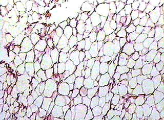
adipose CT
Protects, Insulates, Energy, Storage!!
-subcutaneous layer, surrounds and covers organs
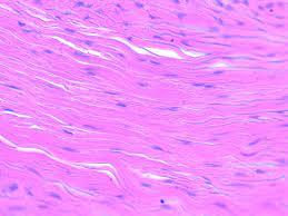
dense regular CT
tightly packed, where stress is applied in a single direction!! only few blood vessels
-tendons (muscle to bone) and ligaments (bone to bone)
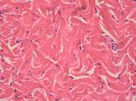
dense irregular CT
resistance to diff directions!
-dermis, periosteum, capsules around internal organs
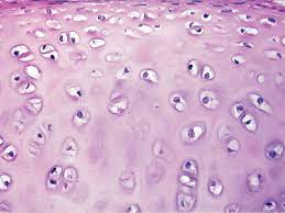
lacunae or lacuna
a cavity in the bone
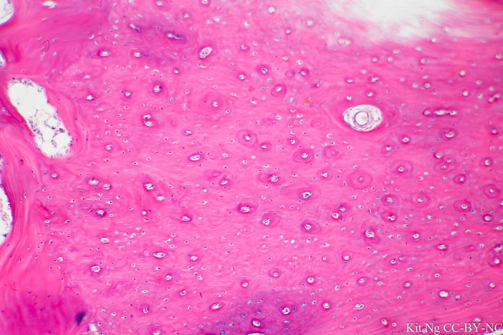
hyaline cartilage
fish eyes!! provides support
-nose, trachea, larynx, ends of long bones
fibrocartilage
weight bearing, resists compression
-in between intervertebral discs, menisci of knee joint
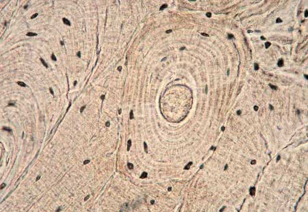
compact bone
cylindrical structures are called osteons, all of the bones in the body!!
COST
compact=osteons
spongy=trabeculae
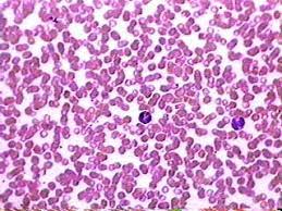
blood
formed elements!!
red (erythrocytes): transport gases
white (leukocytes): infection
has a liquid ground substance
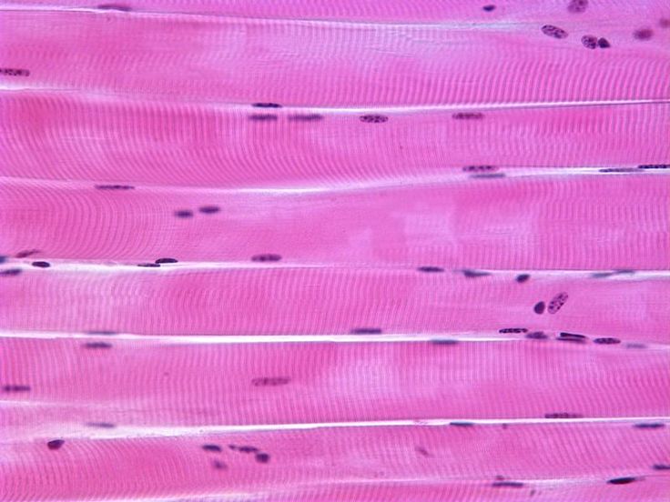
skeletal muscle tissue
voluntary! moves skeletal muscle fibers, very long multiple nuclei
-has striations
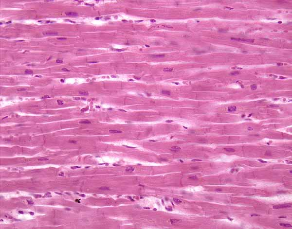
cardiac muscle tissue
involuntary!! cells are connected with intercalated discs, responsible for heart contraction, striated
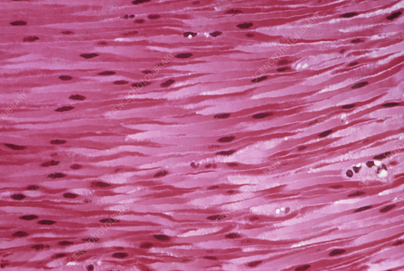
smooth muscle tissue
involuntary!! spindle-shaped, appears smooth
-walls of intestines, stomach, airways, bladder
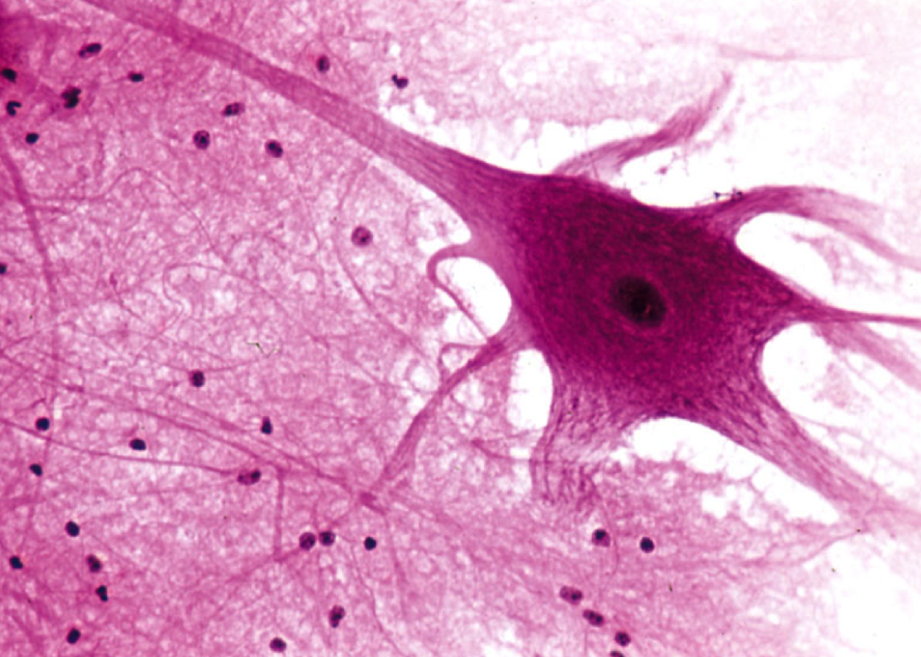
nervous tissue
located in brain, spinal cord, nervous
-neurons: recieve, transmit, processes (dendrite>cell body>axon)
-glial cells: protects neurons awww
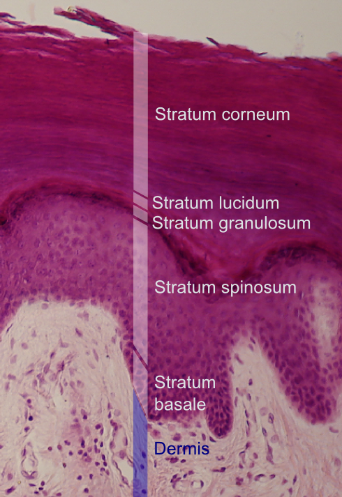
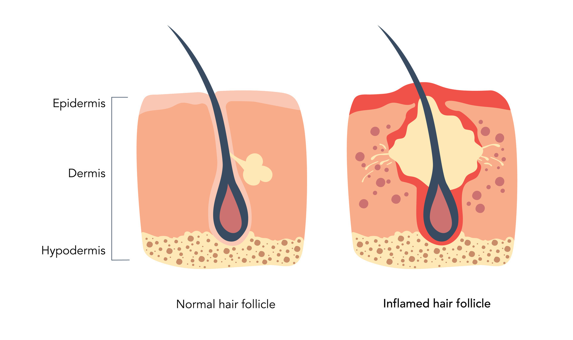
epidermis
epithelium of the integument, is made up of keratinized stratified squamous epithelium!! contains five layers
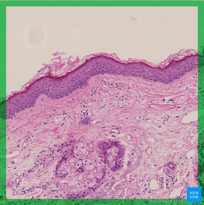
dermis
deep to the epidermis, has a papillary and reticular layer!
papillary layer of dermis: loose areolar CT, dermal papillae (points upwards), and epidermal ridges (points downwards)
reticular layer of dermis: dense irregular CT
hypodermis
aka subcutaneous layer, deep to the dermis not actually part of the integumentary system
-contains adipose CT and adipocytes
stratum basale
the deepest layer of the epidermis, makes up the epidermal ridges!
stratum spinosum
second-most deep layer, right above the epidermal ridges
stratum granulosum
middle layer, typically depicted as a darker line in between the spinosum and lucidum/corneum
stratum lucidum
only present in thick skin! the layer before the cornenum, usually colored lighter or the bottom part of the corneum
stratum corneum
the very top!! has no nucleus, dry to protect abrasion
layers of epidermis
thick skin hint find line between granulosum and cornenum
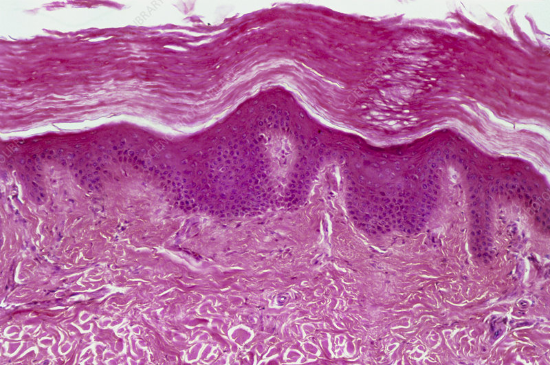
thin skin
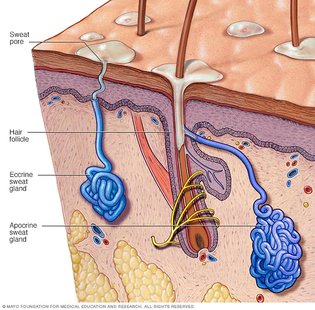
merocrine sweat gland
the leftmost gland aka the most common type of sweat gland not associated with hair
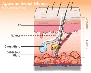
apocrine sweat gland
really big sweat glands attached to hair responsible for making stinky sweat ewww
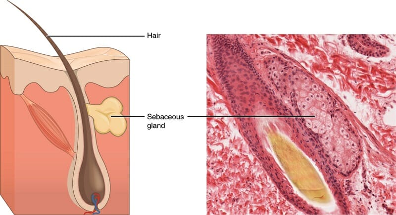
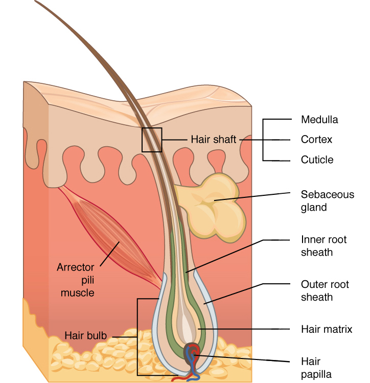
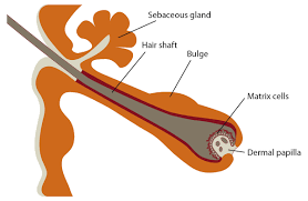
sebaceous gland
attached to the hair follicle causes pimples ewwww
hair shaft
real hair finally
hair root
okay this part is looking a little more like hair now its the beginning!!
hair matrix
a little higher than the hair bulb, before the hair root
hair bulb
the actual ballsack of a hair
hair papilla
aka the dermal papilla, its the area between the ballsack looking part of the hair
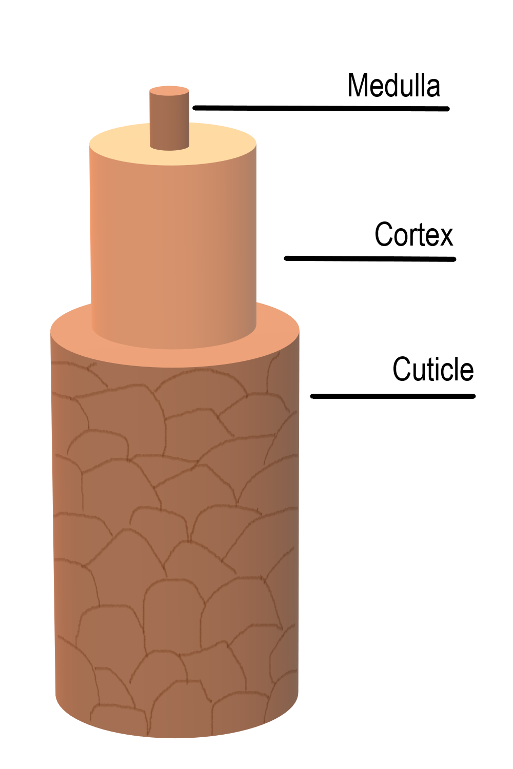
hair medulla
middle most part
hair cortex
the middle layer of hair!
hair cuticle
the outermost layer of the hair
hair follicle
the entirety of a hair, including all structures
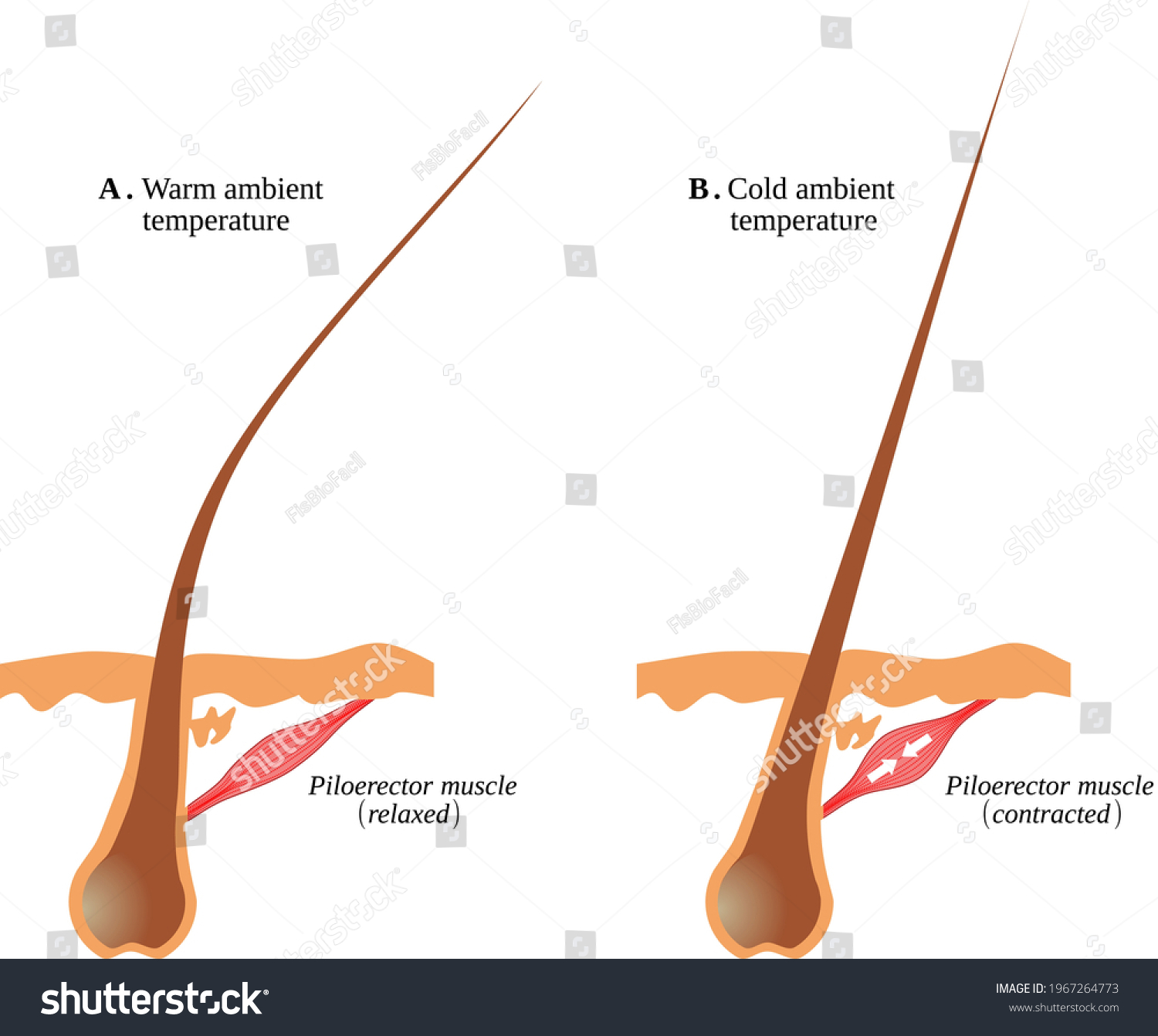
arrector pili muscle
makes goosebumps!! made up of smooth muscle
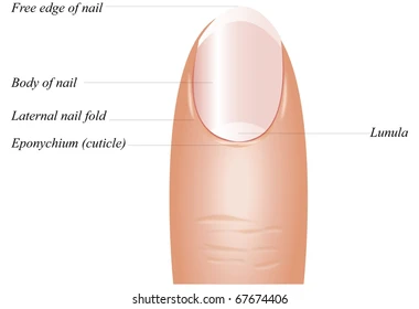
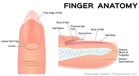
free edge
the part of the nail that hangs out (the thingy you need to cut)
nail body
not including lunula, free edge, just the body itself
nail root