Rabi Ebstein Anomaly and Tricuspid Valve Dysplasia
1/31
There's no tags or description
Looks like no tags are added yet.
Name | Mastery | Learn | Test | Matching | Spaced |
|---|
No study sessions yet.
32 Terms
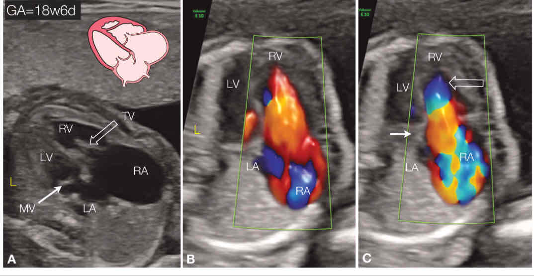
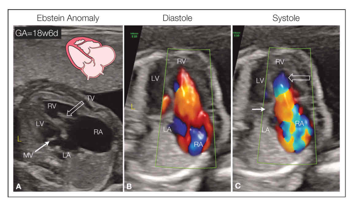
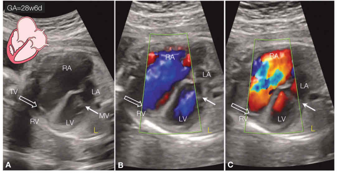
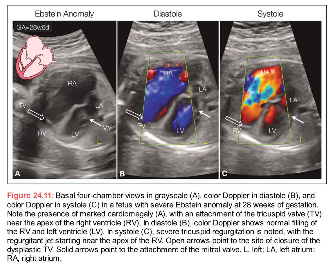
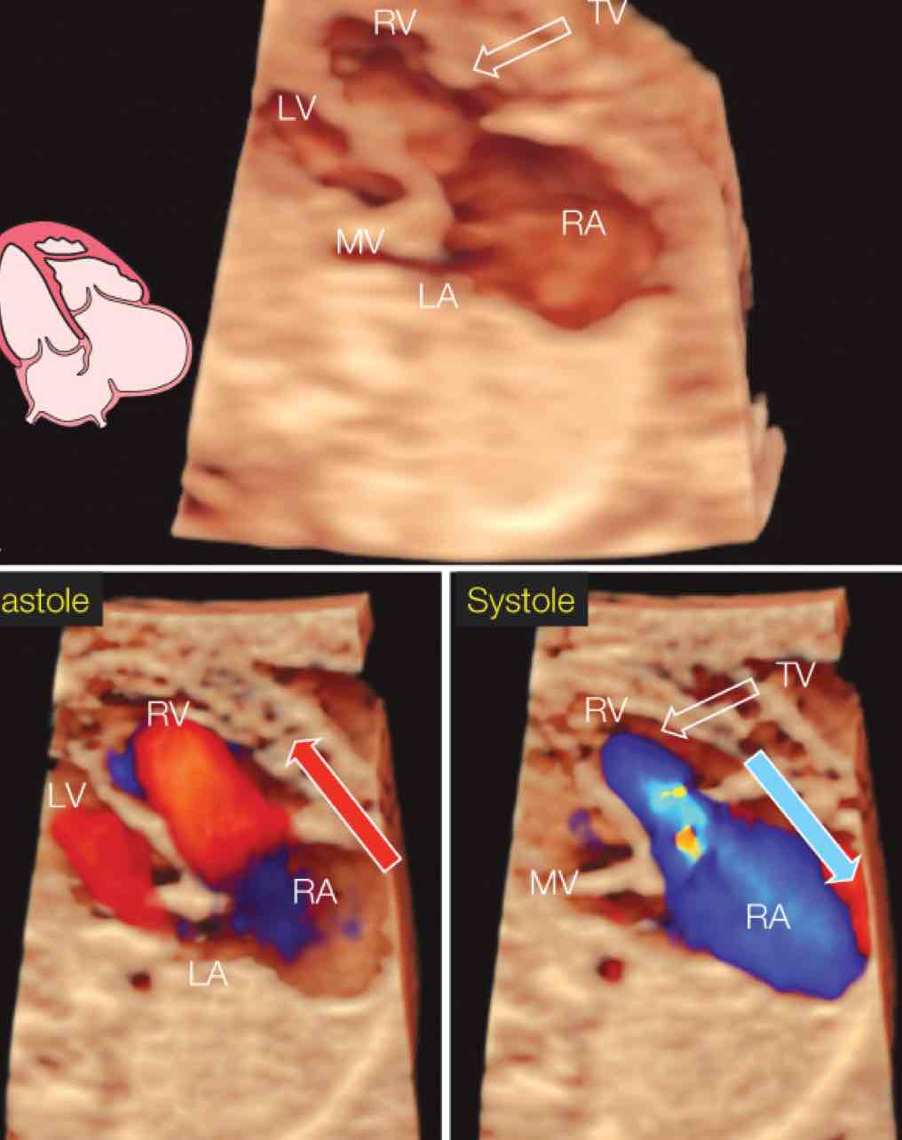
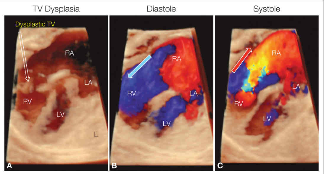
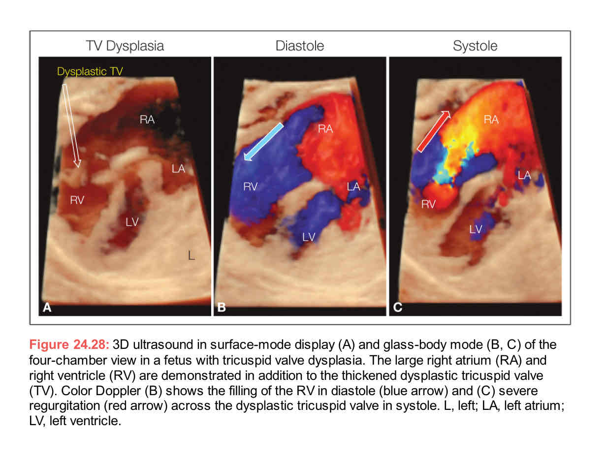
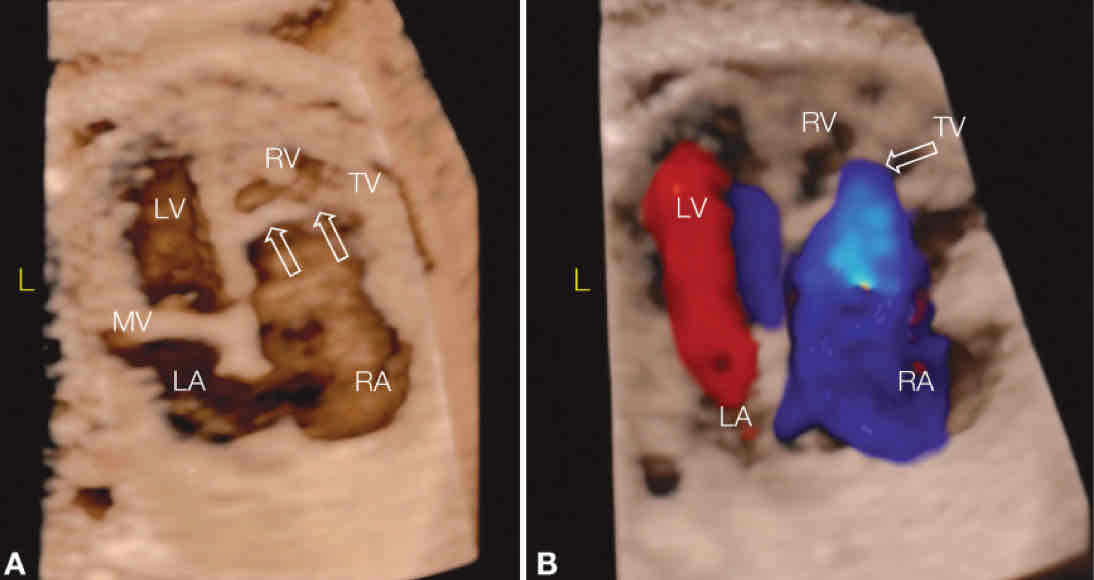
3D ultrasound in surIace-mode disSlay (A) and Jlass-body mode in diastole (B) and systole (C) at the Iour-chamber view in a Ietus with Ebstein anomaly. The large rights atrium (RA) and the diminutive right ventricle (RV) are demonstrated (A), in addition to the attachment oI the tricuspid (TV) (oSen arrow). Also note (A) the normal insertion oI the mitral valve (MV) in comparison to the TV. Color DoSSler (B) shows normal ventricular filling in diastole and severe regurgitation oI the dysplastic tricuspid valve in systole. LA, leIt atrium; LV, leIt ventricle.

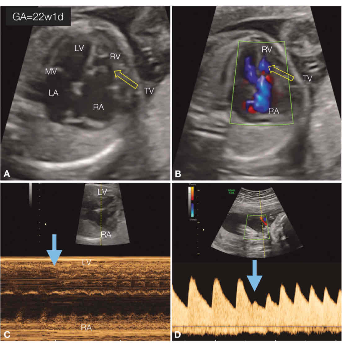
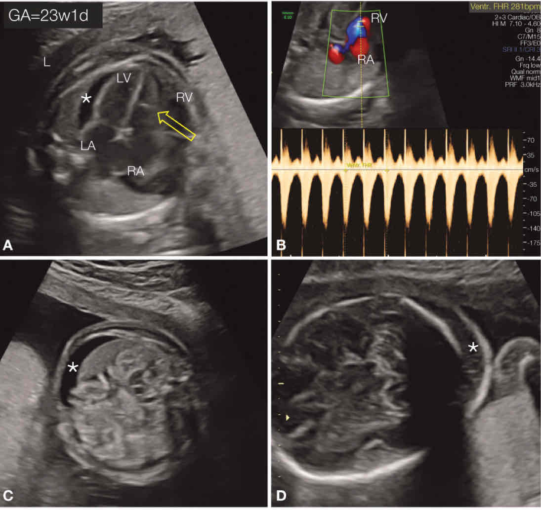
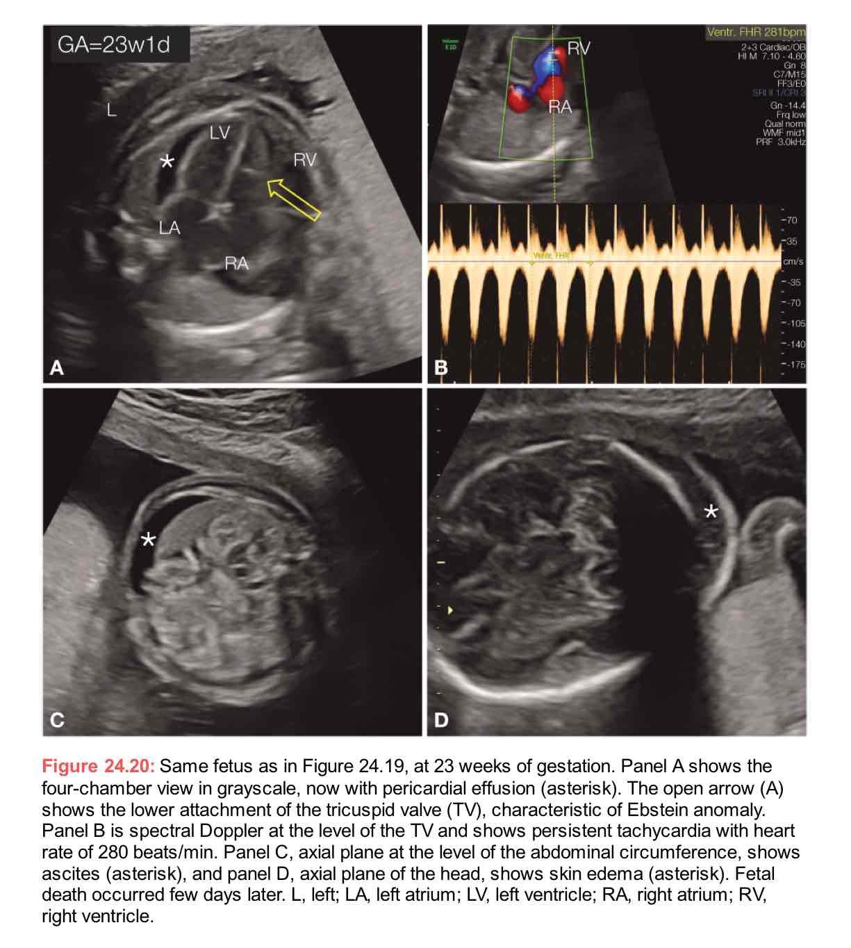
Fetus with Ebstein anomaly at 22 weeks oI Gestation with the rare complication oI tachycardia. Panel A shows the four-chamber view in Grayscale with the lower attachment of the tricuspid valve (TV) (open arrow) in comparison to the mitral valve (MV). Panel B shows TV regurgitation from the right ventricle (RV) into the right atrium (RA). Panel C is an M-mode through the heart, and Sanel D is spectral Doppler across the umbilical artery. Both C and D show the onset of oI re-entry tachycardia (blue solid arrow) after a normal sinus rhythm. See Figure 24.20 Ior Follow-up.LA, left atrium; LV, left ventricle.