Looks like no one added any tags here yet for you.
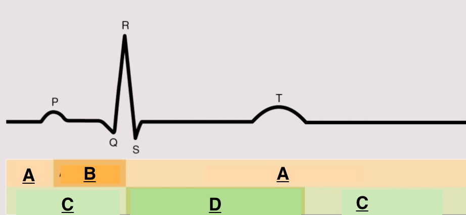
atrial diastole
what is happening in the regions marked A
atrial systole
what is happening in the regions marked B
Ventricular diastole
what is happening in the regions marked C
Ventricular systole
what is happening in the regions marked D
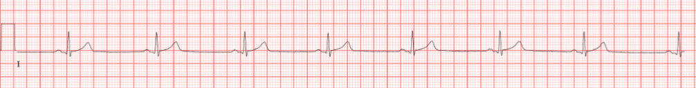
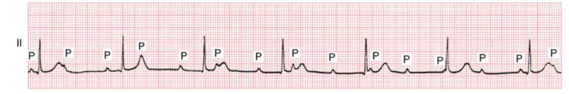
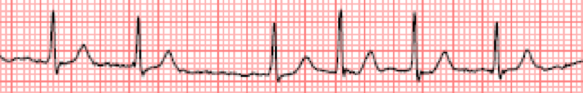
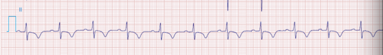
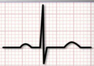
60-100 bpm, positive P wave in lead II, 1 P for every QRS
features of normal sinus rhythm
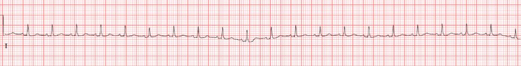
tachycardia
a condition where the heart beats faster than 100 beats per minute
Bradycardia
a condition where the heart beats slower than 60 beats per minute
sinus bradycardia
a type of arrhythmia characterized by a heart rate of less than 60 beats per minute, with a regular rhythm and normal P waves
Sinus tachycardia
a type of arrhythmia characterized by a heart rate of more than 100 beats per minute, with a regular rhythm and normal P waves arrhythmia
3rd degree block/bradycardia
a type of heart block where there is no relationship between the atrial and ventricular rates, leading to a slow heart rate, URGENT CAN BE FATAL
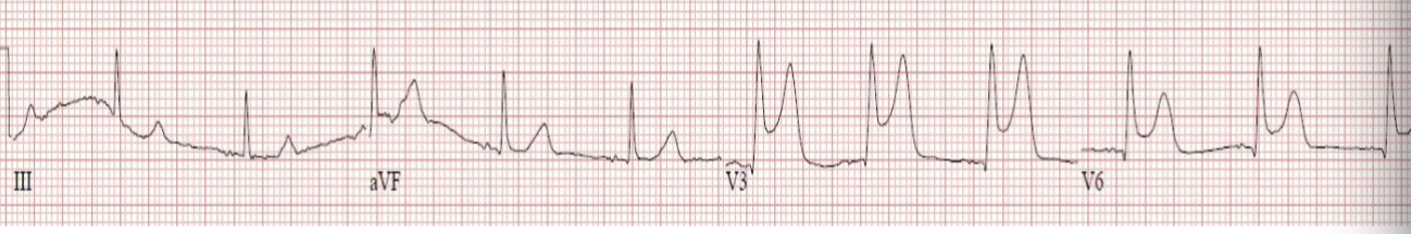
infranodal AV block
a type of heart block that occurs below the AV node, WIDE QRS , potentially LETHAL
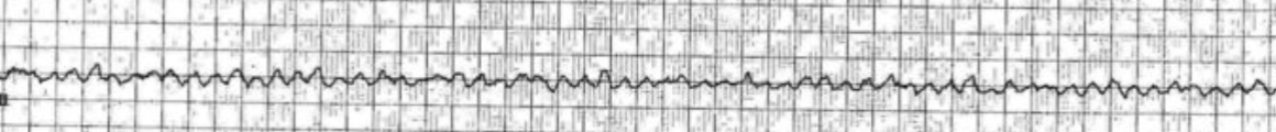
atrial fibrillation
most common pathologic arrhythmia, no p waves
Atrial flutter
saw tooth flutter waves, narrow QRS
ventricular tachycardia
a fast heart rhythm originating from the ventricles, typically characterized by dominant wide QRS complexes and can lead to Vfib or cardiac arrest
ventricular fibrillation
quivering ventricle,no effective contraction, life-threatening without immediate CPR and defibrillation
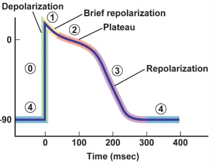
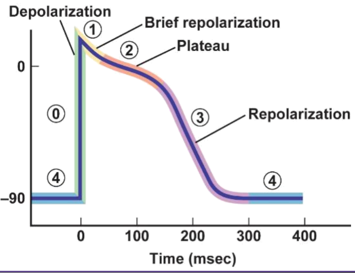
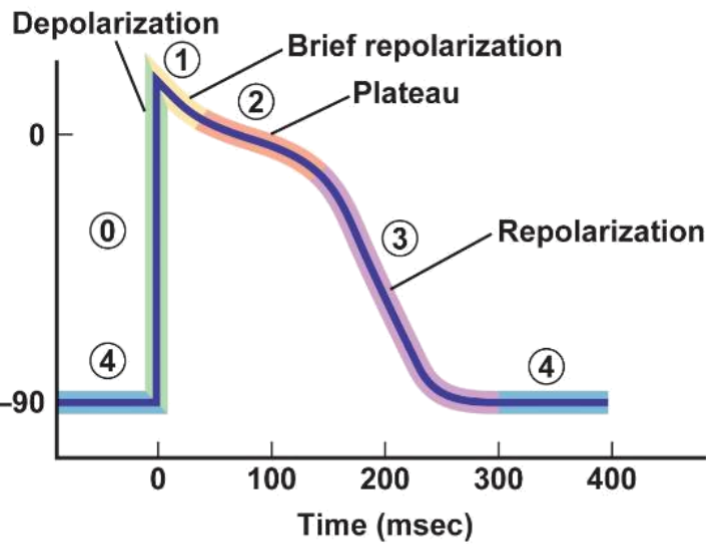
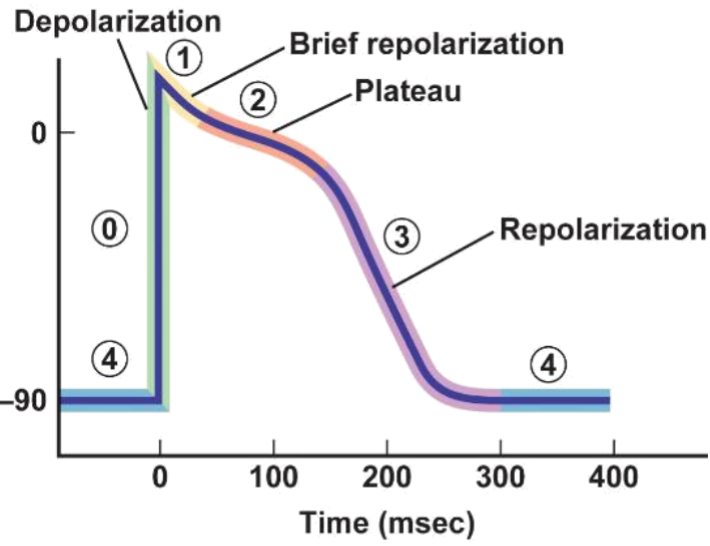
Na in
what is happening in phase 0
K out
what is happening in phase 1
Ca in, K out, myocyte contraction
what is happening in phase 2
K out
what is happening in phase 3
Na/K pumps stabilize resting potential, K in, Na out
what is happening in phase 4
gap junctions
what allows for fast current transduction between myocytes
excitation-contraction coupling
is the physiological process where an electrical stimulus leads to muscle contraction, primarily in cardiac muscle.
phase 2
at what phase of action potential is excitation reaction coupling happening
SA/AV action potential has NO rapid Na influx
difference between action potential of SA/AV nodes and action potential of regular cardiomyocytes
fewer mitochondria, fewer myofibrils, smaller SR
why do SA and AV conduct slower than a regular cardiomyocyte
His bundle, R and L bundle branch, and purkinje fibers
what allows for uniform synchronous contraction of the heart
cell diameter, gap junctions, magnitude and kinetics of depolarizing current
factors that determine cardiac conduction velocity
no summation or tetany
importance of refractory period
norepinephrine
neurotransmitter affecting SA node and AV node, increasing heart rate
acetylcholine
neurotransmitter that decreases heart rate by acting on the SA node and AV node.
epinephrine
neurotransmitter that affects AV node, increasing AV node conduction
high vagal/PSNS tone, endurance athlete, SA node dysfunction
possible causes of sinus bradycardia
patients may be on blood thinners, could increase bleeding risk
why is atrial fibrillation important for dentist to know
coumadin/warfarin, prevents clots formation in left atrium
What particular medication are most people with atrial fibrillation on and why?
syncope
is a temporary loss of consciousness caused by a decrease in blood flow to the brain, often resulting from a drop in blood pressure.
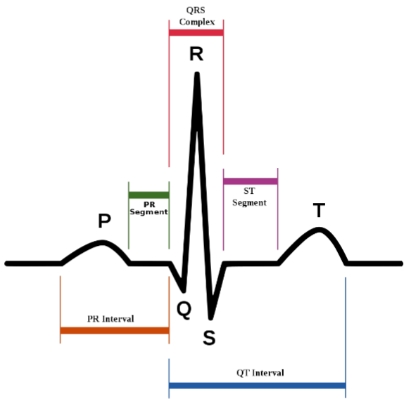
atrial depolarization/ atrial contraction
what is represented by the P wave
time of conduction through AV node
what is represented by the PR interval
ventricular depolarization/contraction
what is represented by the QRS wave complex
time between end of ventricle firing and repolarization
what is represented by the ST interval
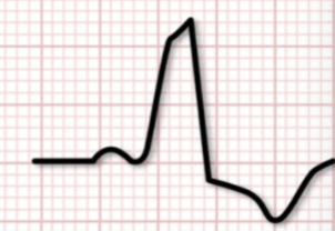
ST
during which interval is ischemia and /or infarction most clearly seen
ventricular repolarization
what is represented by the T wave
purkinje fibers
fastest conductors of signal transduction in thee heart
AV node
slowest conductors of signal transduction in the heart
coordinated contraction
why must the purkinje fibers conduct signals quickly
SA node, AV node, bundle of His, R/L bundle branches, purkinje fibers
route of signal transduction in the heart
filling of ventricles
what does depolarization at the AV node allow for
decreased filling of ventricles
what is the result of an elevated heart rate of 200 bpm
prolonged PR interval
abnormal slowing of conduction in the AV node will result in what ECG abnormality?
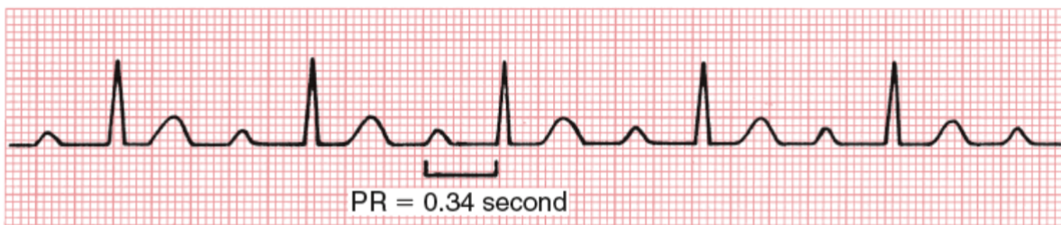
1st degree heart block
prolongation at AV node, prolonged PR, Asymptomatic
angina
a type of chest pain caused by reduced blood flow to the heart muscle (ischemia), often triggered by physical exertion or stress
narrow QRS complex
a finding on an ECG that means electrical signal originated above ventricles
wide QRS complex
a finding on an ECG indicating the electrical signal originate somewhere in ventricle, BAD
assess stroke risk to determine degree of anticoagulation necessary
what is the purposee of CHA2DSVASC scoring system
CHF (congeestive heeart failure), Hypertension, Age (≥75), diabetes, stroke, vascular disease, age (65-74), and sex (female)
what does CHA2DS2-VASc stand for/look at
2
what CHA2DS2-VASc/ CHADS2 score indicated high risk of embolic stroke
warfarin, DOACs (dabigatran, rivaroxaban, apixaban)
common anticoagulants used for patients at risk of stroke
R atrium, R ventricle, SA and AV nodes
heart regions supplied by right coronary artery
L atrium, L ventricle, AV node (10% of pop)
heart regions supplied by left coronary artery
ischemia
a condition characterized by INSUFFICIENT OXYGEN AND NUTRIENTS to tissues, often leading to tissue damage or necrosis, REVERSIBLE
infarction
cell death as a result of prolonged ischemia, IRREVERSIBLE, results in scar
diaphoresis (cold sweat), fatigue, chest pain
symptoms of ischemia
stable angina
angina that occurs predictably with exertion or stress and is relieved by rest or nitroglycerin
unstable angina
chest pain that occurs at rest, medical emergency, IMPENDING INFARCTION
STEMI
complete coronary artery blockagee/occlusion, must be treated immediately
catheterization/stent placement or by-pass
treatments for STEMI
NSTEMI
incomplete coronary artery blockage, positive myocardial markers
unstable angina is negative for myocardial markers
difference between unstable angina and NSTEMI
troponin
best lab marker for diagnosis of myocardial infarction
NSTEMI
ECG with no ST elevation, ST depression sometimes
STEMI
ECG shows ST elevation
Tissue plasminogen activator
A thrombolytic agent used to dissolve blood clots in conditions like myocardial infarction.
shared pathways with branches of vagus and thoracic nerves
why might cardiac pain be felt in the mandible
atherosclerosis
A condition characterized by the buildup of plaque in the arterial walls, leading to narrowed arteries and reduced blood flow, often contributing to cardiovascular diseases.
coronary arteries
where is atherosclerosis located that leads to myocardial infarction
damage to endothelium, cholesterol buildup, plaque hardens, plaque rupture/blood clot formation
process of atherosclerosis plaque formation
blood clot formation
result of atherosclerotic plaque rupture
cell death and scar formation
what reaction happens in the heart after an area has been infarcted
cell death, neutrophil invasion, macrophage cleanup, and scar formation
how does body respond to infarcted tissue
4-10 days (7 days)
time of maximal weakness of heart tissue after an MI
mural thrombus
blood clot formation in wall of artery or heart chamber that can lead to obstruction of blood flow or embolism
atherosclerosis
number 1 cause of coranary artery disease
unstable atherosclerotic plaque
a type of plaque that can rupture, leading to thrombosis and acute coronary syndromes.
stable atherosclerotic plaque
a type of plaque that is LESS likely to rupture, gradual growth and vessel narrowing
thrombus formation and potential heart attack
what happens when lipid pool of thin-capped atherosclerotic plaque ruptures into the lumen of the vessel
surface of plaque erodes
how does stable plaque result in thrombus development and lead to myocardial infarction
stroke or bleeding
major risks for fibrinolytic agents (TPA)
beta-blockers and nitroglycerin
common anti-anginal medications used in ACS (acute coronary syndromes)
aspirin and P2y12 inhibitor
common antiplatelet medications used in ACS (acute coronary syndromes)
heparin
common anticoagulation medication used in ACS (acute coronary syndromes)
unstable angina, NSTEMI, and STEMI
examples of acute coronary syndromes (ACS)
oxygen demand/supply, coronary blood flow, myocardial oxygen consumption
What are the determinants of myocardial ischemia?
arrhythmias, cardiogenic shock, stroke
complications of MI that can happen within FIRST FEW DAYS
ventricular septal rupture, LV wall rupture, ischemic MR, papillary muscle rupture, pericarditis
what are complications after MI that can happen with first 2 WEEKS
SCAD (spontaneous coronary dissection)
a condition where a tear forms in the coronary artery wall, leading to reduced blood flow and potential heart attack.
first degree block
degree of AV block with prolonged PR interval, not complete block
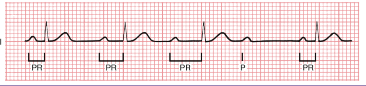
2nd degree type 1 (nodal)
AV block in which signal has PROGRESSIVE difficulty traversing the AV node, dropped QRS
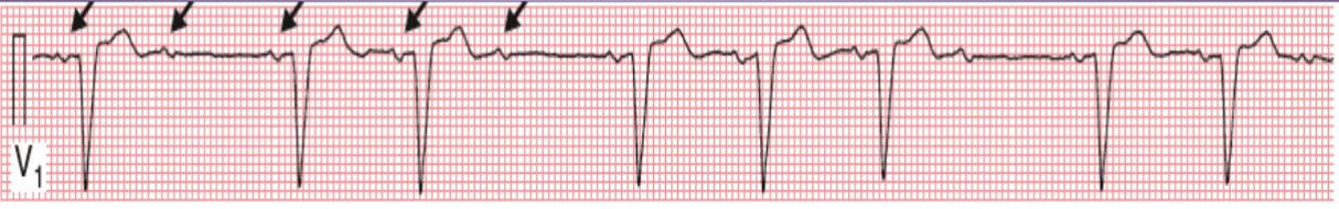
2nd degree type 2 (infranodal)
AV block with WIDE QRS, stable PR, abruptly dropped QRS
sinus tach, Afib, and Aflutter
examples of narrow QRS complex tachycardias
ventricular tachycardia and ventricular fibrillation
examples of wide QRS complex tachycardias