WSU Bio 315 Digestive System
1/102
There's no tags or description
Looks like no tags are added yet.
Name | Mastery | Learn | Test | Matching | Spaced | Call with Kai |
|---|
No analytics yet
Send a link to your students to track their progress
103 Terms
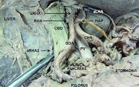
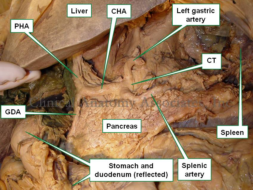
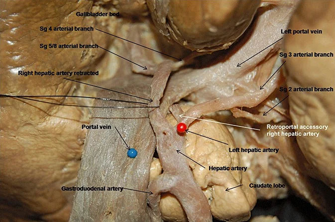
liver
large cut up part
oblong horseshoe crab shaped
structure
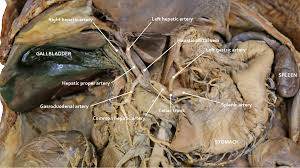
gallbladder
blueish green sack below liver
structure
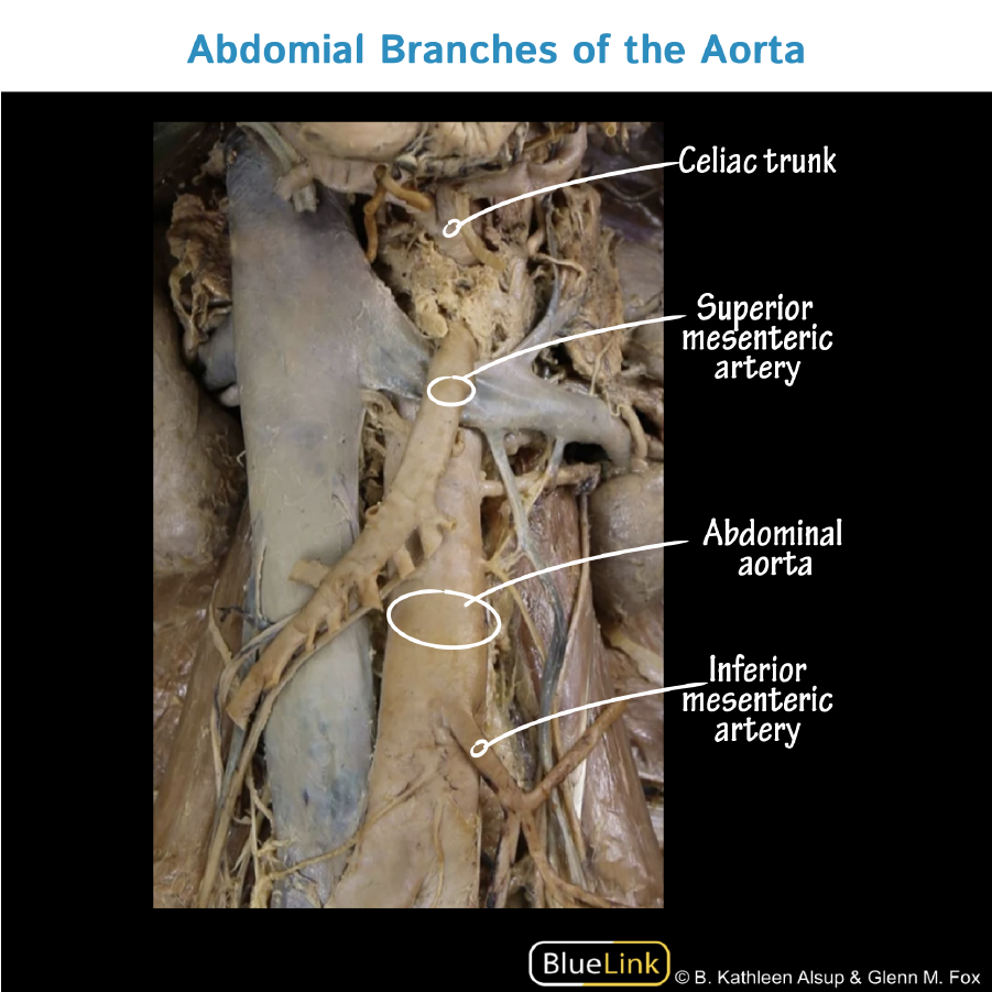
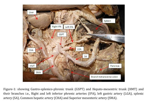
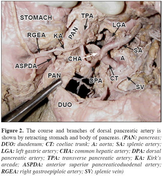
celiac trunk
1st branch of the abdominal aorta
leaves abdominal aorta just below T-12 vertebrae
structure
left gastric artery
loops to the left up towards the esophagus then passes L to R along the lesser curvature of the stomach
first branch of the celiac trunk
structure
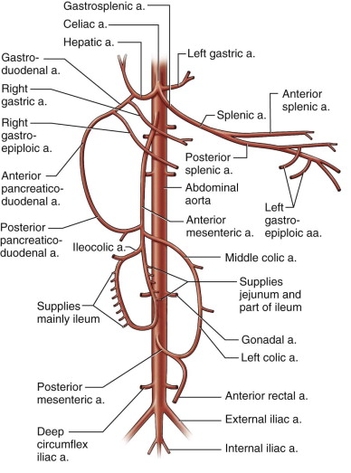
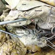
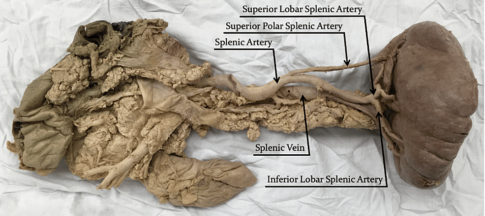
splenic artery
behind stomach along upper boarder of pancreas
TA will flip stomach up and it is the second branch off of the celiac trunk, she is thick wide and flat for an artery
structure
pancreatic branches (of the splenic artery)
major blood supply to pancreas
small branches that come off of the splenic artery (there are many of them)
structure
L gastro-omental artery
the more inferior branch of the Y
supplies the greater curvature of the stomach
structure
common hepatic artery
branches to the right
3rd branch of the celiac trunk
structure
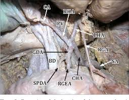
gastroduodenal artery
supplies part of the stomach and duodenum
comes down and splits, goes downward off of the common hepatic artery
right gastro-omental artery
runs from right to left along greater curvature of the stomach
horizontal looong, almost webbed in a way
structure
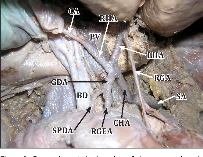
superior pancreaticoduodenal artery
supplies the head of the pancreas
branch off to the bodies R side
structure
R gastric artery
courses from right to left in the lesser curvature of the stomach
smallest vessel , upper portion of the hepatic artery proper
structure
hepatic artery proper
branch (inferior) of the hepatic artery that goes to the right
structure
R hepatic artery
branch of the hepatic artery proper that supplies the right lobe of the liver
split of the Y that goes to pt R
structure
cystic artery
the lower inferior branch coming of the R hepatic artery
structure
L hepatic artery
branch that goes from hepatic artery proper to the left
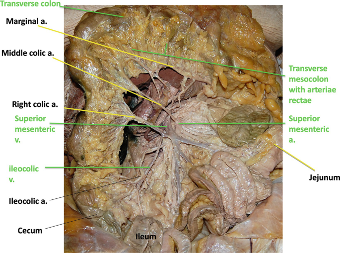
structure
ileocolic artery
looks like a part of a X
passes from down and to the right
goes across belly and down
structure

R colic artery
extends to R colic flexure
looks like a big Y
structure

middle colic artery
passes upward to the transverse colon
structure

inferior mesenteric artery
third branch off of the abdominal aorta
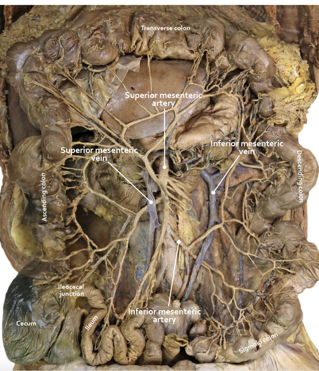
marginal artery of the colon
along the inner margin of the colon
ta will flip descending colon
dark line on colon inner boarder
structure
splenic vein
right below the splenic artery
very thin walled
superior mesenteric vein
thin little flap above/next to partial flap
hepatic portal vein
right below the common hepatic artery, but split in half kina
diaphragmatic surface of the spleen
superior surface that touches the diaphragm
visceral surface of the spleen
lower inferior surface
superior border of the spleen
top bump of spleen kinda shiny
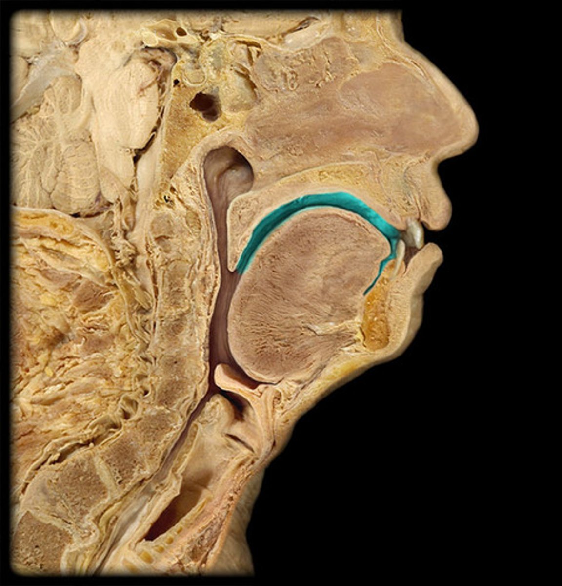
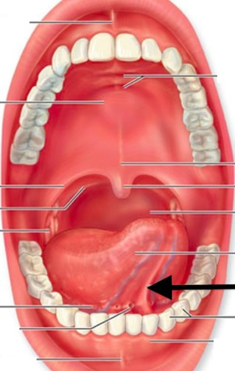
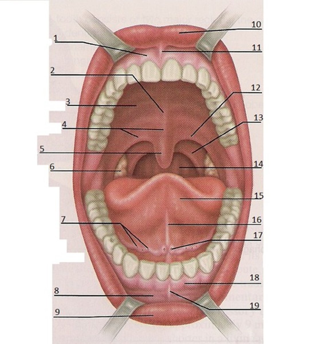
Upper lip
Structure

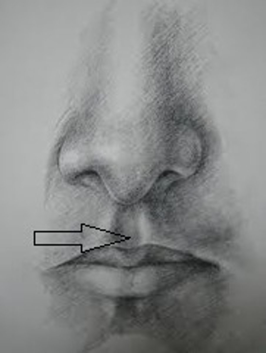
Philtrum
Depression
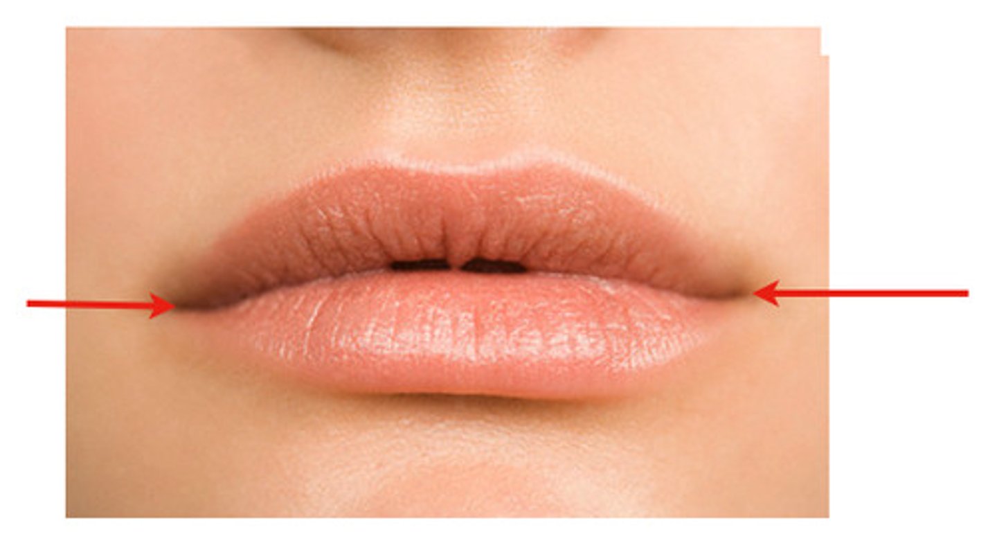
Angles of the mouth
Feature
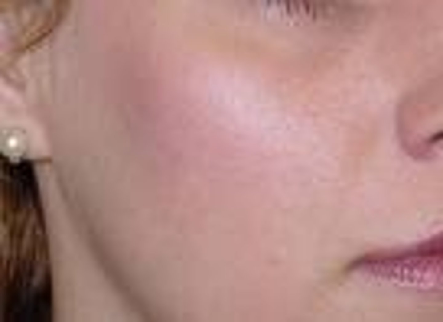
Cheeks
feature
Oral vestibule
Space
between teeth and lips
Oral cavity proper
Space
behind teeth
Frenulum of the tongue

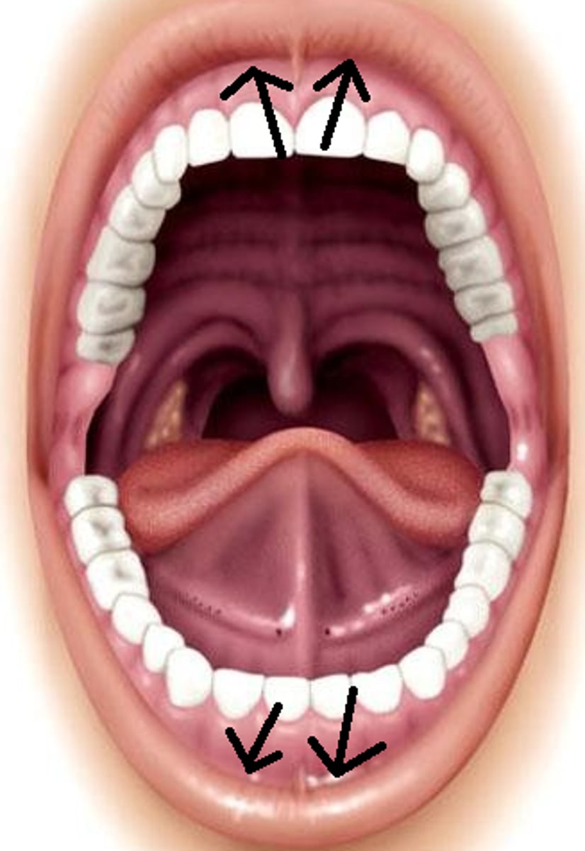
Hard Palate
Purple part
Soft palate
Red part

Gingivae
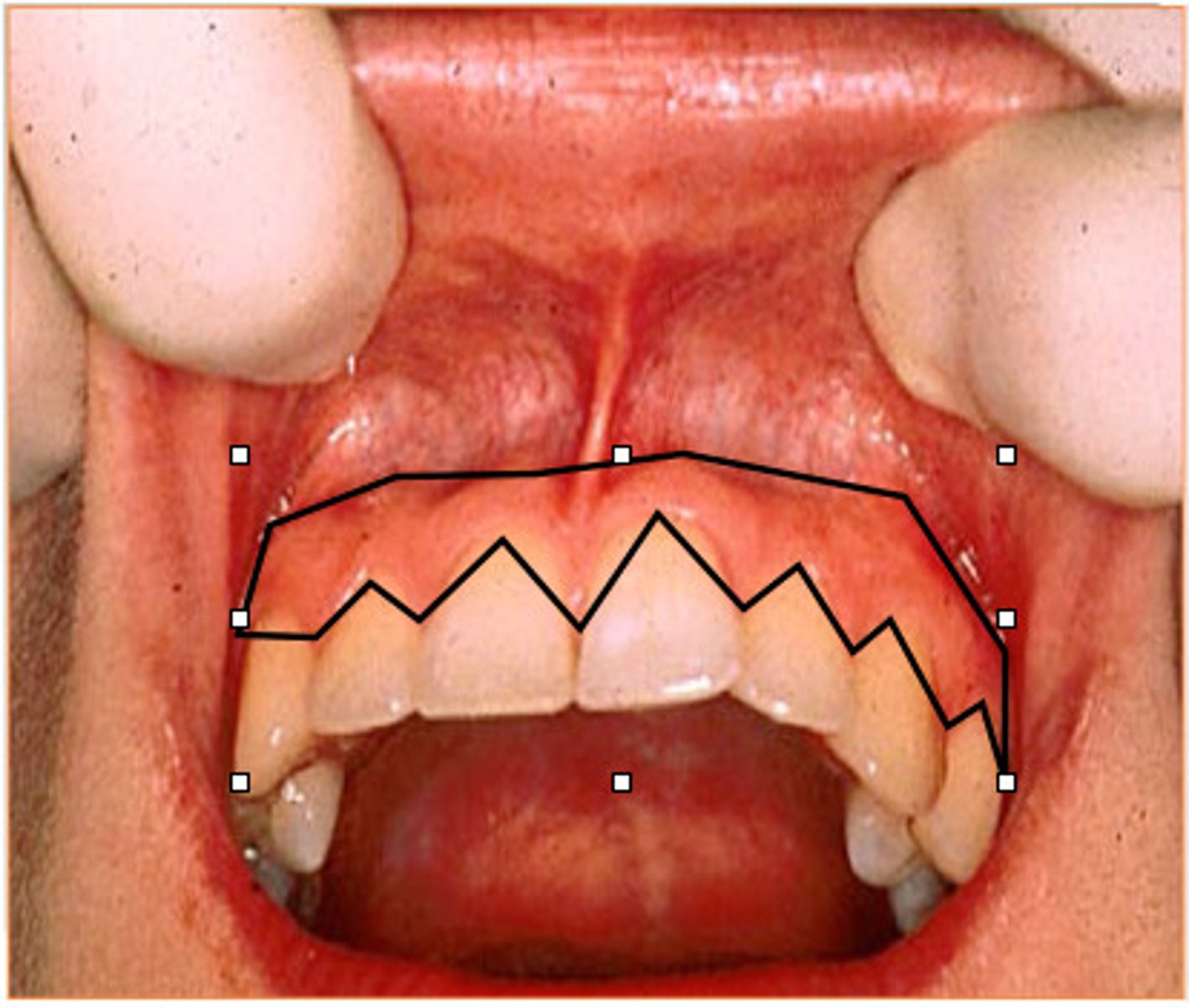
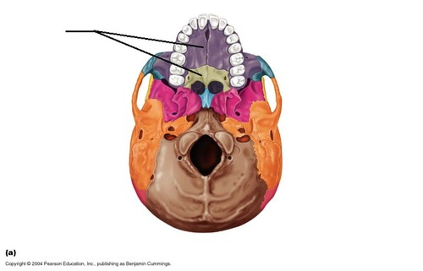
Maxillary dental arch
Feature
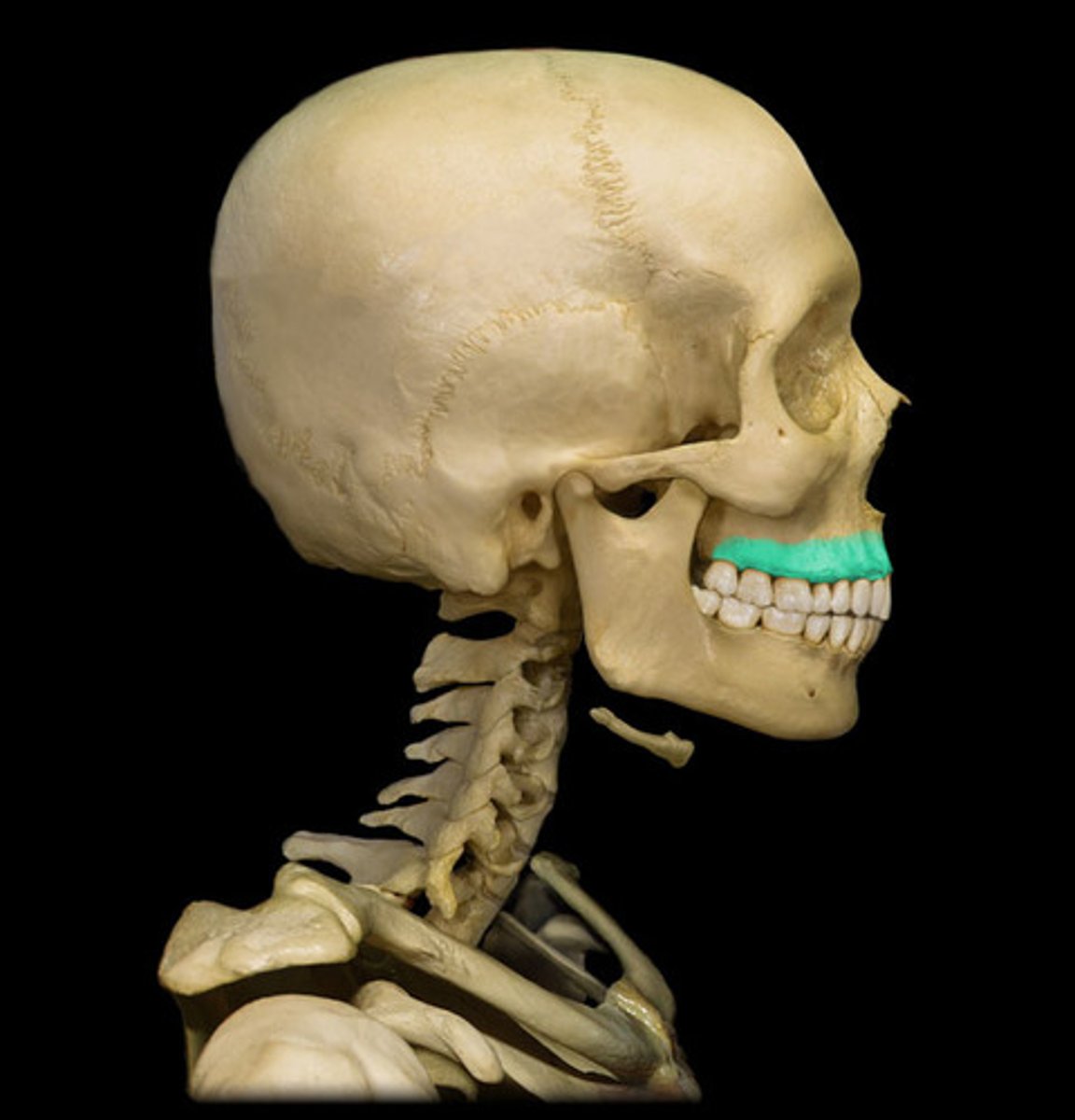
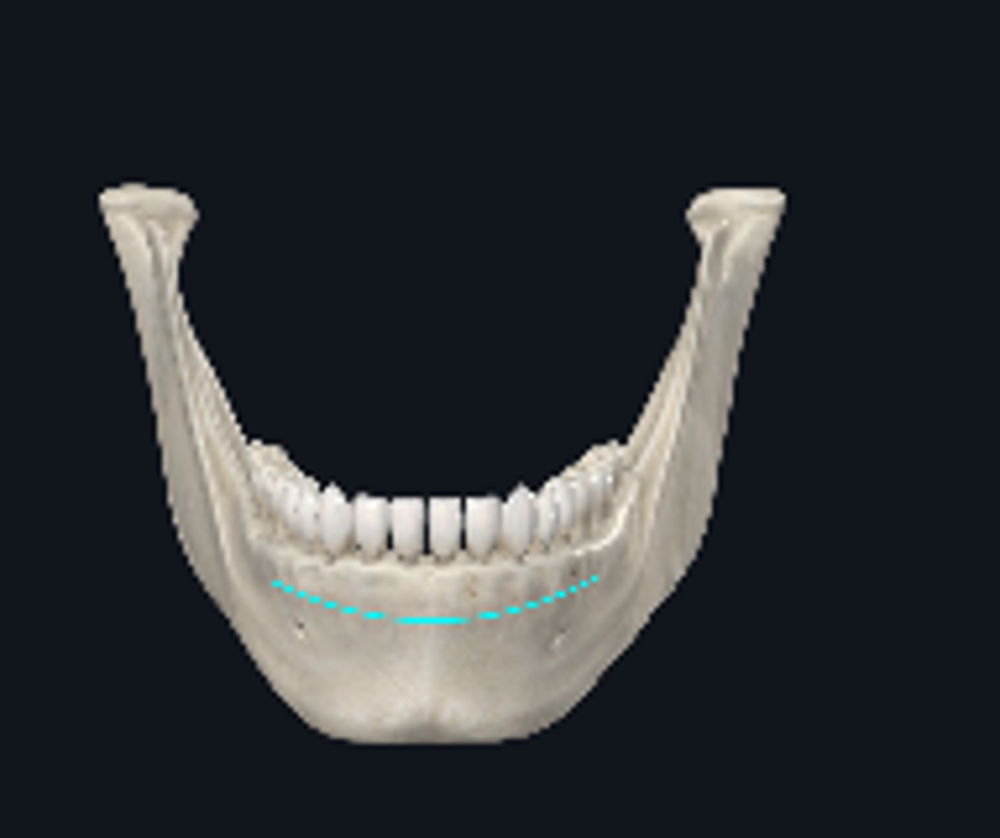
Mandibular dental arch
freature
Incisor teeth
Canine teeth
Premolar teeth
Molar teeth
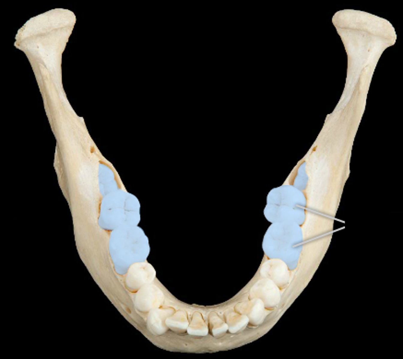
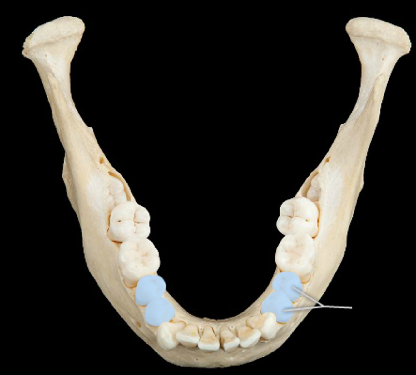
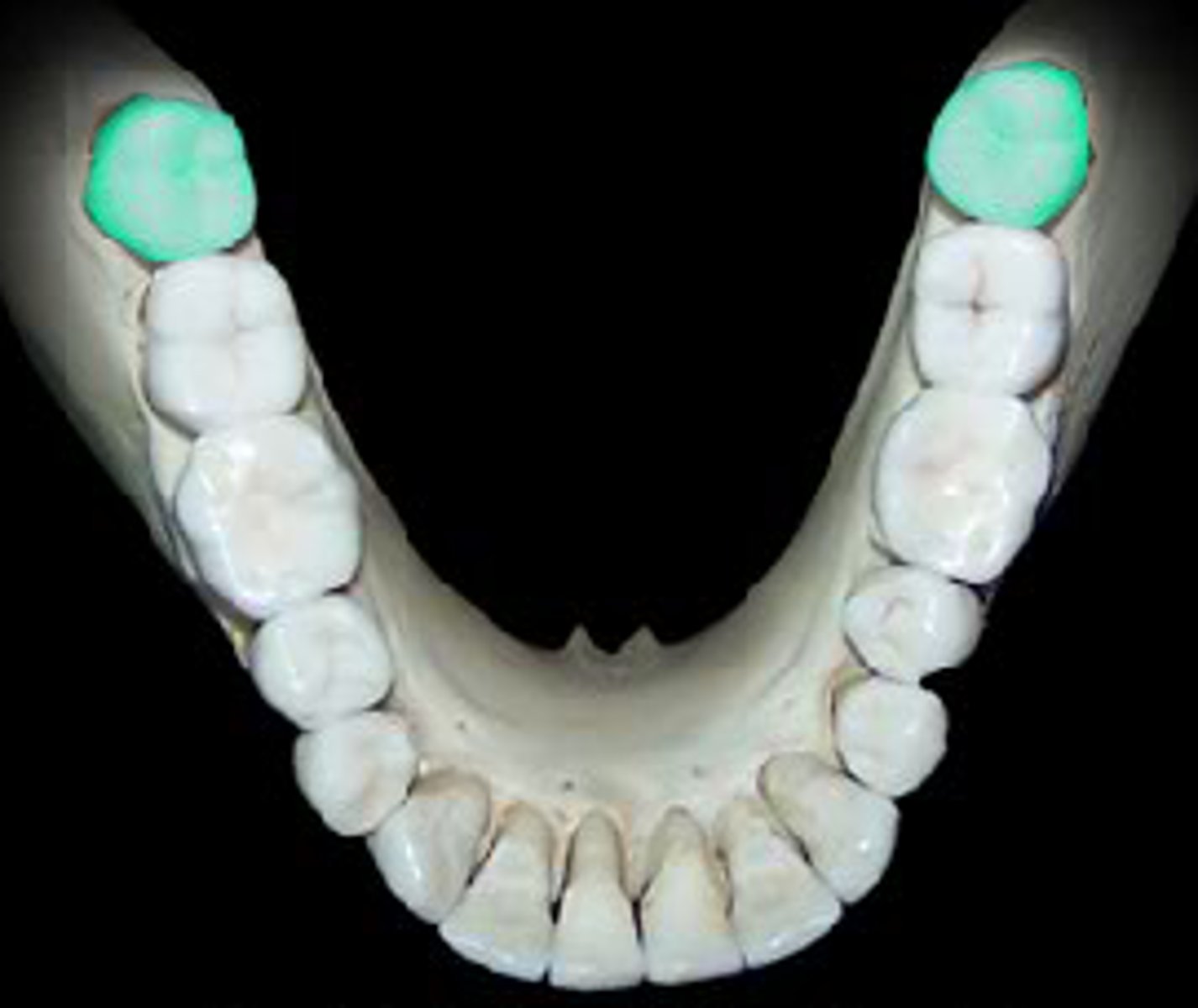
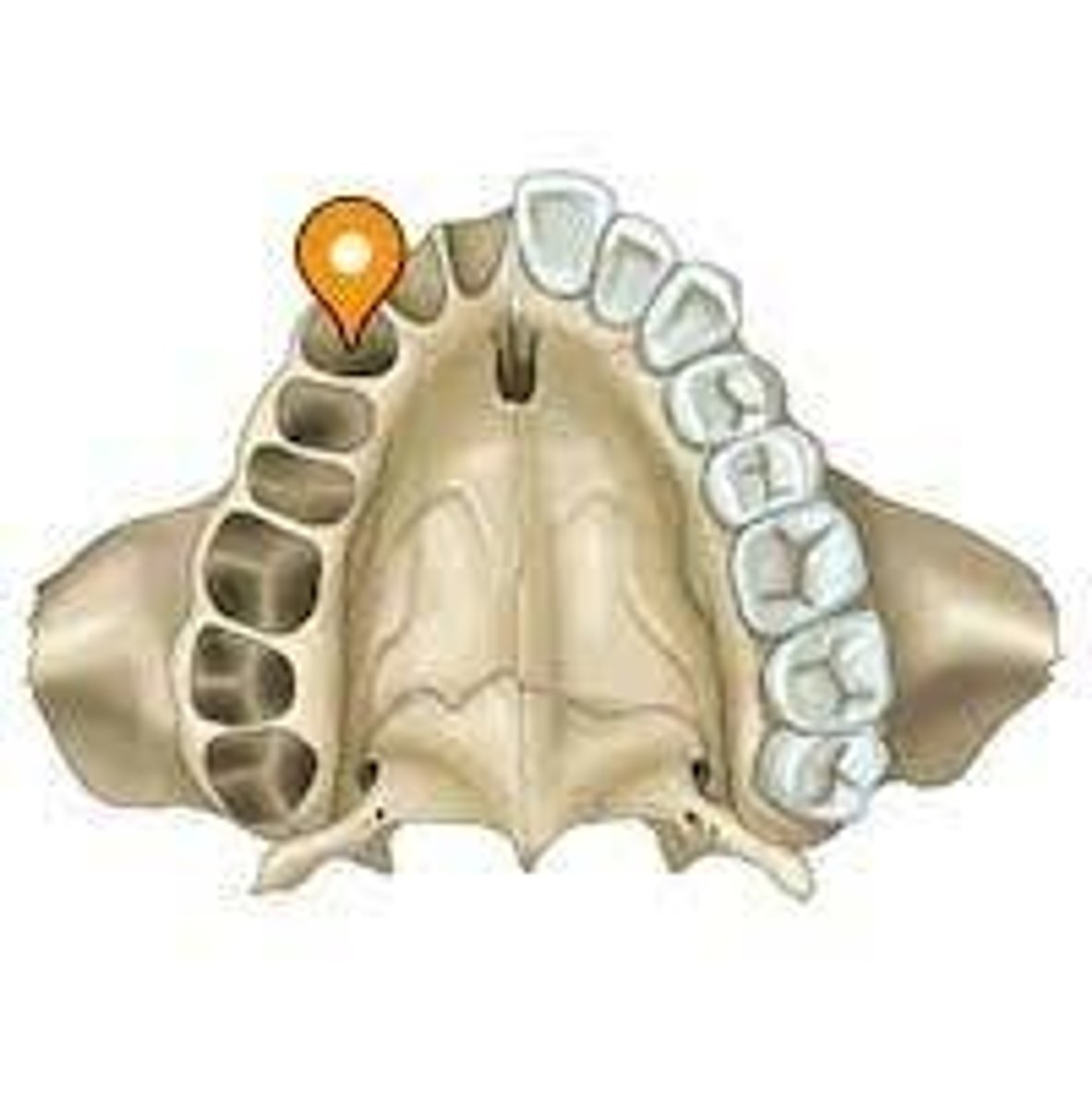
Wisdom teeth
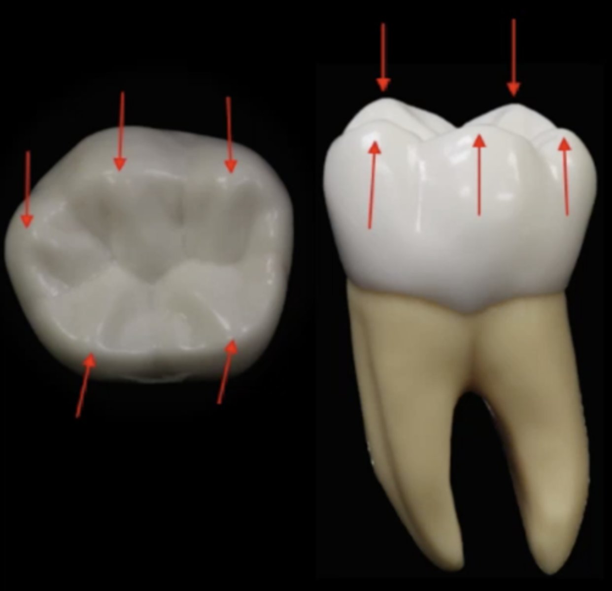
Crown of the tooth
Feature
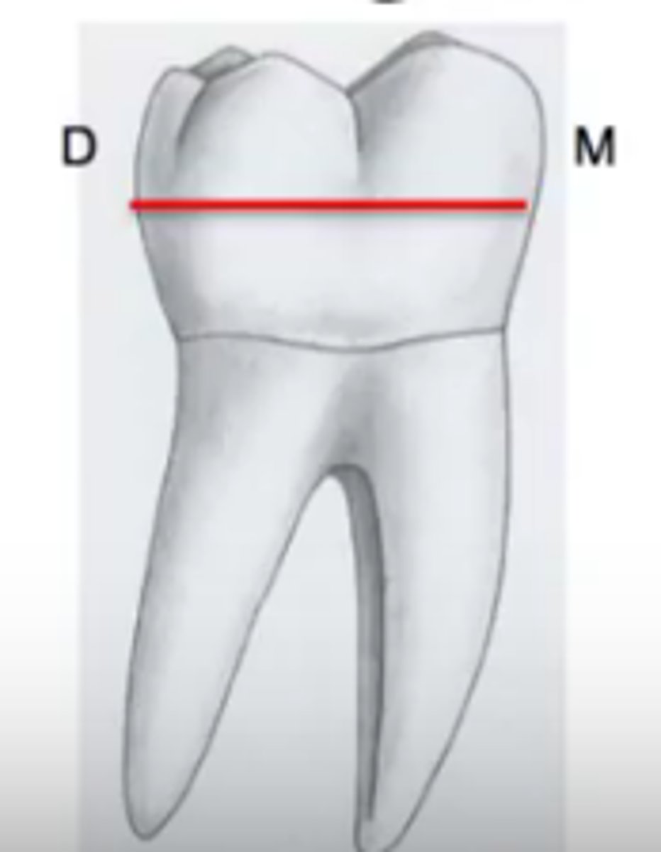
Cusps of the tooth
Feature
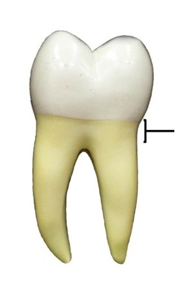
Neck of the tooth
Boundary
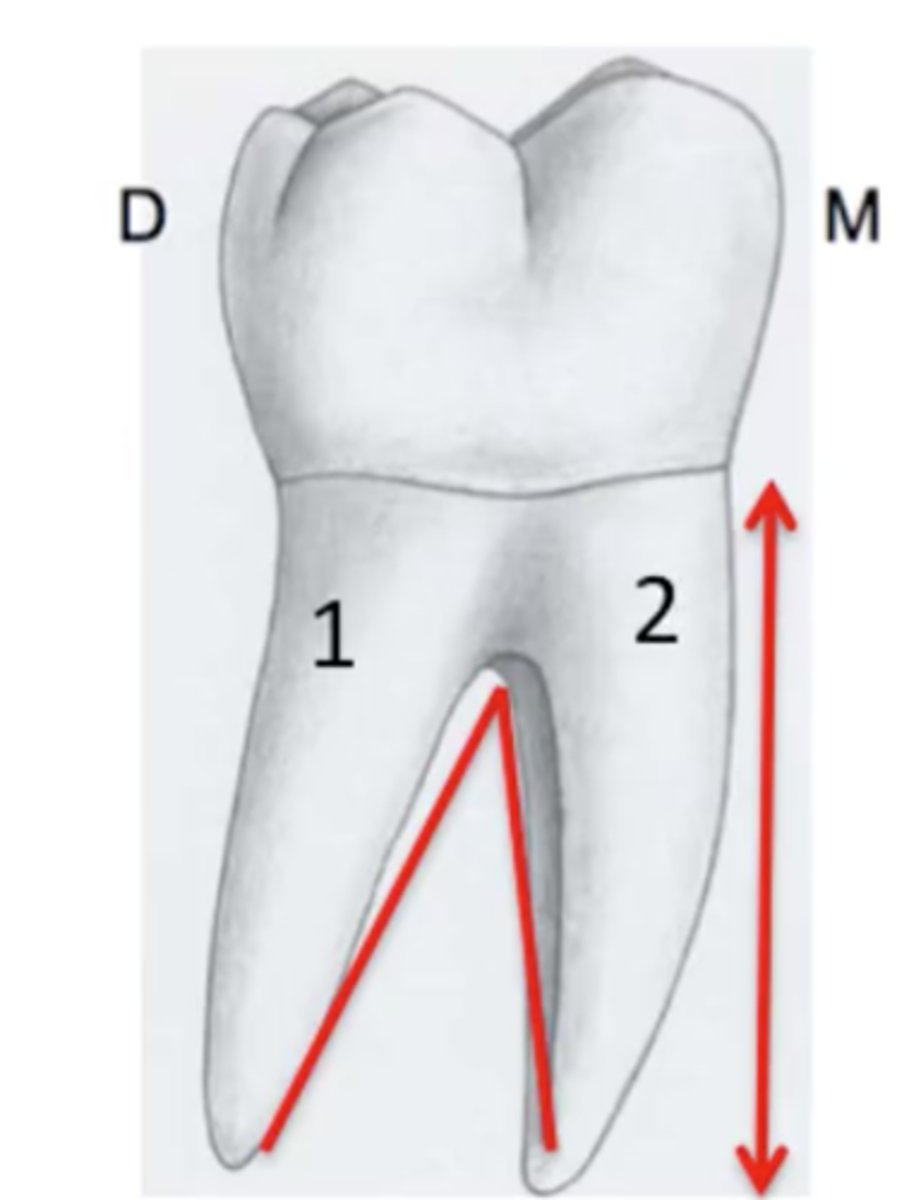
Root of the tooth
Feature
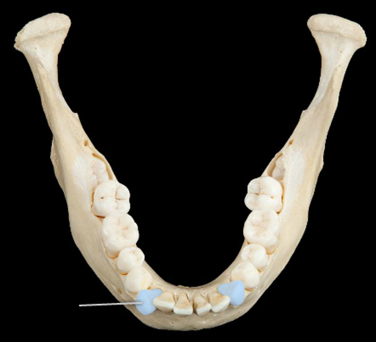
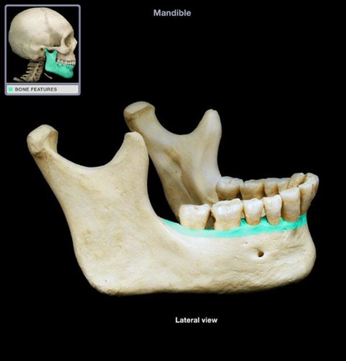
Dental Alveoli of the mandible
Sockets
Dental Alveoli of the maxillae
Sockets
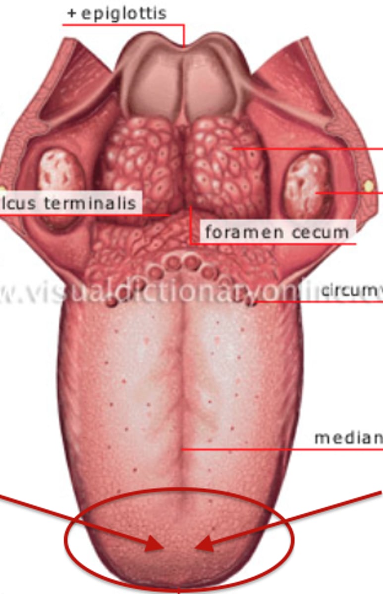
Root of the tongue
Black line under tongue
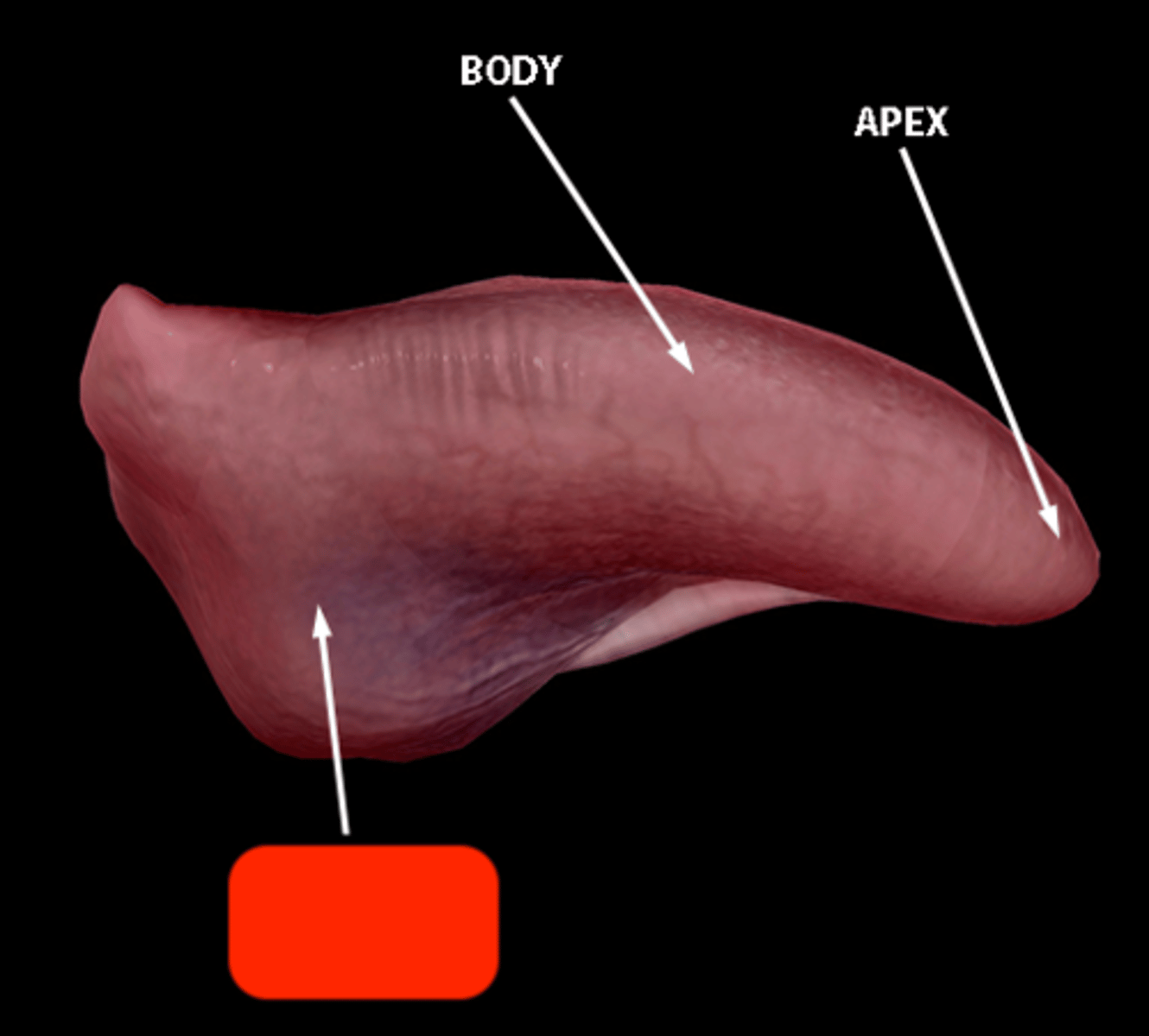
Tip of the tongue
Structure
Body of the tongue
Whole pink underside
Dorsum of the tongue
Surface
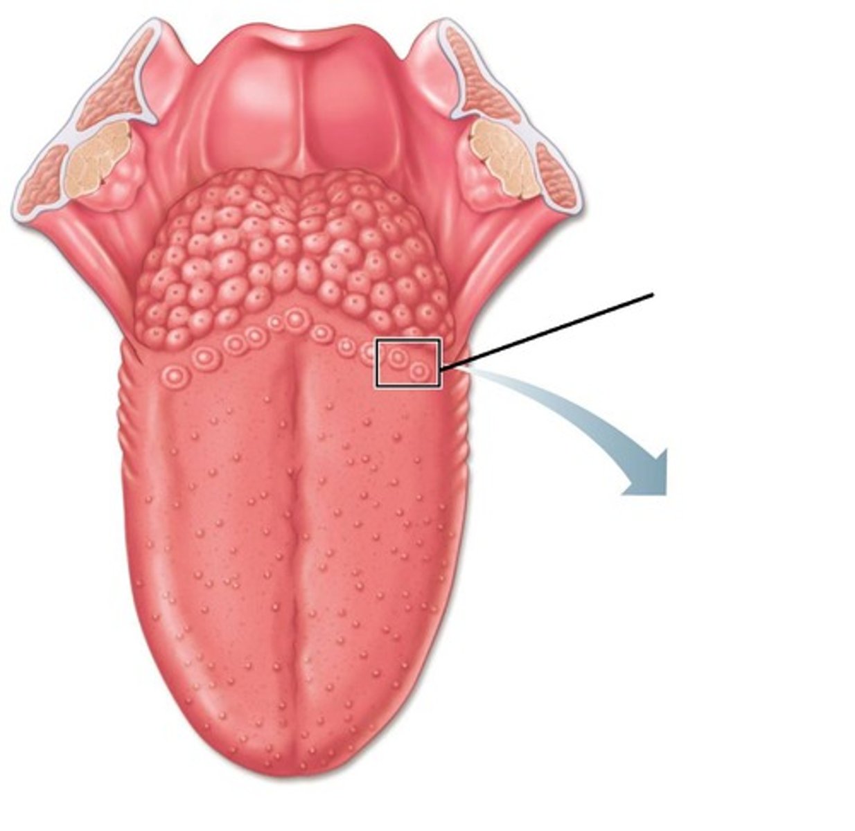
Terminal sulcus
Depression
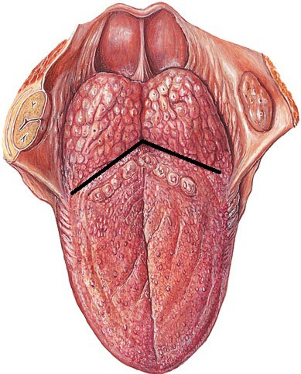
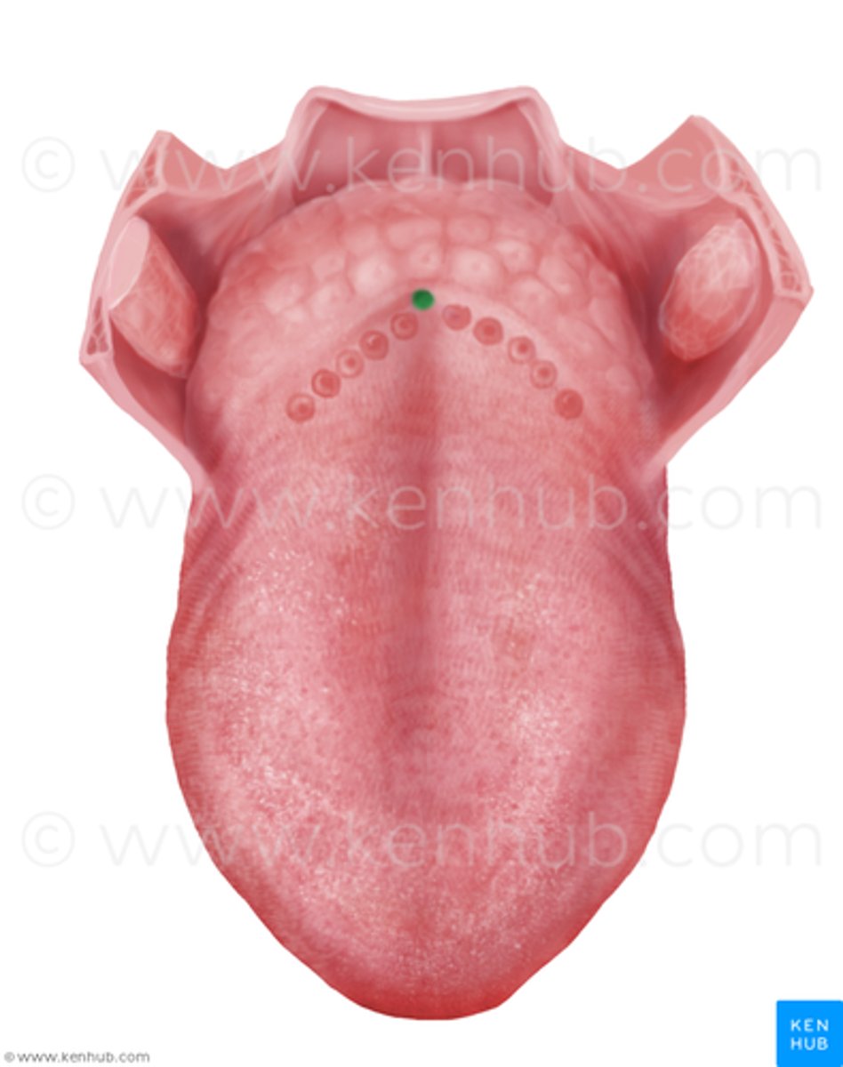
Foramen cecum
Hole
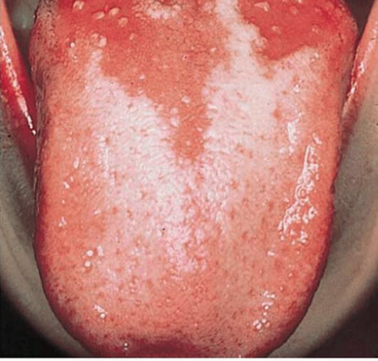
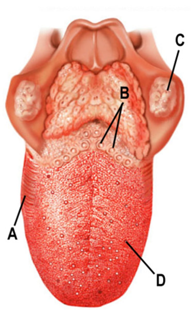
Lingual tonsil
Black bumps at base
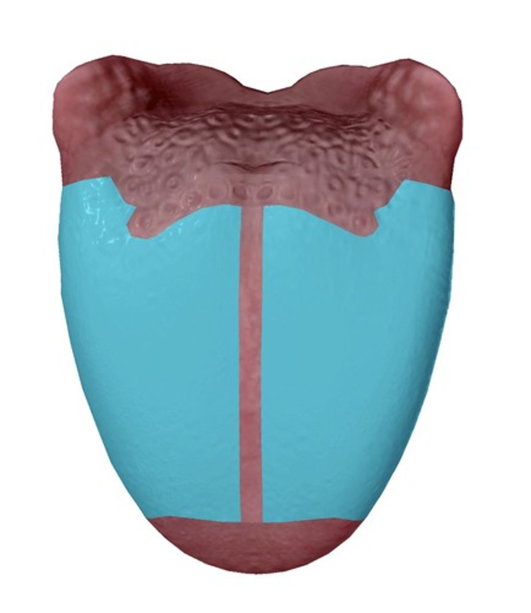
Filiform papillae
First 1/3
Fungiform papillae
Middle bumps
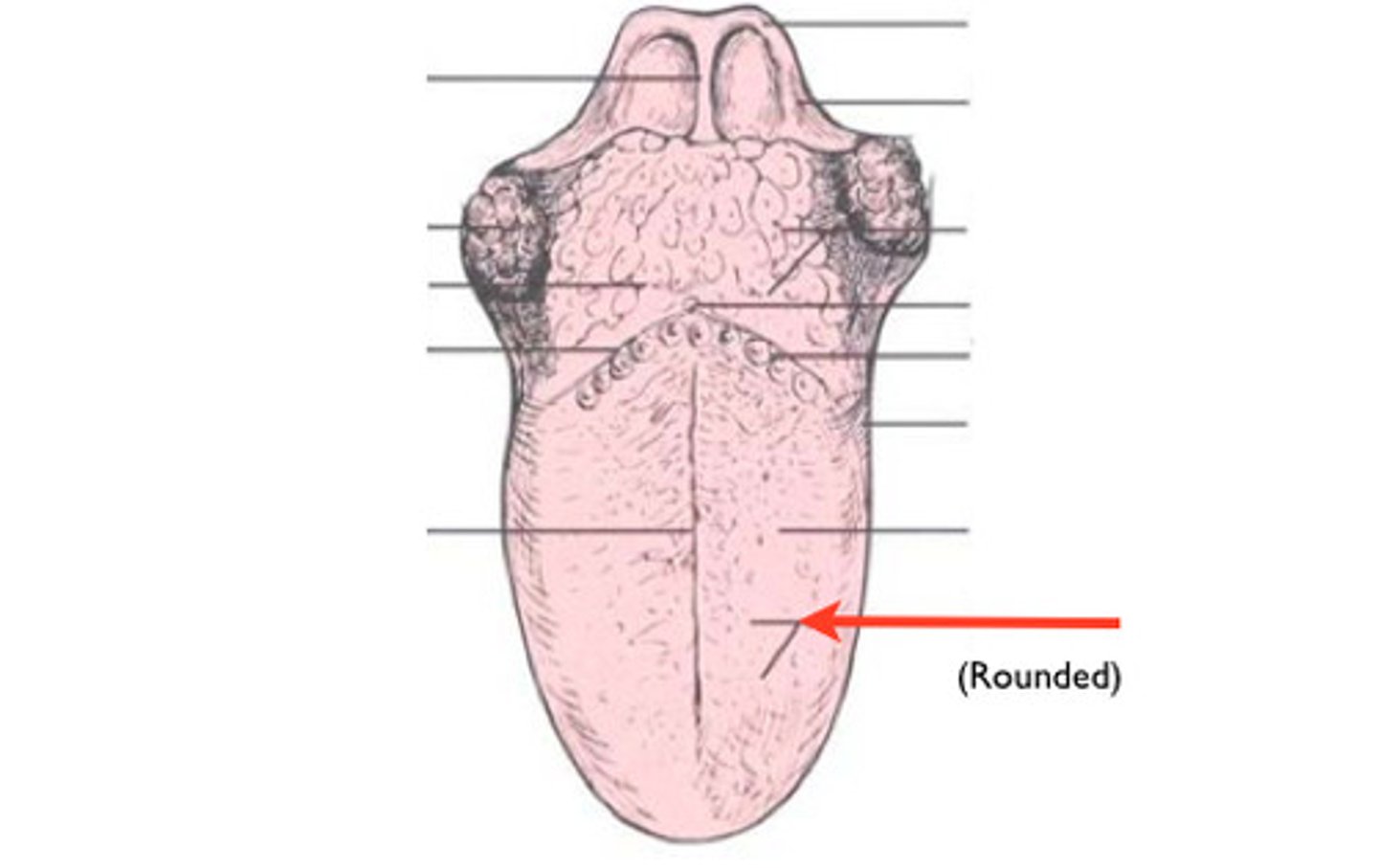
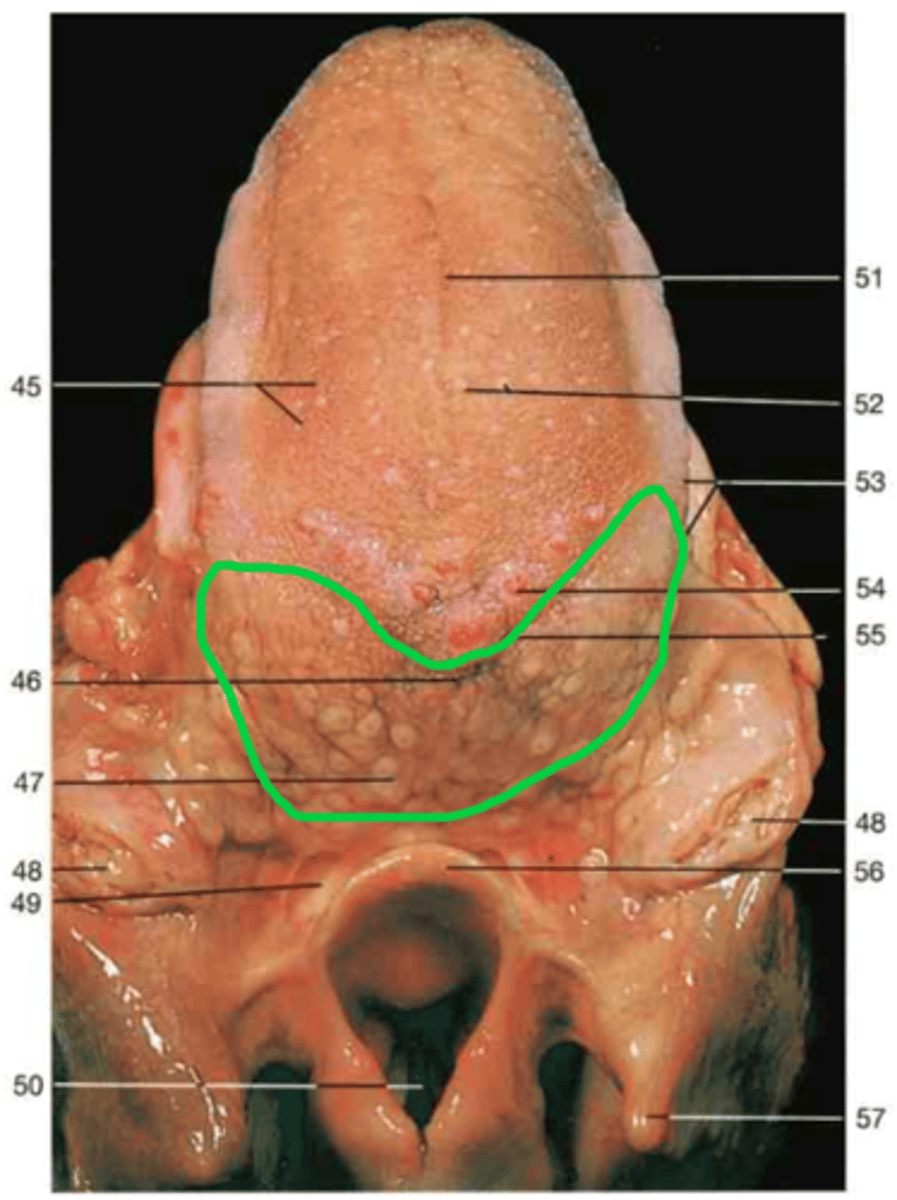
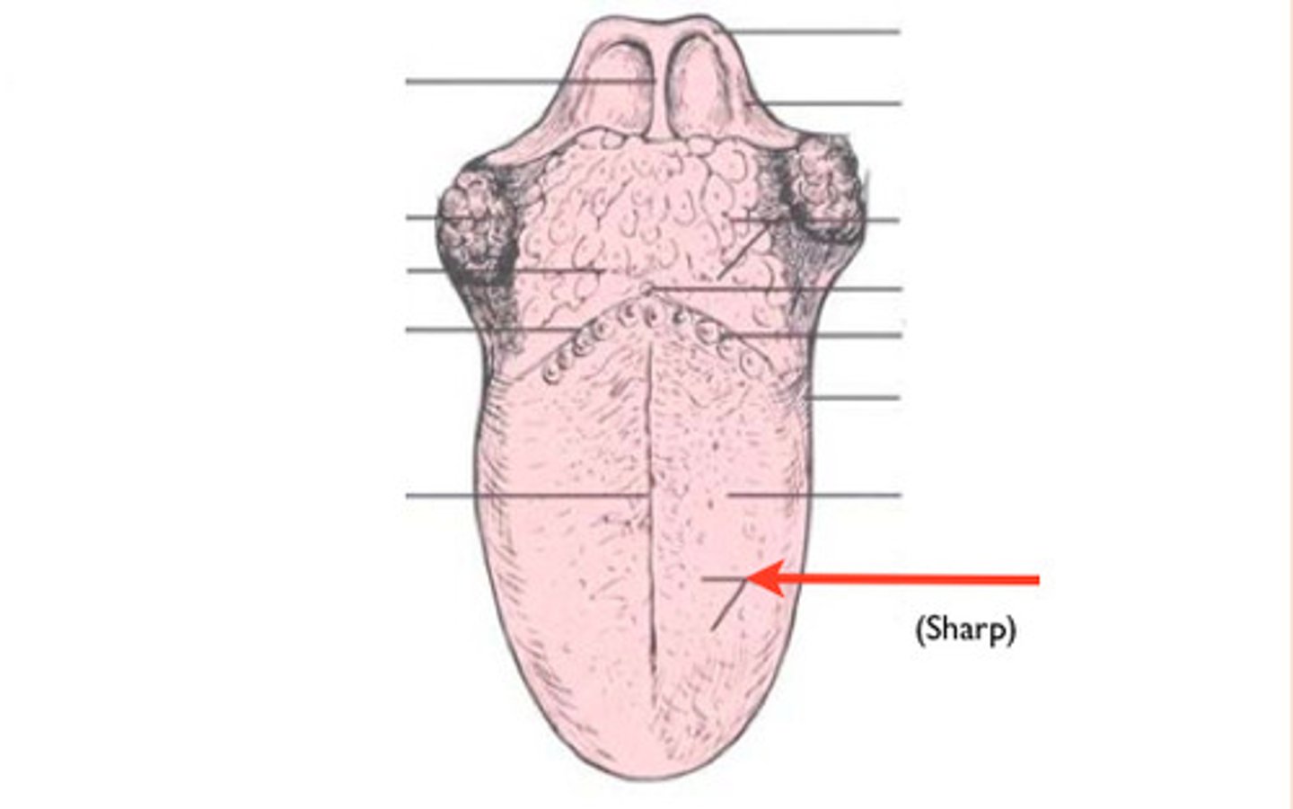
Vallate papillae
Back bumps before lingual tonsil
Inferior surface of tongue
surface
Frenulum of tongue
Feature
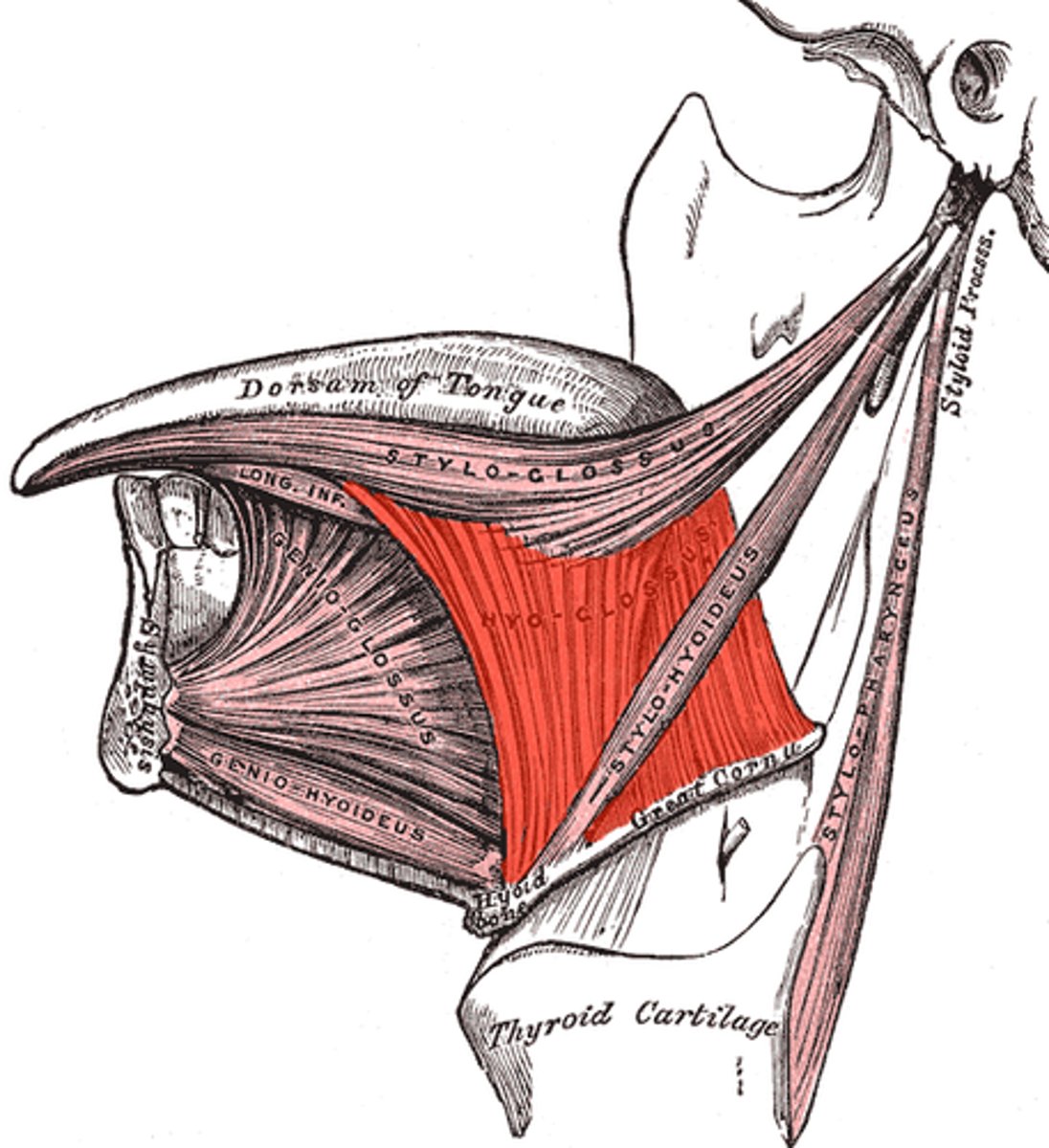
Genioglossus m.
Under tongue struecture
Hyoglossus m.
Side of tongue structure
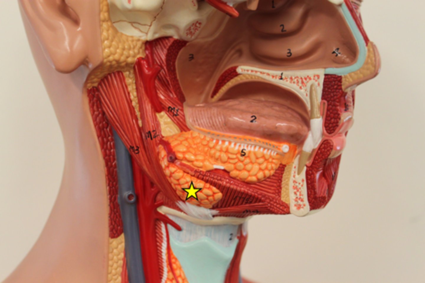
Submandibular gland
Structure
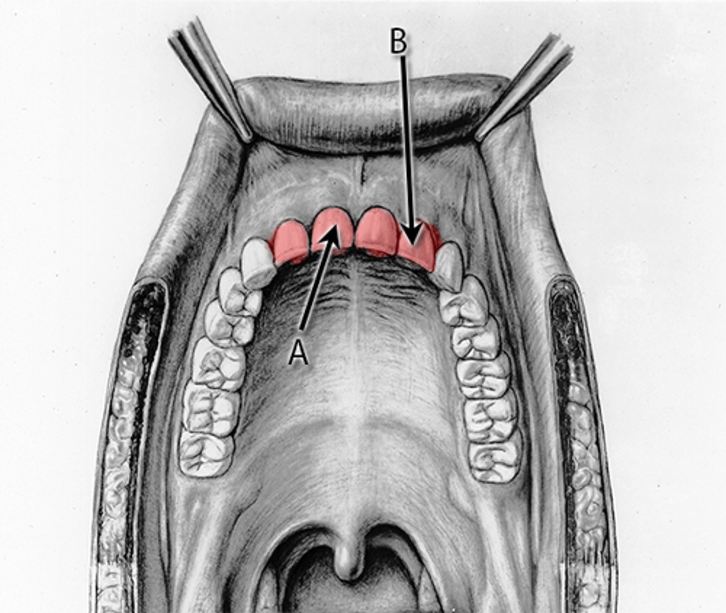
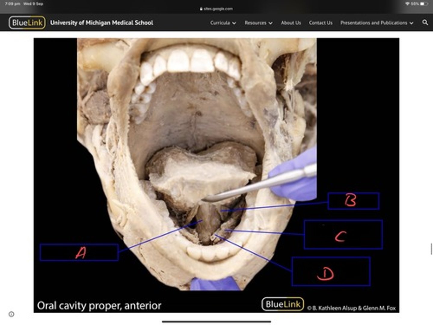
Palatoglossal arch
Closest to tongue (12) "arch"
Palatopharyngeal arch
"Arch"
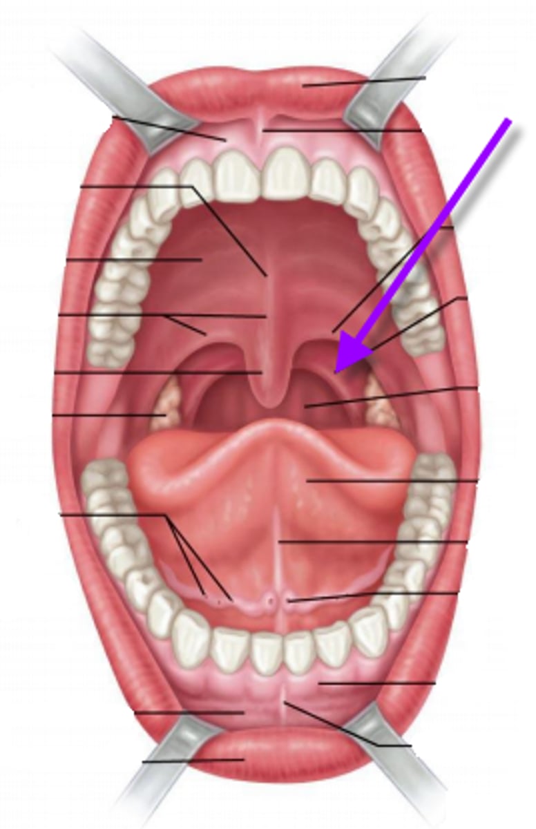
Tonsillar fossa
Depression
Between arches
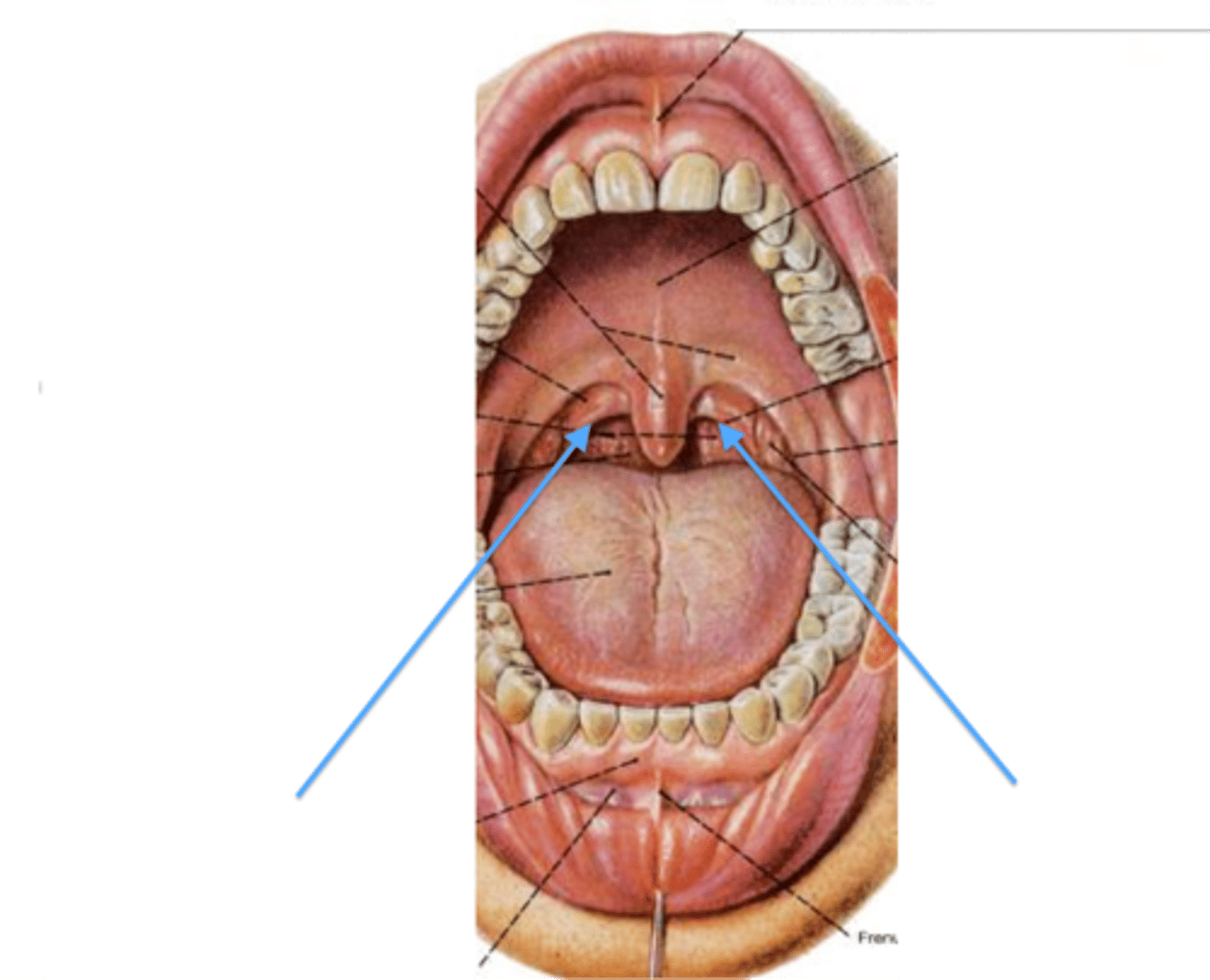
Palatine tonsil
Behind black bumps
C
Nasopharynx
Boundary
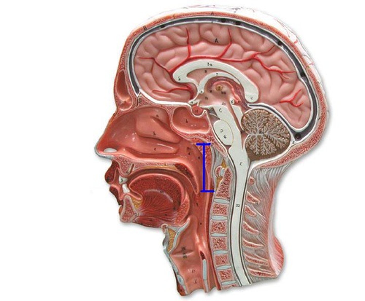
Oropharynx
Boundary
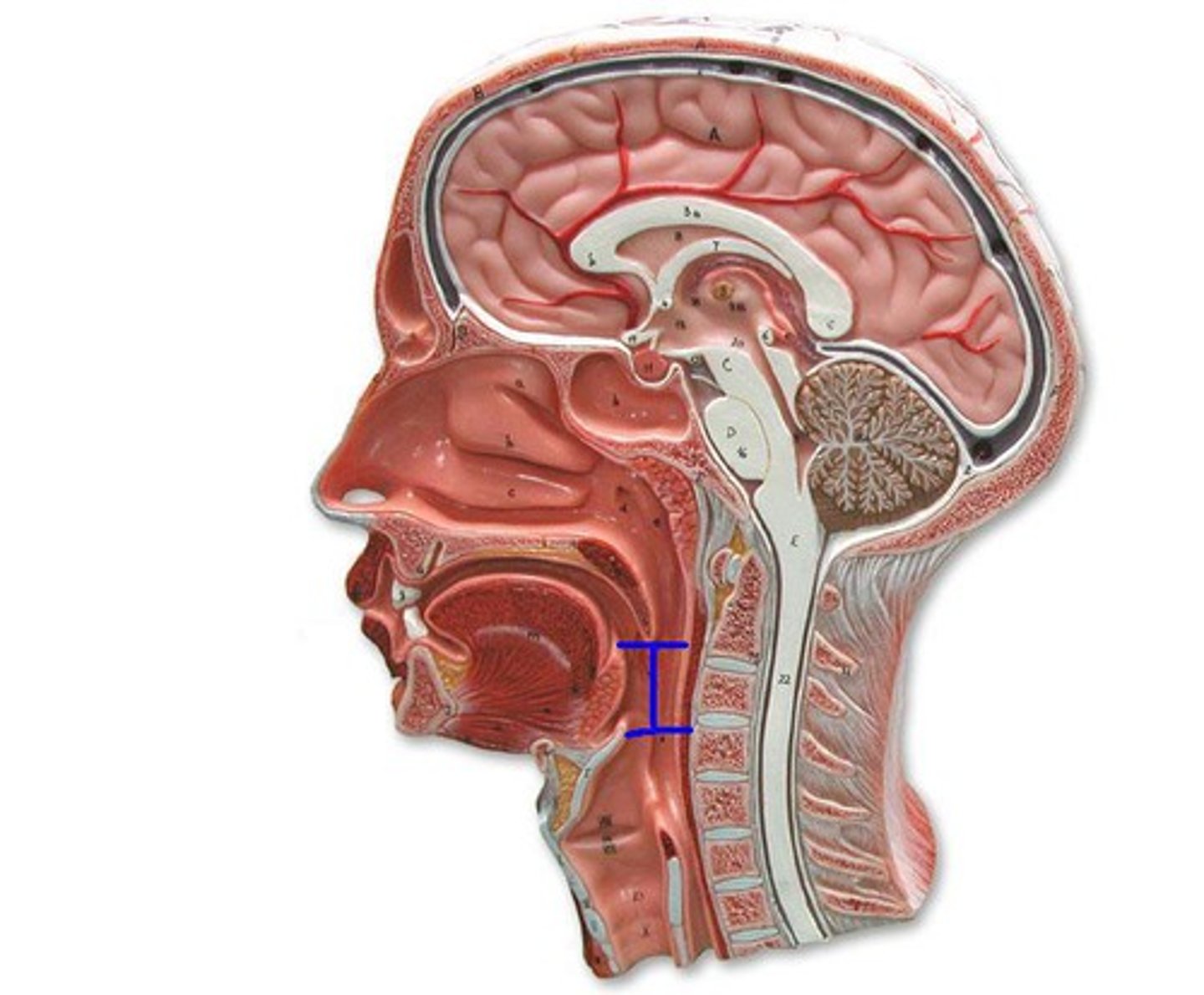
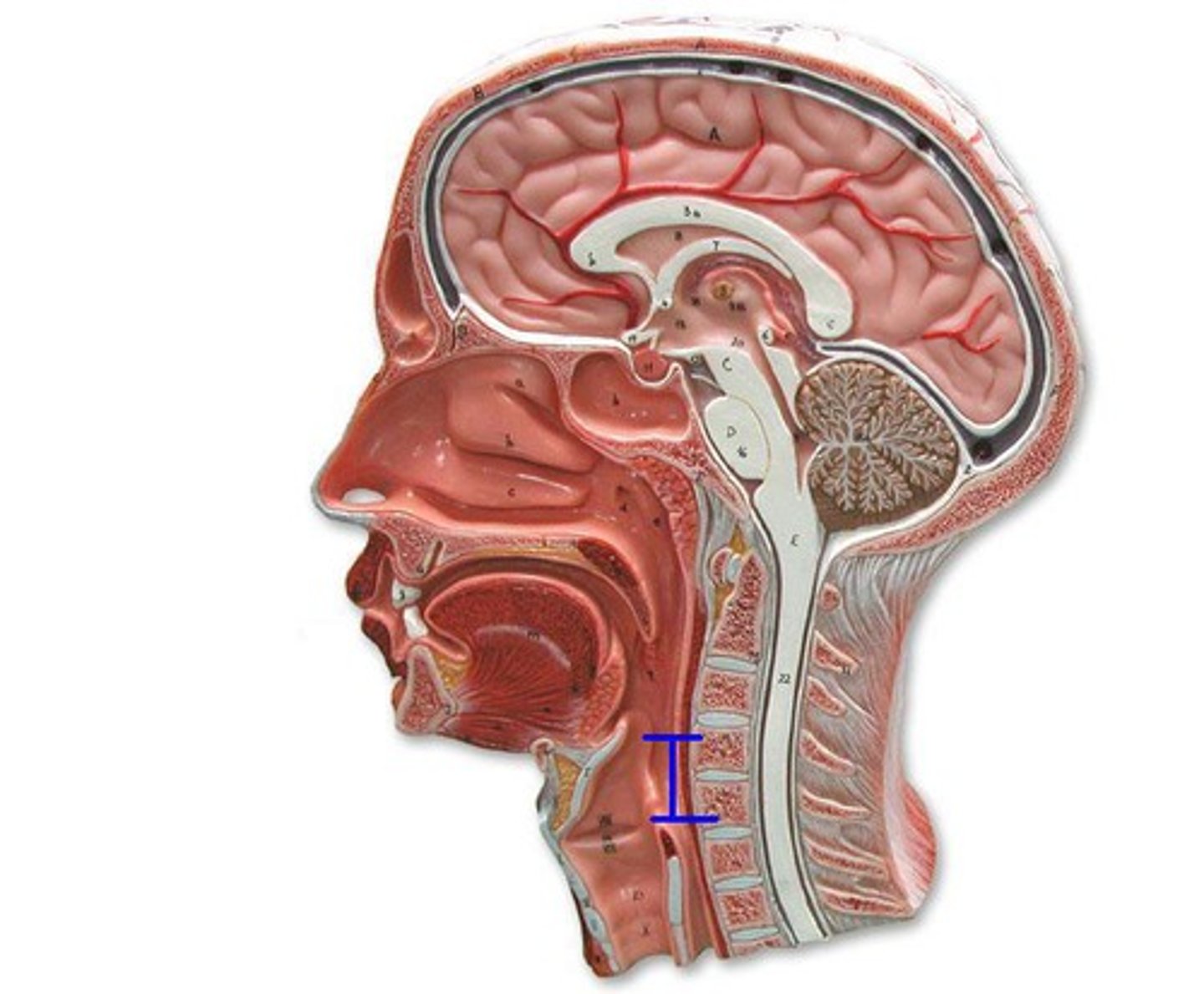
Laryngopharynx
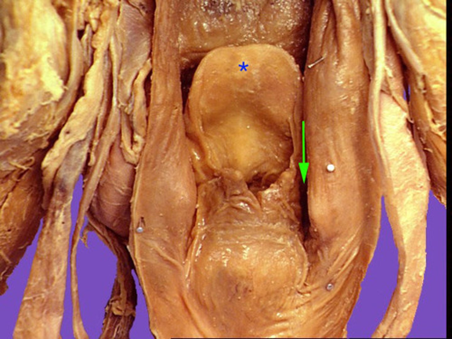
Piriform Recesses
depression
on either side of the epiglottis
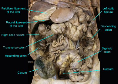
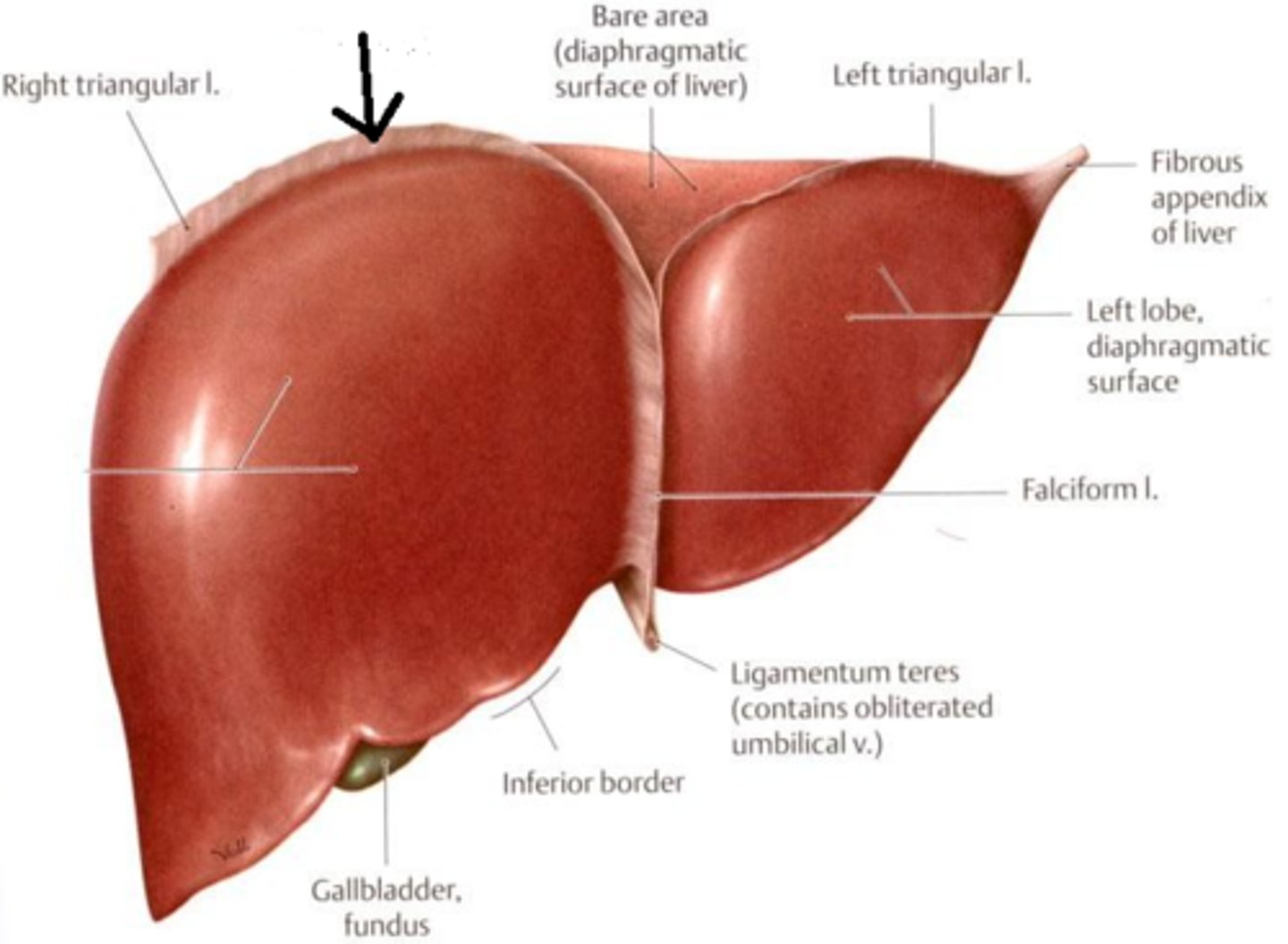
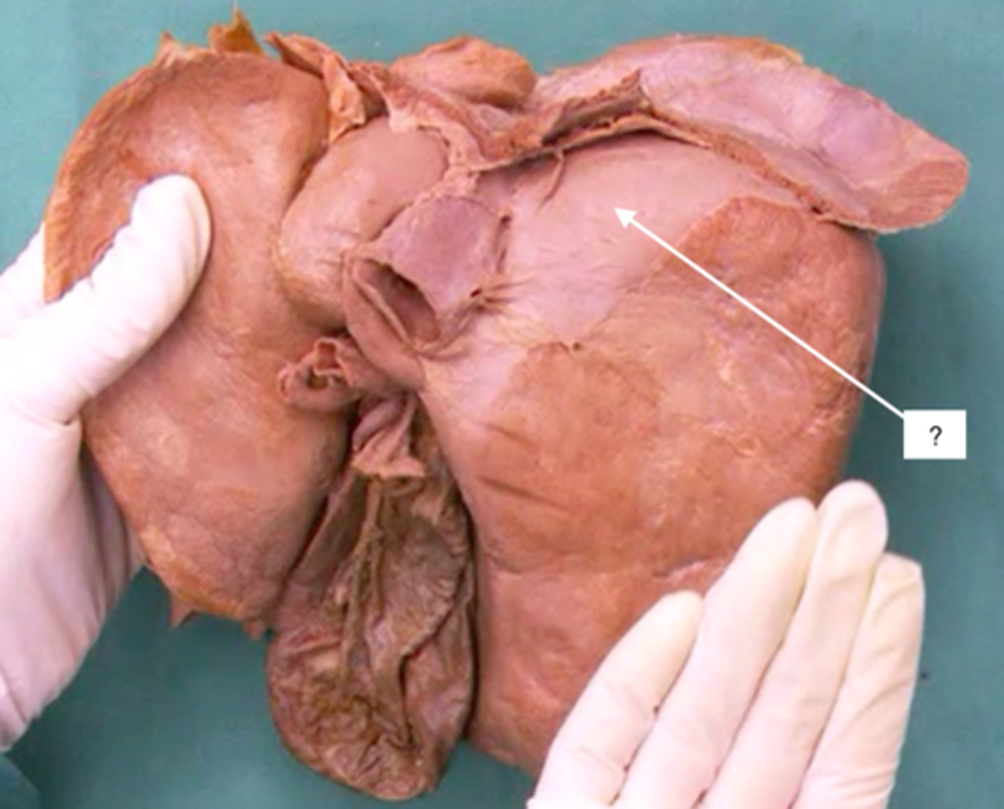
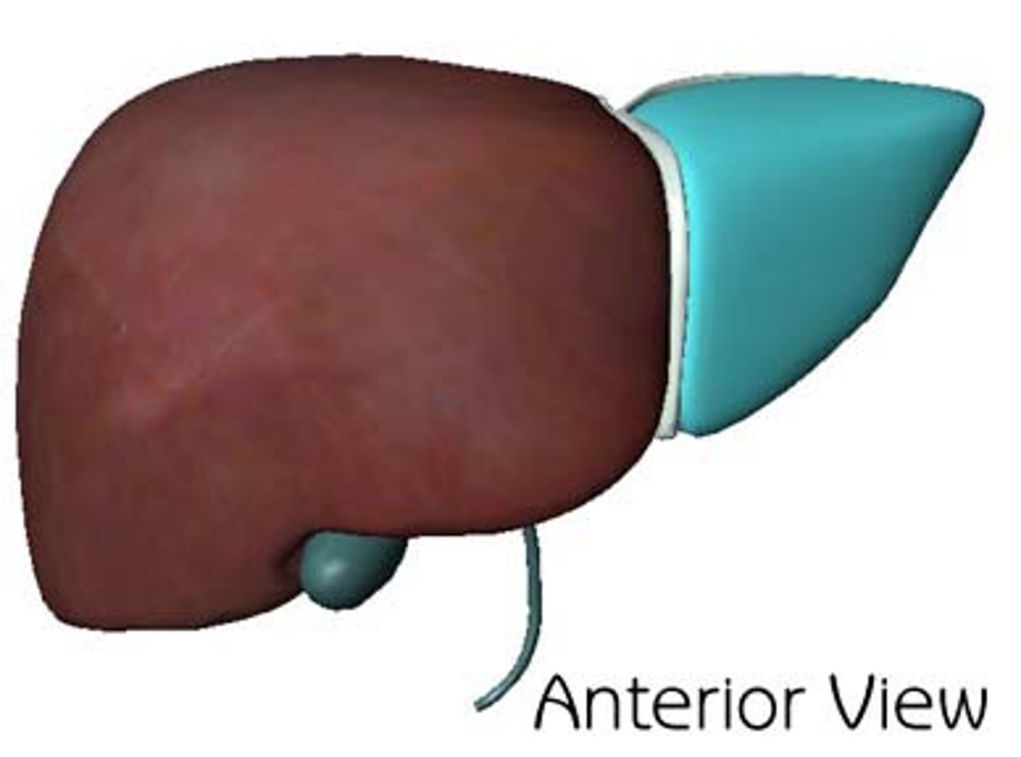
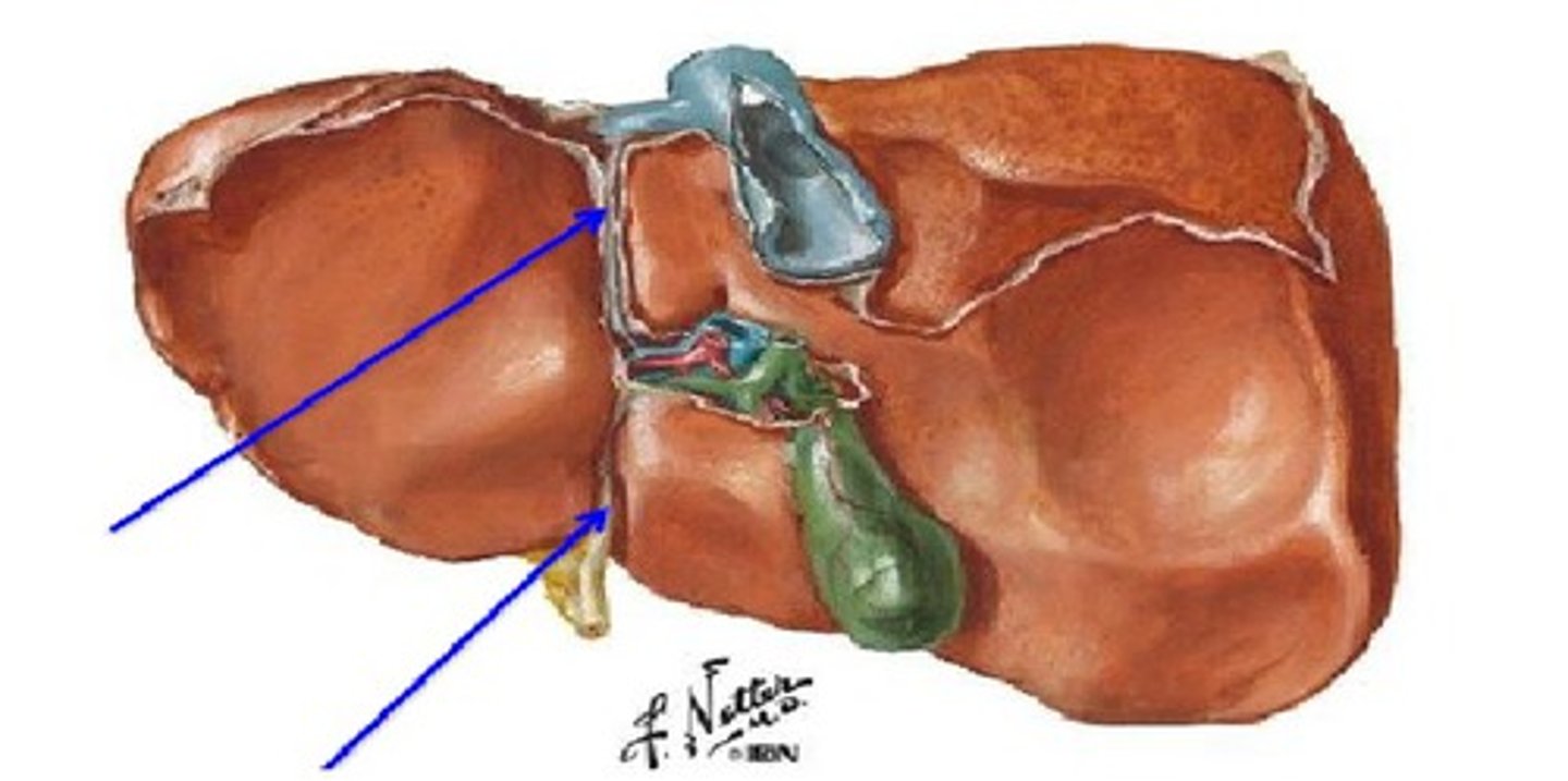
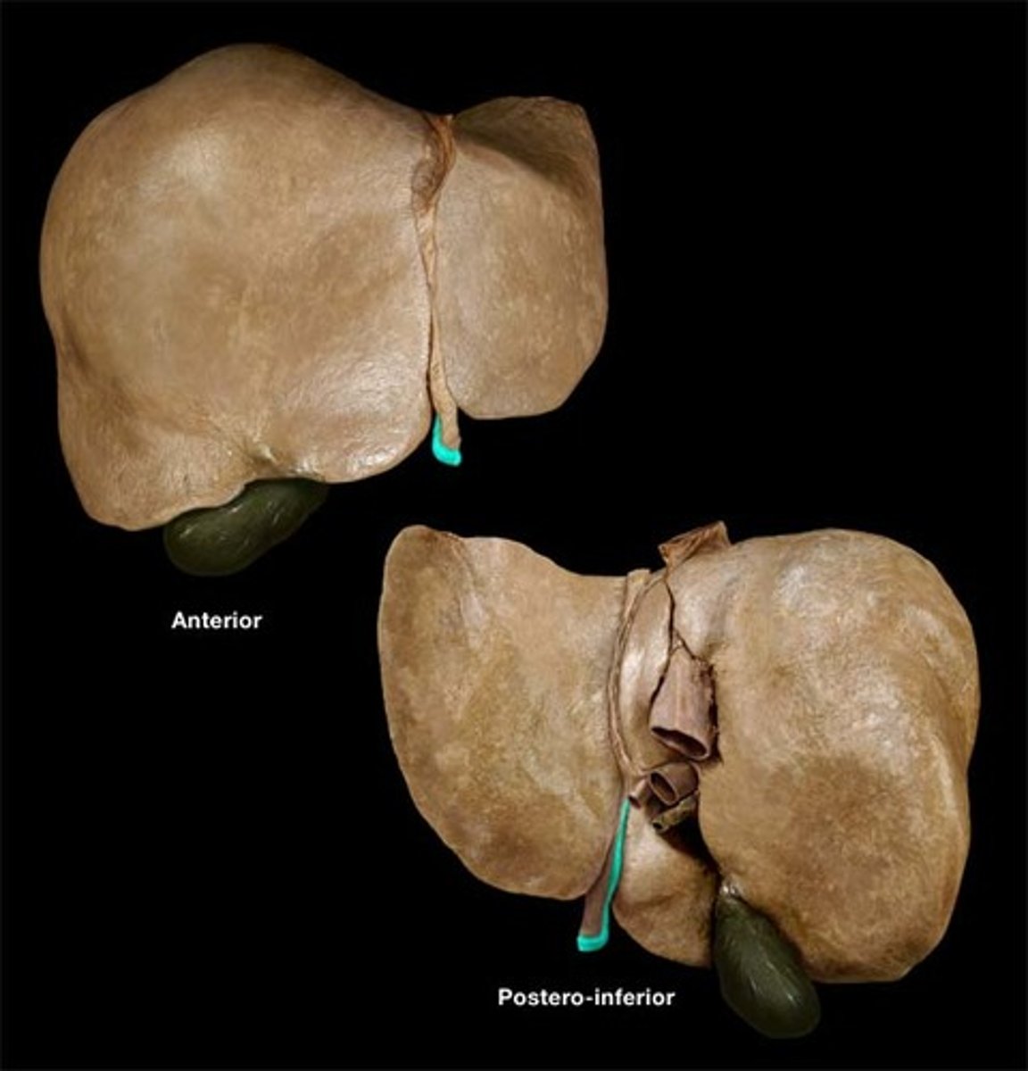
Diaphragmatic surface of the liver
Region
where diaphragm touched liver
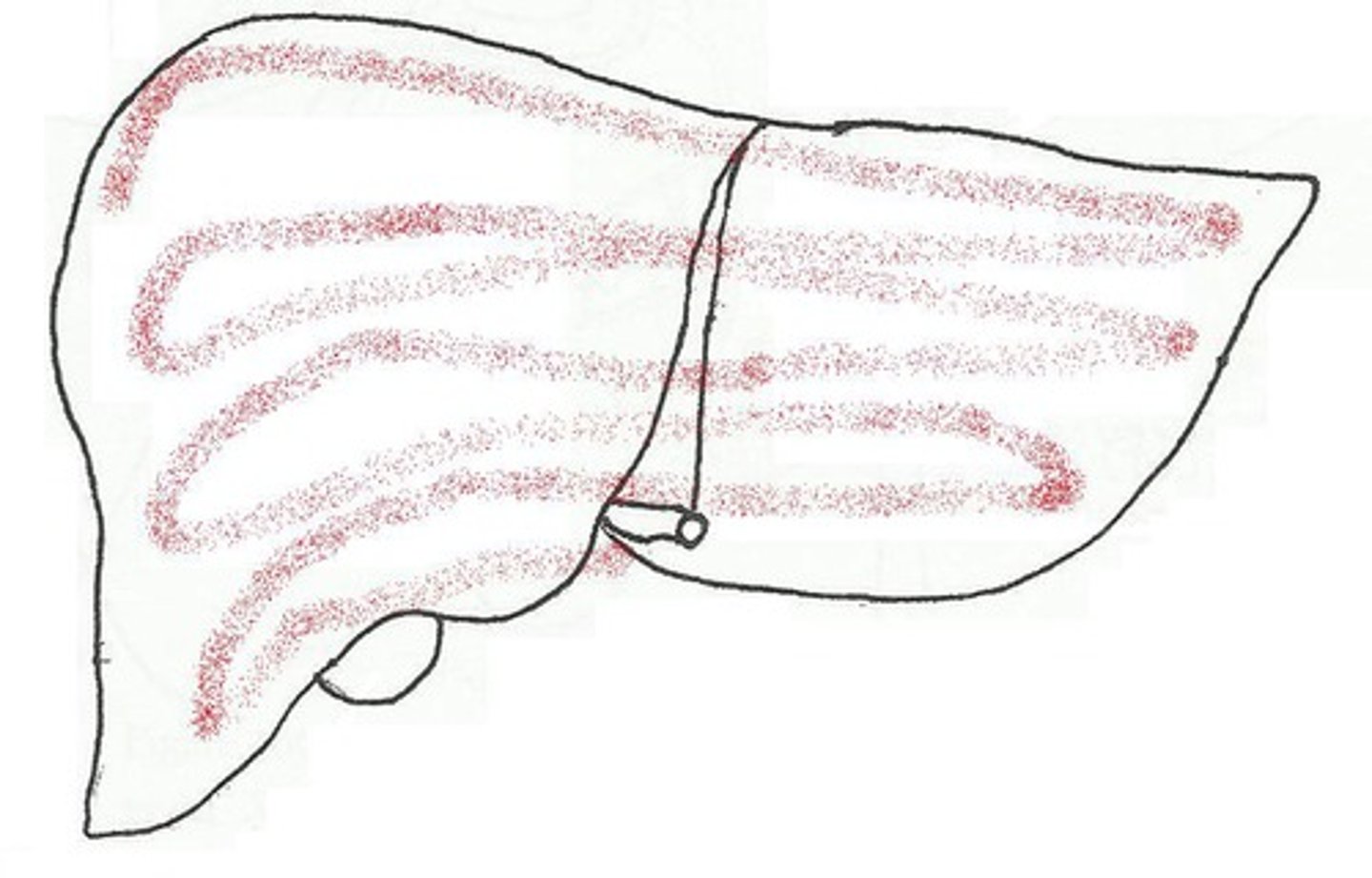
Visceral surface of the liver
Region
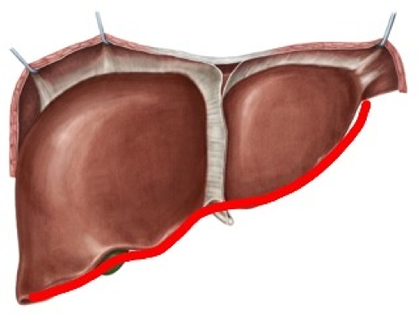
Inferior border of the liver
Boundary
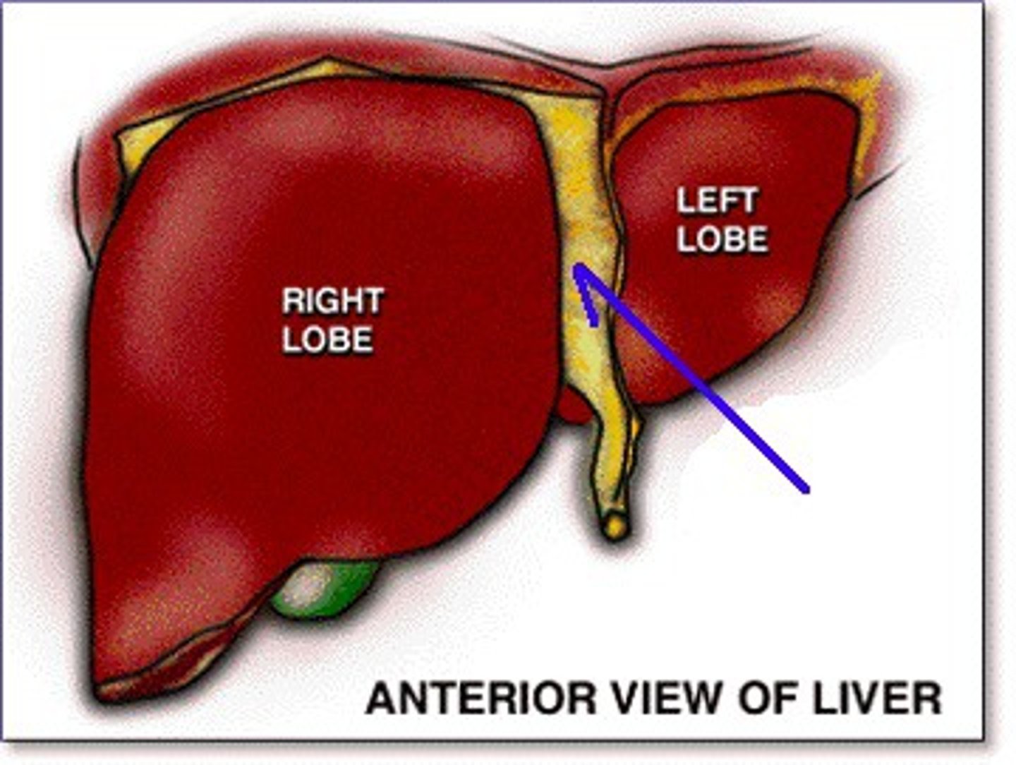
Falciform ligament
Tissue between lobes
anterior
Anterior layer of the coronary ligament
Posterior layer of the coronary ligament
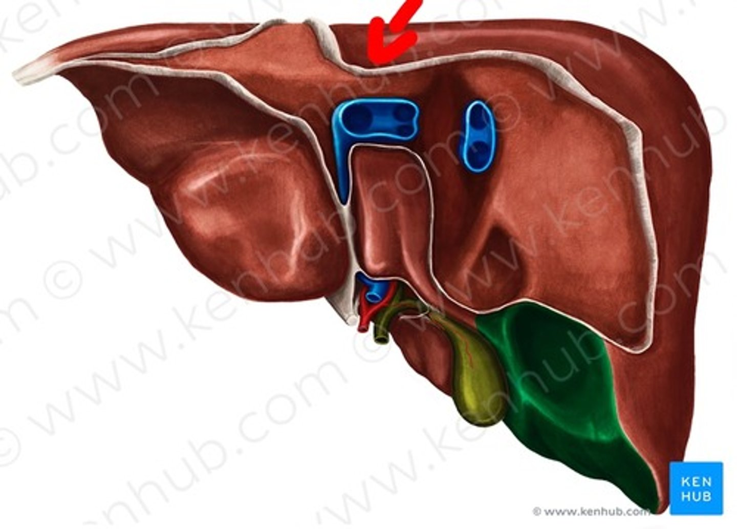
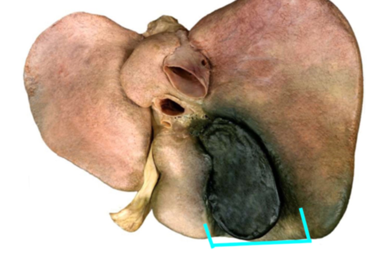
Bare area of the liver
Area
Between split on diaphragm
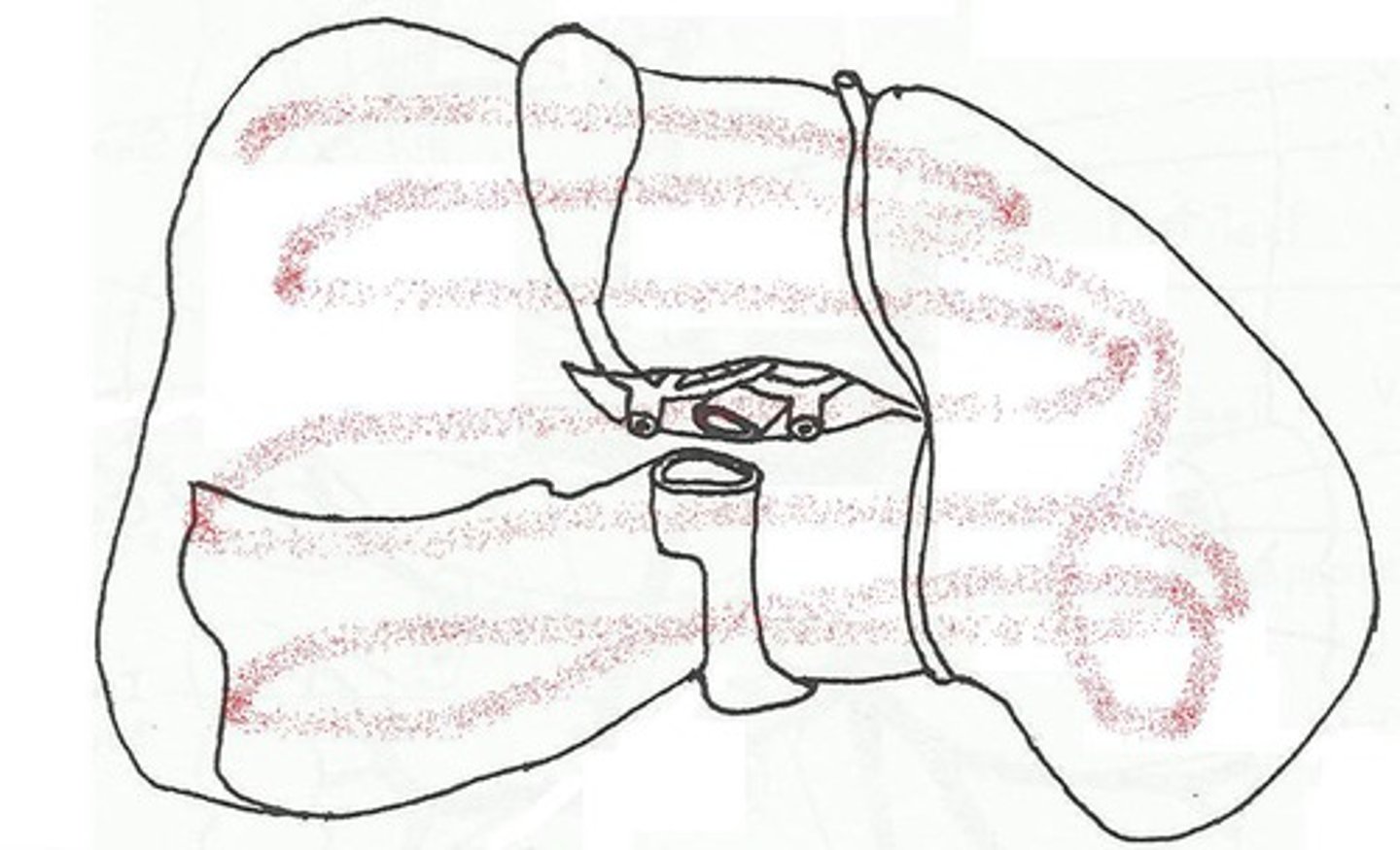
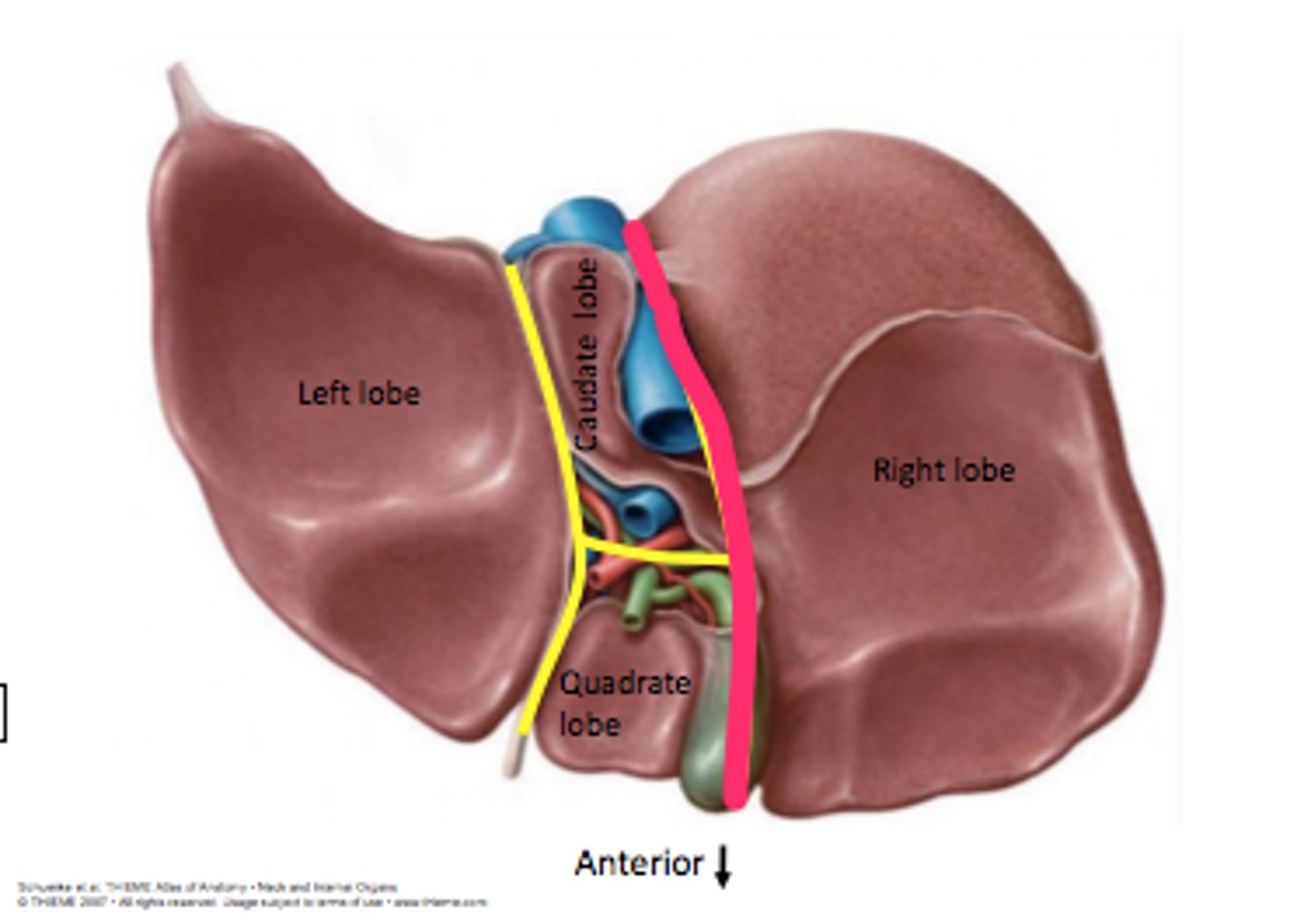
Right lobe of the liver
Left lobe of the liver
Quadrate lobe of the liver
Caudate lobe of the liver
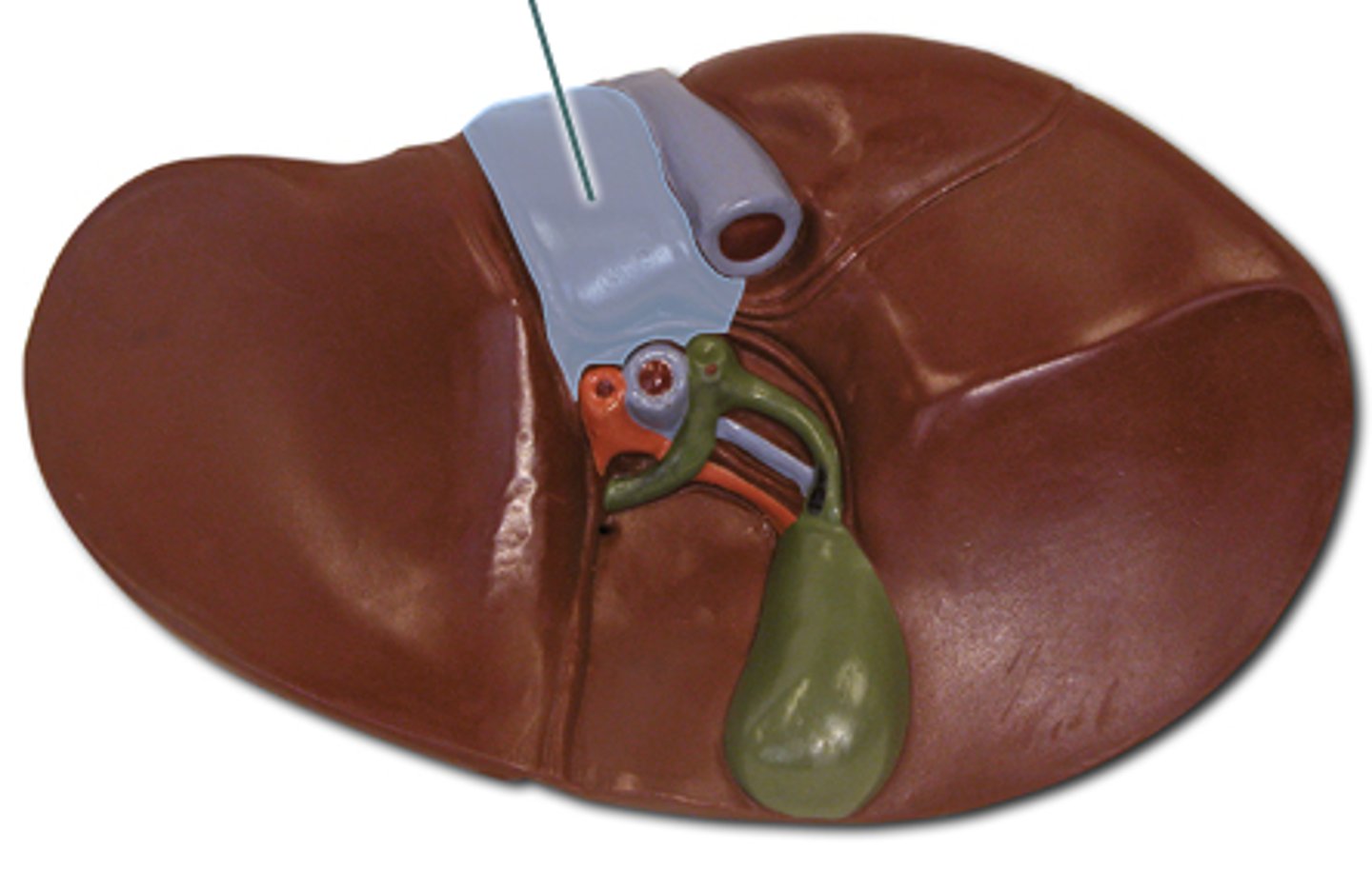
Right sagittal fissure
Division
Sulcus for the inferior vena cava
Depression
top right of the H
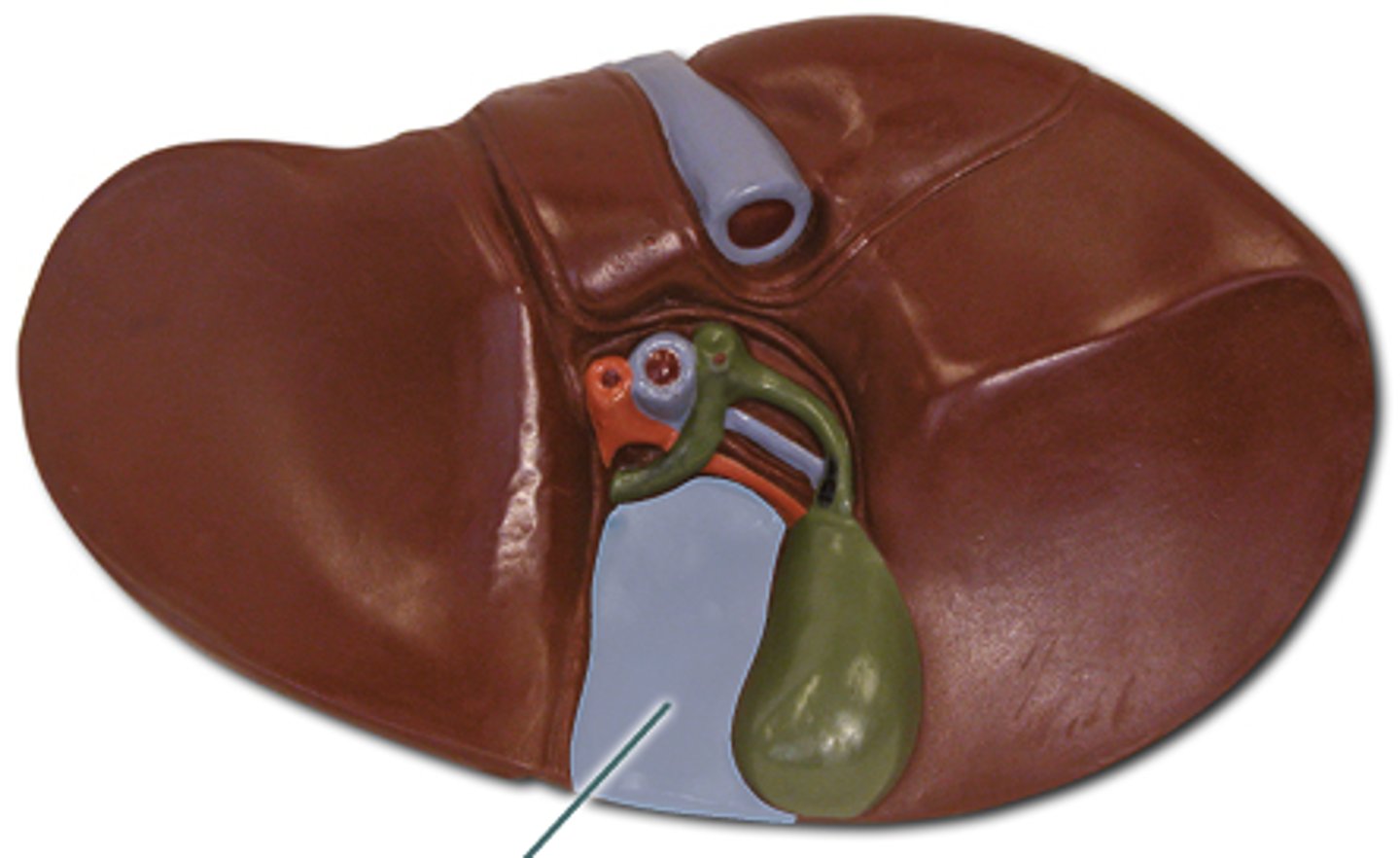
Fossa for the gallbladder
Depression
bottom right of the H
Left sagittal fissure
Division
Fissure for the round ligament
Depression
Bottom left 1/2 of H
Round ligament of the liver
Structure
Bottom left 1/2
Fissure for ligamentum venosum
Depression

Ligamentum venosum
Structure
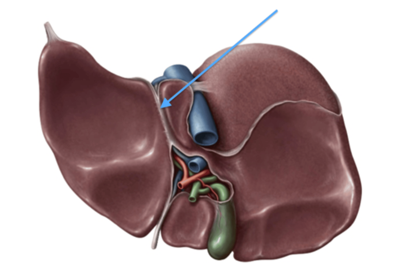
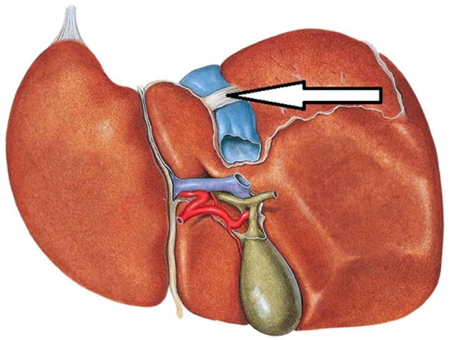
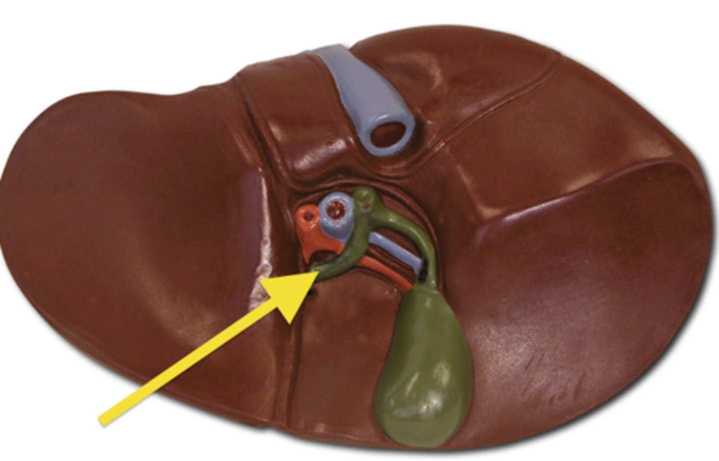
Porta hepatis
Division
horizontal part of H

The hepatic H
formed by the two sagittal fissures and the porta hepatis
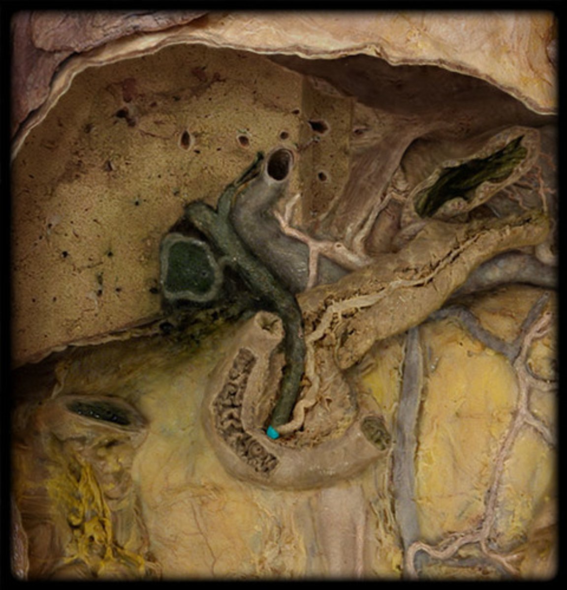
Gallbladder
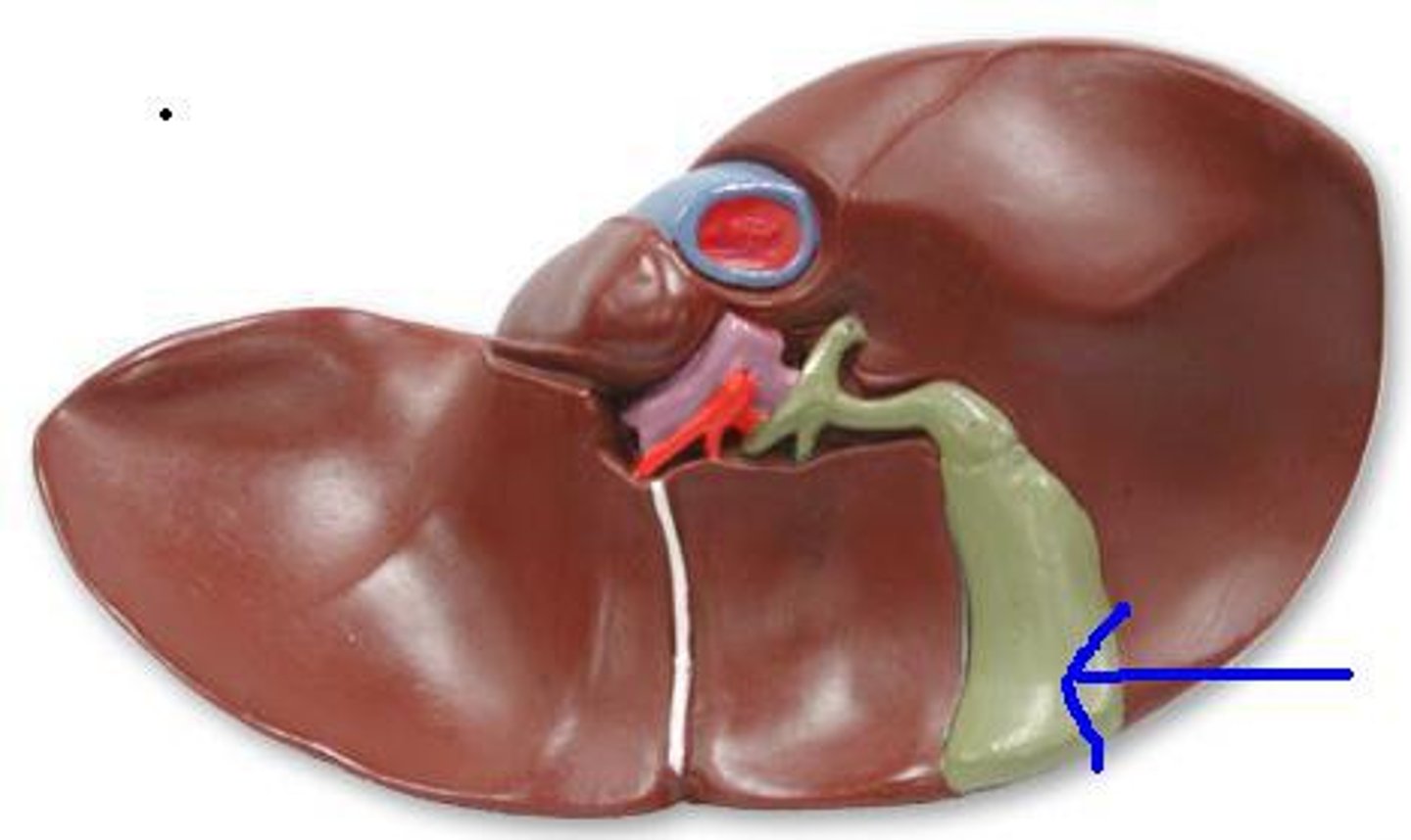
Fundus of gallbladder
Feature
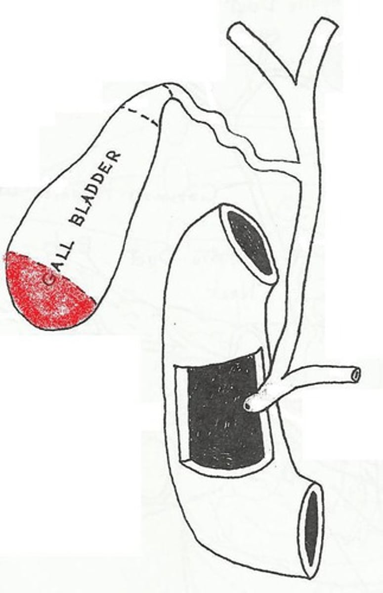
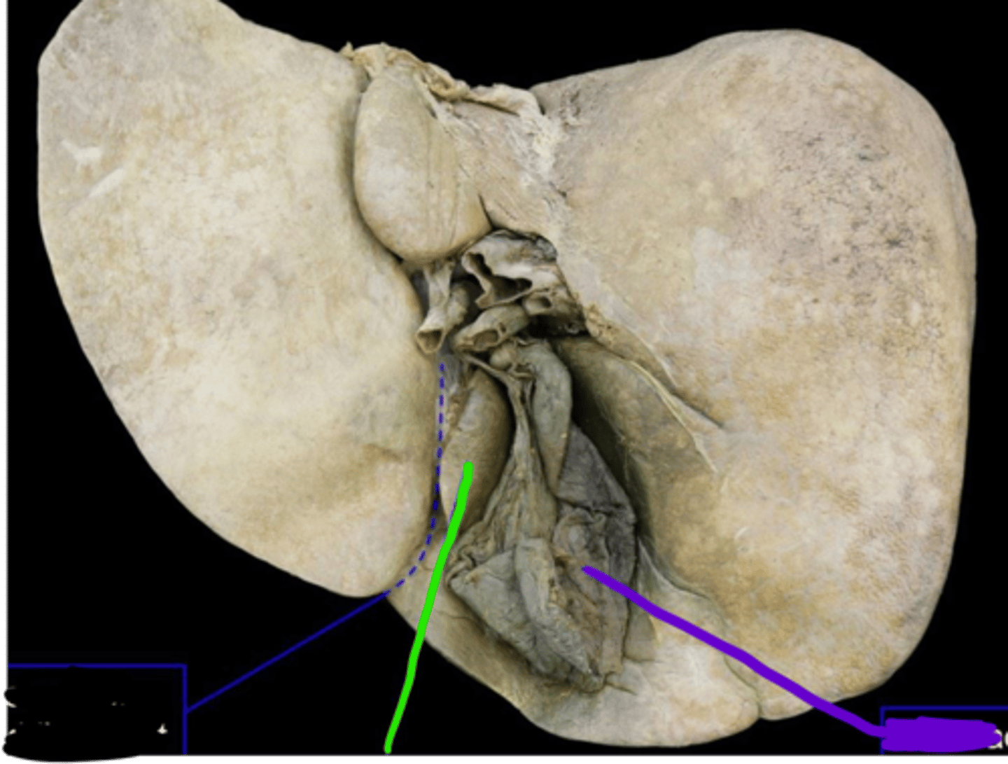
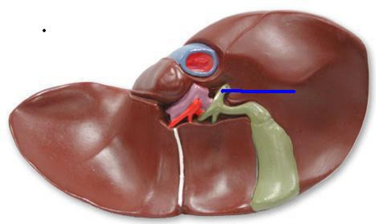
Cystic duct
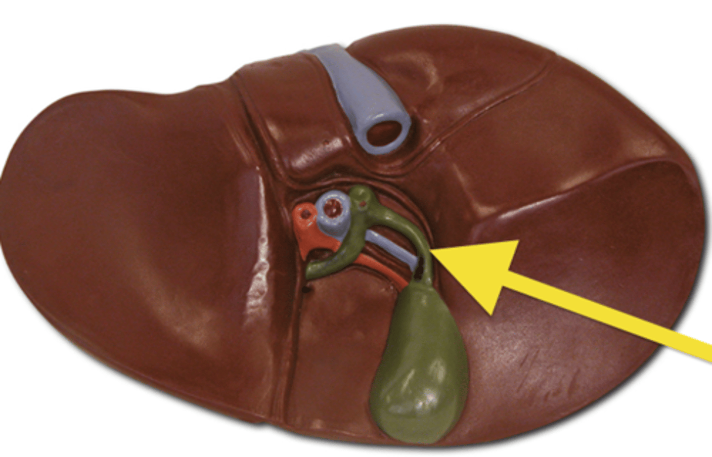
Common hepatic duct
Bile duct
See diagram
Hepatopancreatic ampulla
end of bile duct