you got this girl!!
1/348
There's no tags or description
Looks like no tags are added yet.
Name | Mastery | Learn | Test | Matching | Spaced | Call with Kai |
|---|
No analytics yet
Send a link to your students to track their progress
349 Terms
Describe what is meant by an "emergent property"
a characteristic of a complex system that is not present in its individual components but arises from the interactions and relationships between those components at a higher level of organization
Place the levels of biological organization in sequence from molecule to ecosystem
Molecule, organelle, cell, tissue, organ, organisms, populations, communities, ecosystem, biosphere.
Identify examples of each level of biological organization
Molecule (carbon atoms), organelle (mitochondria), cell (plant cell), tissue (muscular tissue), organ (heart), organisms (fox), populations (group of foxes), communities (foxes and grass), ecosystem (non living and living), biosphere (everything).
Identify the elements that compose living matter. Specifically, know which 4 elements make up >95% of the body mass of a human.
Main 4= (O (65%), C(18.5%), H(9.5%), N(3.3%))
Others=Ca (1.5%), P (1%), K (0.4%), S(0.3%), Na (0.2%), Cl (0.2%), (Mg 0.1%), and trace elements
Polar covalent
Electrons are shared, but it is unequal, which leads to partial charges.
Nonpolar covalent
electrons are evenly shared, molecules exhibiting this are hydrophobic
Ionic bond
electrons are "given" which leads to two ions, this occurs between a metal and nonmetal.
Hydrogen bond
bond between partial positive hydrogen and a partial positive between another molecule.
Understand how differences in electronegativity can lead to polar bonds, and how polar molecules and partial charges are related to hydrogen bonds.
Partial bonds are created when there is a difference in electronegativity, which leads to pulling and unequal sharing in a polar bond, giving the atoms partial charges. These can then be used to make hydrogen bonds.
Explain why differences in electronegativity lead to polar covalent bonds within water molecules.
Certain atoms have more of an affinity for electrons, they "want" them more, so they will pull the electrons to their side, leading to a polar covalent bond.
Explain how polar covalent bonds within water molecules are related to partial charges.
Partial charges allow hydrogen bonding to occur.
Explain how attraction between partial charges leads to hydrogen bonding among water molecules, as well as hydrogen bonding between water and other molecules
Partial charges allow hydrogen bonding to occur.
Identify the four emergent properties of water essential for life and provide an example of each.
C- cohesive, water sticks together
A-ability to moderate temperature
V- versitality as a solvent
E- expansion upon freezing
What role do hydrogen bonds play in these emergent properties?
hydrogen bonds allow these properties to even exist, they contribute to surface tension, cohesion, adhesion, etc.
Four macromolecules
carbohydrates, lipids, proteins, nucleic acids
Monomer, polymer, and bond of carbohydrates
monosaccharides, polysaccharides, glycosidic bond
Monomer, polymer, and bond of lipids
N/A
Monomer, polymer, and bond of proteins
Amino acids, polypeptides, peptide bond
Monomer, polymer, and bond of nucleic acids
Nucleotides, nucleic acids, phosphodiester bonds
Which macromolecule does not have monomers and polymers
Lipids
What major roles does each type of macromolecule play in the cell?
Lipids= Storage, protection, signaling
Carbohydrates= Storage, structure
Nucleic Acids= Reproduction, DNA, RNA
Proteins= speed up chemical reactions, play a role in defense, storage, transport, cellular communication, movement, or structural support.
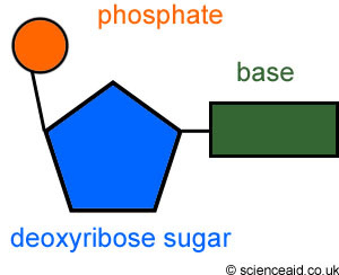
Describe the structures of nucleotides and nucleic acid polymers. What parts of the nucleotides make up the "backbone"? What type of bond connects nucleotides?
5 carbon sugar, nitrogenous base, phosphase group. Sugar phosphate backbone by strong phosphodiester bonds.
Describe the basic structure of an amino acid. Use the terms: amino group, carboxyl group, R group. Which of these groups are involved in forming peptide bonds?
a central carbon atom (called the alpha carbon) connected to an amino group (NH2), a carboxyl group (COOH), and a variable side chain called an R group. The amino group and carboxyl group are the parts of an amino acid that are involved in forming peptide bonds.
Define/describe primary, secondary, tertiary, and quaternary protein structure. What parts of the molecule are involved in each level of structure? What types of bonds?
Primary- Sequence of amino acids, joined together via dehydration synthesis (peptide bonds).
Secondary- Coils and folds from hydrogen bonding, alpha helix, beta pleated sheet.
Tertiary- Interactions between R groups, hydrophobic interactions, hydrogen bonding, ionic bonding, disulfide bridge
Quaternary- multiple polypeptide chains
How are secondary and tertiary structures directly dependent on primary structure?
Primary is the sequence, and the sequence determines what bonds are even possible, which impacts the other structures.
Where do alpha-helices and beta-pleated sheets come into play?
Secondary structure
Distinguish the diversity of roles that proteins play in the cell and recognize examples.
Enzymatic proteins- Selective acceleration of chemical reactions (digestive enzymes)
Defensive proteins- Protection against disease (antibodies)
Storage proteins- Storage of amino acids (casein, protein in milk)
Transport proteins- Transport substances (hemoglobin)
Hormonal proteins- Coordination of an organisms activities (insulin)
Receptor proteins- Response of cell to chemical stimuli (receptors in membrane)
Contractile/ Motor proteins- Movement (cilia)
Structural proteins- Support (keratin)
Define the terms prokaryotic and eukaryotic and identify which groups of organisms fall into each category
Prokaryotes (simple) do not have membrane bound organelles, where as eukaryotes (complex) do. They also lack a nucleus and instead have a nucleoid.
Differences of prokaryotic cells and eukaryotic cells
DNA location- Eukaryotes have DNA in the nucleus, prokaryotes have in a nucleoid region
Size-Eukaryotes are bigger
Complexity- Eukaryotes contain more complex membrane-bound organelles.
Cell structure
Similarities of prokaryotes and eukaryotes
Plasma membrane, cytosol, chromosomes (DNA), and ribosomes
Describe the structure and function of the cell components that are common to all cells(plasma membrane, cytosol, chromosome, ribosomes)
Plasma membrane- selectively permeable membrane that encloses the cell. Made of phospholipids
Cytosol- Gel inside of cells, provides means of transport, and suspension.
Chromosome- Contains DNA, genetic blueprint.
Ribosome- synthesizes proteins.
Describe the structure and function of the cell components that are part of the eukaryotic endomembrane system (the rough and smooth ER, Golgi, lysosomes, vesicles)
Rough ER- Has bound ribosomes, allowing for creation and secretion within the rough ER
Smooth ER- Synthesis of lipids, metabolism of carbs, detoxification of drugs and poisons, storage of calcium ions.
Golgi- Synthesis, modification, sorting, secretion of cell products
Lysosomes- Cell digestion
Vesicles- Transport, fluid storage
Describe the structure and function of the nucleus and the role of nuclear pores
Nucleus holds most of the genetic information. Enclosed in a double membrane. Nuclear pores allow for transport of molecules such as mRNA.
Describe the structural and functional differences of the rough and smooth ER
Smooth ER- Lacks ribosomes, lipid synthesis, detoxification of drugs and poisons, carbohydrate metabolism, and calcium ion storage.
Rough ER- bound ribosomes involved in protein synthesis of secretory proteins.
Identify in general terms the structure and function of the Golgi
The golgi is responsible for receiving, sorting, shipping, and manufacturing proteins and lipids.
Path of proteins and lipids
ER -> Golgi -> Final destination
Identify functions associated with mitochondria and chloroplasts
Mitochondria= cellular respiration
Chloroplasts= photosynthesis
Different membrane proteins
Transport, Enzymatic Activity, Signal transduction, Cell- cell recognition, Intercellular joining, Attachment to the Cytoskeleton and ECM
Describe the function of transport proteins
Facilitate the movement of substances across the membrane.
Function of Enzymatic Activity proteins
Act as enzymes, catalyzing reactions at the membrane surface.
Function of proteins that are involved in signal transduction
Receptor proteins bind to signaling molecules, triggering a response inside the cell.
Function of proteins involved in cell- cell recognition
Glycoproteins serve as identification tags, recognized by other cells.
Function of proteins that are involved in intercellular joining
Proteins form junctions between cells, providing structural connection
Function of proteins that attach to the cytoskeleton and ECM
Proteins anchor the membrane to the cytoskeleton and extracellular matrix, maintaining cell shape and stability.
Given a particular molecule you should be able to predict if it can diffuse through the lipid bilayer or if it will require other methods for transport
Understand the concept of a semipermeable membranes as it relates to cell membranes
Small molecules can, large molecules cannot
Hydrophobic molecules can, hydrophilic get stuck in center. Small uncharged polar molecules can, charged and large require assistance.
Hydrophobic and hydrophilic areas of phospholipid bilayer
Hydrophobic interior, hydrophilic exterior. Interior repels hydrophilic molecules.
Predict the direction of solute net diffusion across a semipermeable membrane
Molecules diffuse depending on the concentration gradient. It also depends if the solute can even diffuse through the membrane.
Predict the direction of water net diffusion (osmosis) across a semipermeable membrane
Water would go with a concentration gradient, would have to be able to even enter membrane.
Use the concept of 'tonicity' to predict the movement of water into or out of cells in hypertonic solutions.
Means the cell is shrinking. This is due to an increased solute concentration on the outside of the cell, causing water to diffuse out.
Use the concept of 'tonicity' to predict the movement of water into or out of cells in hypotonic solutions.
Means the cell is swelling. There is an increased solute concentration inside of the cell, so water flows in to even out the concentration.
Use the concept of 'tonicity' to predict the movement of water into or out of cells in isotonic solutions.
This stage is equal, there is no net diffusion, molecules flow back and forth at an equal rate.
Passive transport
the movement of substances across a cell membrane without the use of energy by the cell. goes down the concentration gradient.
Explain how membrane proteins (transporters, channels) allow facilitated diffusion tohappen
The movement of materials through a cell membrane using energy goes against the concentration gradient.
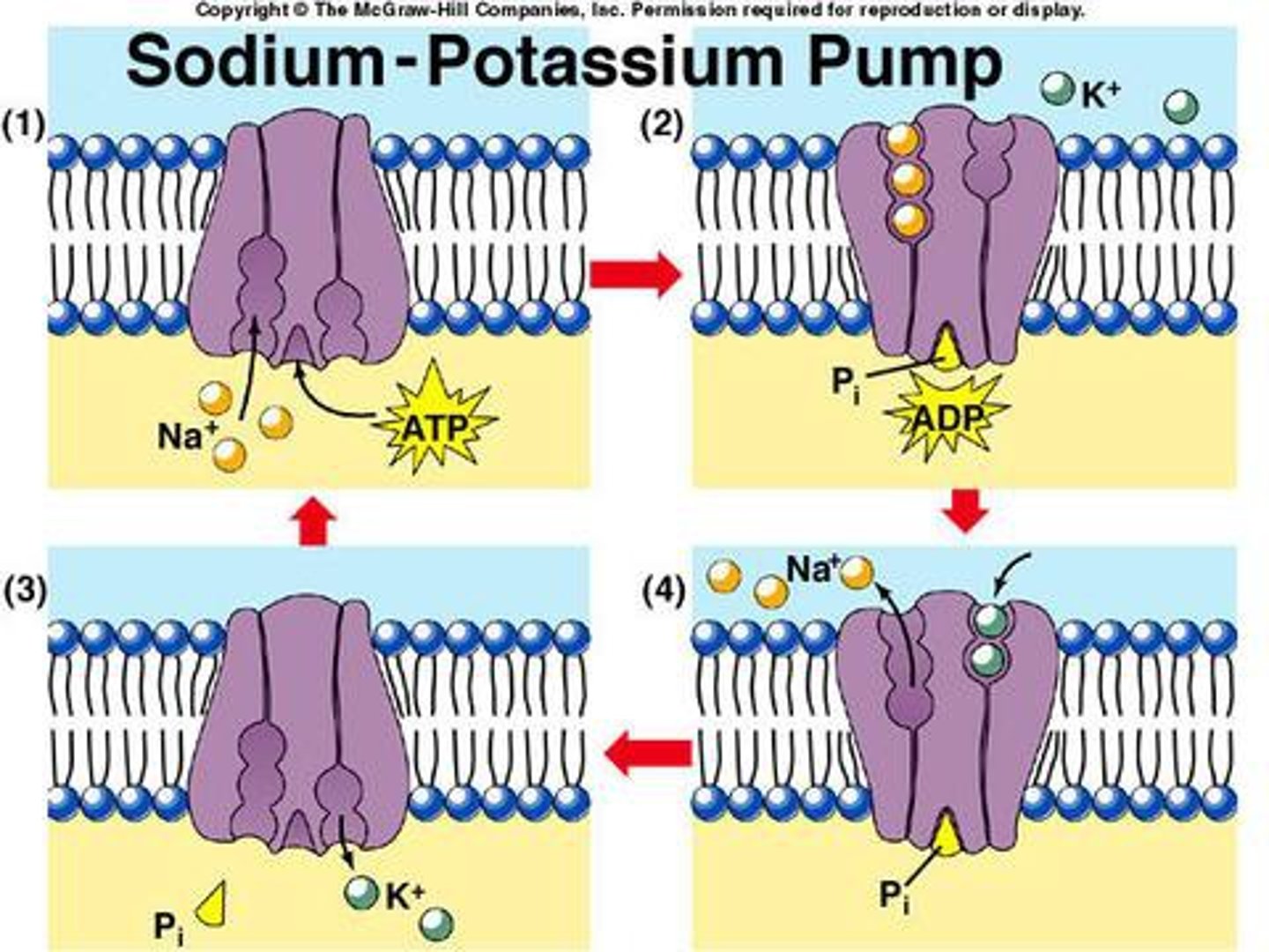
Describe the action of the sodium-potassium pump. Explain the role of ATP in its operation.
Sodium ions bind on the inside of the cell, ATP provides energy for the pump to change shape. Sodium is released outside of the cell, then potassium binds to outside. Original shape is restored and potassium is brought inside.

Exocytosis
The process by which a cell releases large amounts of material, requires ATP, increase membrane size.
Endocytosis
A process in which a cell engulfs extracellular material through an inward folding of its plasma membrane, requires ATP Decrease membrane size.
Identify reasons that a cell may engage in cell signaling.
Specific Responses, Pathway Complexity, Regulation and Coordination, Gene Regulation, Apoptosis.
Simple: talk to neighbors, supplies, grow, divide, die, produce, contract, response.
Endocrine signaling
Specialized cells release hormone molecules into vessels of the circulatory system, by which they travel to target cells in other parts of the body. Long distance.
Paracrine signaling
Signal released from a cell has an effect on neighboring cells. Local
Synaptic signaling
a nerve cell releases neurotransmitter molecules into a synapse, stimulating the target cell, local
Three stages of cell signaling
reception, transduction, response
Reception
Target cells detection of signaling molecule. Signal detected when a signaling molecule binds to a receptor protein at the cell surface.
Transduction
The shape of the receptor protein is altered, converting the signal to a form that can bring about a specific cellular response.
Response
Transduced signal triggers cell response, ensuring signals occur in right cell and right time.
Explain the role of transmembrane proteins in cell signaling
Transmembrane proteins act as receptors for the signals. These receptors change shape, causing them to interact with intracellular proteins, then the signal is cascaded.
Define signal transduction and explain the purpose of signal transduction
The binding changes the receptor, initiating a cascade of molecular interactions. These interactions often involve relay molecules, which pass the signal through the cell. This stage can amplify the signal and provide opportunities for regulation.
Identify examples of responses (to cell signals) within a cell.
A single response pathway is an example. This involves binding of a signaling molecule to a receptor, cascades, and then response.
Reception (endocrine)
First stage, where a signaling molecule (ex hormone) binds to a specific receptor on the target cell's surface or inside the cell. This binding is highly specific and causes a change in the receptor's shape, begins signaling process
Transduction (endocrine)
After reception, the signal is relayed through a series of steps inside the cell. This often involves relay proteins and second messengers that amplify and transmit the signal to the appropriate cellular machinery.
Response (endocrine)
The final stage is the cellular response, where the signal leads to a specific action within the cell. This could involve changes in gene expression or the activation of enzymes, such as the breakdown of glycogen into glucose in response to epinephrine.
Define 'hormones' and describe what they are used for
A chemical signal that is secreted into thecirculatory system that communicates regulatorymessages within the body. They are used for regulation, communication, coordination, and feedback.
Pathway water soluble hormones arrive in cell
Transport: These hormones are secreted by exocytosis and travel freely in the bloodstream.
Receptor Binding: They cannot pass through the cell membrane, so they bind to receptors on the cell surface.
Response Pathway: This binding triggers a signal transduction pathway, leading to changes in cytoplasmic molecules or gene transcription.
Pathway lipid-soluble hormones arrive in cell
Transport: These hormones diffuse out of endocrine cells and bind to transport proteins in the blood.
Receptor Binding: They can pass through the cell membrane and bind to receptors inside the cell, either in the cytoplasm or nucleus.
Response Pathway: The hormone-receptor complex directly influences gene transcription, leading to a cellular response.
Be able to identify the organs involved in hormone production.
Hypothalamus, pituitary gland, anterior pituitary, posterior pituitary, thyroid gland, parathyroid glands, adrenal glands, pancreas, gonads(testes and ovaries)
Neurosecretory cells
These are specialized neurons in the hypothalamus that produce hormones, known as neurohormones. They play a crucial role in linking the nervous system to the endocrine system.
Neurohormones
These are hormones released by neurosecretory cells. In the hypothalamus, they include antidiuretic hormone (ADH) and oxytocin, which are stored in the posterior pituitary and released into the bloodstream in response to nerve impulses.
how the nervous system and hormone pathways are coordinated in the hypothalamus.
Posterior Pituitary Pathway: Neurosecretory cells in the hypothalamus synthesize ADH and oxytocin. These hormones travel down axons to the posterior pituitary, where they are stored and released upon nerve signal stimulation.
Anterior Pituitary Pathway: The hypothalamus releases hormones into portal vessels that reach the anterior pituitary, stimulating or inhibiting hormone release, which then acts on various body tissues
Positive feedback
Feedback that tends to magnify a process or increase its output.
Negative feedback
A regulatory mechanism in which a response reduces the initial stimulus, helping maintain homeostasis.
Describe examples of the role of negative feedback in regulatory pathways
Blood Glucose Regulation:
After eating, blood glucose levels rise, stimulating the pancreas to release insulin.
Insulin prompts body cells to absorb glucose and the liver to store it, lowering blood glucose levels.
As glucose levels decrease, the stimulus for insulin secretion is reduced, shutting off the pathway.
Tropic hormone
hormone that stimulates the secretion of another hormone
Non-tropic hormone
hormone that directly influences non-endocrine tissues
Relate cell body to function of neuron
Contains the nucleus and most organelles, serving as the neuron's control center.
Relate dendrites to function of neuron
Highly branched extensions that receive signals from other neurons. They increase the surface area for receiving information.
Relate axon to function of neuron
A long extension that transmits signals to other cells. The axon can be very long, allowing signals to travel over significant distances.
Relate Axon Hillock to function of neuron
The cone-shaped base of the axon where signals are generated.
Relate synapse to function of neuron
The junction where the axon terminal meets another cell, allowing for signal transmission via neurotransmitters.
Relation of membrane potential to voltage
Voltage is the measure of electrical potential energy, and in the context of cells, it is referred to as the membrane potential.The membrane potential acts like a battery, influencing the movement of charged substances across the membrane.
How is voltage measured in membrane potential
Electrophysiologists measure membrane potential using intracellular recording techniques. A microelectrode is inserted into the cell, and the voltage difference between the inside and outside of the cell is recorded.
Explain the role of the sodium/potassium pump in resting membrane potential.
Ion Concentration Gradients: The pump actively transports three sodium ions (Na⁺) out of the cell and two potassium ions (K⁺) into the cell, using energy from ATP. This creates a higher concentration of Na⁺ outside and K⁺ inside the cell.
Membrane Potential: This ion exchange results in a net export of positive charge, contributing to a small change in membrane potential. However, the main factor in establishing the resting potential (around -60 to -80 mV) is the movement of K⁺ through open potassium channels, leading to a net negative charge inside the cell.
Selective Permeability: The membrane's selective permeability allows K⁺ to diffuse out more readily than Na⁺ can enter, further contributing to the negative resting potential.
Describe 'leak' channels and their role in maintaining resting potential.
Selective Permeability: Leak channels are always open, allowing K⁺ ions to move freely across the cell membrane. This selective permeability is crucial because it permits K⁺ to diffuse out of the cell, following its concentration gradient.
Concentration Gradient: Inside the cell, the concentration of K⁺ is about 140 millimolar (mM), while outside it is only 5 mM. This gradient favors the outflow of K⁺, which is a key factor in establishing the resting potential.
Membrane Potential: As K⁺ ions exit the cell, they leave behind a net negative charge inside. This buildup of negative charge is the primary source of the resting membrane potential, typically between -60 and -80 millivolts (mV).
Equilibrium Potential: The movement of K⁺ through leak channels continues until the electrical gradient balances the chemical gradient, reaching an equilibrium potential
Explain the role of ligand-gated ion channels in nerve signal conduction.
Activation by Neurotransmitters: These channels are located on the postsynaptic membrane and open in response to neurotransmitters released from the presynaptic neuron. The neurotransmitter acts as a ligand, binding to the receptor and causing the channel to open.
Ion Flow and Graded Potentials: Once open, these channels allow specific ions to flow across the membrane. For example, channels permeable to both Na⁺ and K⁺ can cause depolarization, leading to an excitatory postsynaptic potential (EPSP). Conversely, channels that allow K⁺ or Cl⁻ to pass can cause hyperpolarization, resulting in an inhibitory postsynaptic potential (IPSP).
How ligand gated ion channels relate to graded potential
The changes in membrane potential due to these ion flows are called graded potentials. They vary in magnitude and can summate to influence whether the neuron reaches the threshold to fire an action potential.
Depolarization (Excitatory)
When a stimulus causes sodium (Na⁺) channels to open, Na⁺ flows into the neuron, making the inside less negative. This is called depolarization and can lead to an excitatory postsynaptic potential (EPSP).
Hyperpolarization (Inhibitory)
If potassium (K⁺) channels open, K⁺ exits the neuron, making the inside more negative. This hyperpolarization results in an inhibitory postsynaptic potential (IPSP).
How hyperpolaization and depolarization can cause a neuron to reach threshold potential (at the axon hillock) and fire an action potential.
Threshold and Action Potentials:
Threshold Potential: If depolarization reaches a critical level, known as the threshold (about -55 mV), it triggers an action potential.
Action Potential: This is a rapid, all-or-none response that propagates along the axon, allowing the neuron to communicate over long distances.
Summation:
Temporal and Spatial Summation: Multiple EPSPs can combine to reach the threshold, while IPSPs can counteract this effect. The axon hillock integrates these inputs to decide if an action potential will fire.
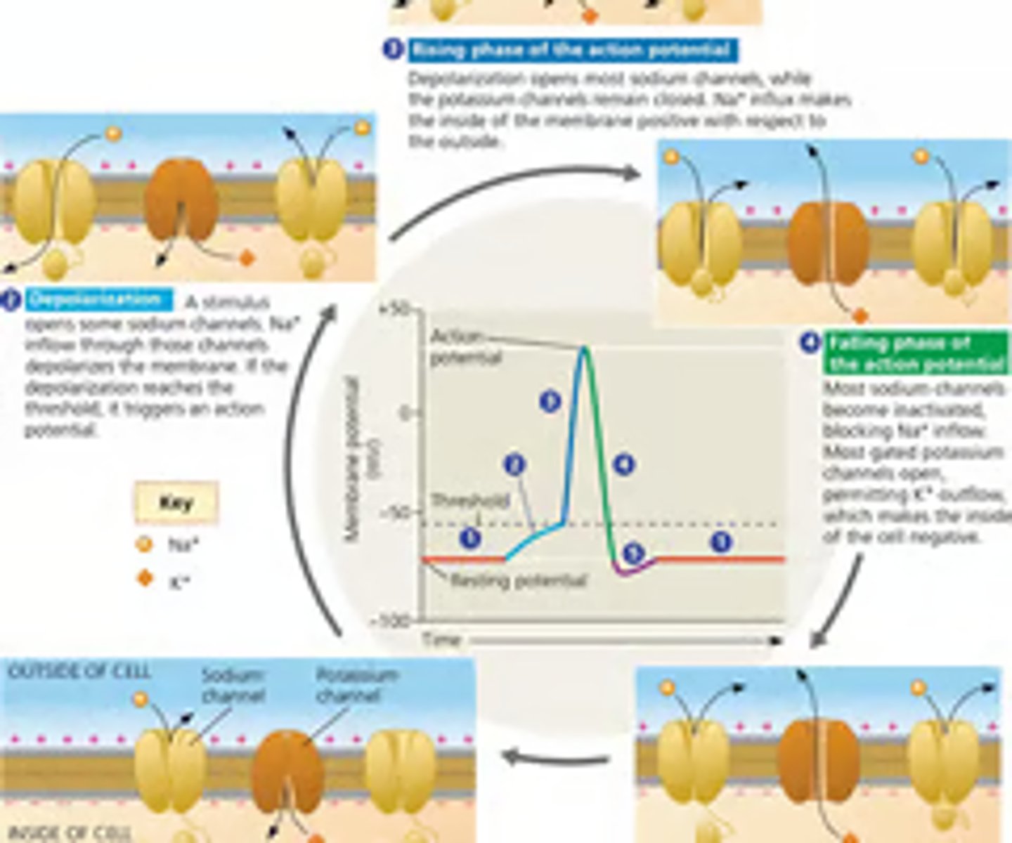
Explain the role of voltage-gated ion channels in nerve signal conduction
Resting Potential:
At rest, most voltage-gated sodium (Na⁺) channels are closed, while some potassium (K⁺) channels remain open, maintaining the resting membrane potential.
Depolarization:
A stimulus causes some Na⁺ channels to open, allowing Na⁺ to enter the cell, leading to depolarization. If the stimulus is strong enough, more Na⁺ channels open, further depolarizing the membrane.
Rising Phase:
Once the threshold is crossed, a positive-feedback loop rapidly increases Na⁺ inflow, generating an action potential.
Falling Phase:
Na⁺ channels inactivate, and K⁺ channels open, allowing K⁺ to exit the cell, repolarizing the membrane.
Undershoot:
The membrane potential temporarily becomes more negative than the resting potential before returning to normal.
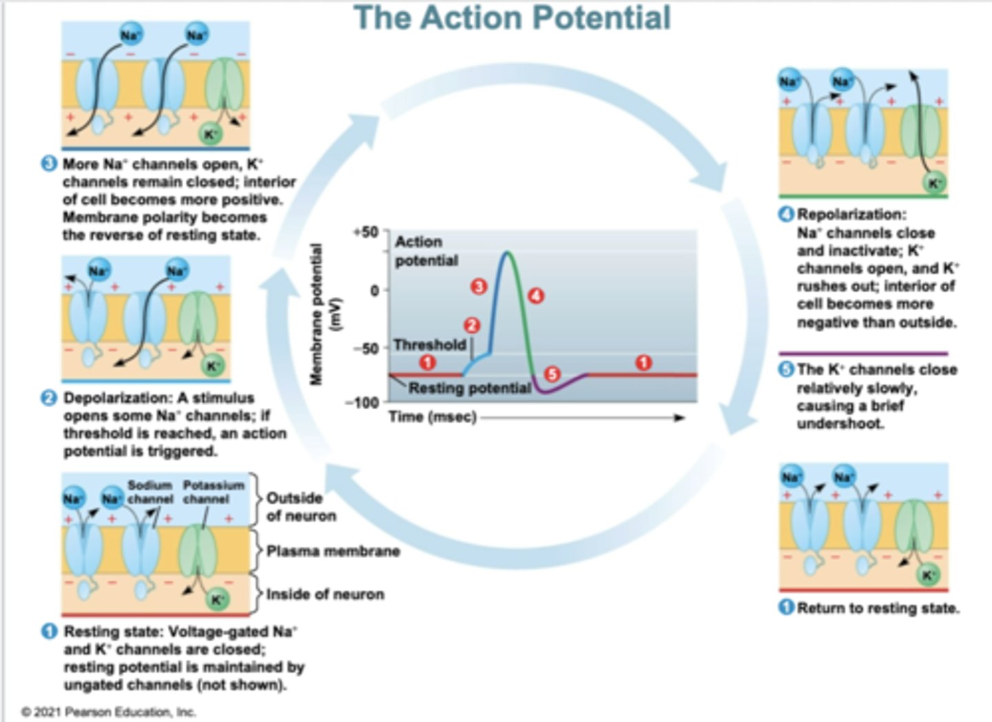
Sequence the process of action potential generation, including describing how membrane potential changes and how ions move during different phases of the membrane potential
Resting State:
The neuron is at rest with a membrane potential of about -70 mV. Voltage-gated sodium (Na⁺) and potassium (K⁺) channels are closed, maintaining this potential.
Depolarization:
A stimulus opens some Na⁺ channels, allowing Na⁺ to enter the cell. This inflow depolarizes the membrane, reaching a threshold of -55 mV, triggering an action potential.
Rising Phase:
Most Na⁺ channels open, causing a rapid influx of Na⁺, making the inside of the membrane positive, peaking at about +35 mV.
Falling Phase:
Na⁺ channels inactivate, and K⁺ channels open, allowing K⁺ to exit the cell, repolarizing the membrane back to resting potential.
Undershoot:
K⁺ channels remain open longer, causing the membrane potential to dip below resting potential before stabilizing.
Interpret a graph of membrane potential (at rest, during graded potential(s), and during an action potential)
Graph Attached