General Anatomy & Physiology (Online, Pelletier): Exam 4 (Final)
1/28
There's no tags or description
Looks like no tags are added yet.
Name | Mastery | Learn | Test | Matching | Spaced |
|---|
No study sessions yet.
29 Terms
What gross structures are part of the upper respiratory tract, and which of the lower respiratory tract?
Upper respiratory tract: Nasal cavity, pharynx, larynx
Lower respiratory tract: Trachea, main bronchi, lungs and alveoli within
What are the overall functions of the upper and lower respiratory tracts?
Upper: air passageways
Lower: Passageways for air but also purify, humidify, and warm air
What is special about these structures of the nasal cavities: the mucosa, structural adaptations, and paranasal sinuses?
Mucosa: The mucosa of the nasal cavity contain olfactory receptors, and produce mucus that moisturizes, traps and neutralizes bacteria, and is ciliated to move mucus
Structure: Has conchae formed by raised nasal meatuses that increase the SA and air turbulence to trap more particles
Paranasal sinuses: Include the frontal, sphenoidal, ethmoid, and maxillary sinuses; serve to lighten the skull, produce mucus, and are resonance chambers for speech
What are these structures within the pharynx: posterior nasal aperture, regions of the pharynx, pharyngotympanic tubes, and tonsils?
Posterior nasal aperture: connects the nasal cavity and the nasopharynx
Regions: Nasopharynx, oropharynx, and laryngopharynx
Pharyngotympanic tubes: drain inner ear fluid into the pharynx
Tonsils: include the pharyngeal, palatine, lingual, and tubal tonsils; clusters of lymphatic tissue that are protection from infections
What is the larynx, and what are these structures of the larynx: epiglottis, thyroid cartilage, vocal cords, vestibular folds, and glottis?
Larynx: separates air and food passages and helps in the formation of speech
Epiglottis: cartilaginous flap covering the glottis that separates the trachea and esophagus
Thyroid cartilage: also known as the Adam’s apple
Vocal folds/cords: inner mucous membrane folds of the glottis that produce speech through vibration and air flow
Vestibular folds: outer mucous membrane folds that support the vocal cords
Glottis: the passage between the vestibular and vocal folds
What is the trachea and these structures of the trachea: cartilaginous rings, carina, trachealis muscle, cells that function to move debris away from lungs?
Trachea: the windpipe anterior to the esophagus
Cartilaginous rings: C-shaped rings that support the trachea and keep it open but are open posteriorly to allow for esophageal expansion
Carina: the last, key-shaped tracheal cartilage before the bronchi
Trachealis muscle: lies between the trachea and esophagus; constriction allows for tracheal constriction important in coughing
Cells for debris removal: Pseudostratified ciliated columnar epithelium and goblet cells produce respiratory mucus
What are the main bronchi, and what are the differences between the right and left main bronchi?
Main bronchi: the two divisions of the trachea
The right main bronchus is wider, shorter, and more horizontal than the left main bronchus (more common for trapping objects)
How many orders of branching are there of the bronchi? What is the function of all this branching?
23 orders of branching
Functions to increase SA for greater respiration
What are these structures of the lungs: apex, base, lobes (and how many), coverings (pulmonary/visceral pleura, parietal pleura, pleural space/cavity, pleural fluid)?
Apex: top point just deep to clavicle
Base: bottom that rests on diaphragm
Lobes: Divided by fissures; Left has 2 lobes and is smaller because of cardiac notch, right has 3 lobes
Pulmonary/Visceral pleura: visceral serosa that covers the surface of the lung
Parietal pleura: visceral serosa that lines the walls of the thoracic cavity
Pleural space/cavity: very tight space between the pulmonary and parietal pleura filled with pleural fluid
Pleural fluid: serous fluid that allows the lungs to harmlessly brush against the thoracic cavity walls without injury
Name the basic construction of the bronchial/respiratory tree
Primary bronchi → secondary/lobar bronchi → tertiary/segmental bronchi → bronchioles → terminal bronchioles
What structural changes occur in the transition between the trachea and the bronchi?
Cartilaginous rings become elastic fibers
Pseudostratified columnar cells become cuboidal cells
Capillaries wrap around alveolar sacs
Smooth muscle increases
23 orders of bronchial branching
What is the difference between the respiratory and conducting zones?
Respiratory zone: Sites of gas exchange; includes the respiratory bronchioles, alveolar ducts, alveolar sacs, and alveoli
Conducting zone: all other respiratory structures that are air passages
What are the alveoli and what is the stroma?
Alveoli: gas-filled air spaces (the lungs are mostly air spaces)
Stroma: lung tissue; mostly elastic connective tissue for expansion during breathing
What is the air-blood barrier?
In the respiratory membrane; site of gas exchange/respiration
Composed of alveoli and capillaries between
Alveoli exchange their O2 with the CO2 from deoxygenated blood in the capillaries
What are these structures between alveoli: Type II or surfactant secreting cells, Type I alveolar cells, and alveolar macrophages?
Type II cells or surfactant secreting cells: secretes lipid molecules that coat gas-exposed alveolar surfaces; moisturizer
Type I alveolar cells: simple squamous epithelium
Alveolar macrophages: pick up debris, carbon particles, bacteria, etc.
What are the 4 steps of respiration, and which do the respiratory system and the circulatory system do?
Respiratory system:
Pulmonary ventilation: physical process of breathing
External respiration: gas exchange
Circulatory system:
Transport of O2 and CO2 through blood
Internal respiration: occurs in body tissues → carbon dioxide taken away and tissue is oxygenized
What are the 2 types of mucosa present in the nasal cavity?
Olfactory mucosa: pseudostratified columnar cells, contain sensory nerve endings involved that trigger the sneeze reflex, Bowman’s gland produces mucus that absorbs chemicals related to smell
Respiratory mucosa: pseudostratified ciliated columnar and goblet cells to move mucus and debris, contains plexus of capillaries and veins that warms are
What are the overall functions of the urinary system?
Filter and excrete waste, regulate blood volume and acid-base balance, regulate BP through renin, stimulate RBC production, activate Vit D
What are the gross structures of the urinary system?
Kidneys, ureters, urinary bladder, urethra
What are these structures of the kidneys: renal hilum, adrenal gland, 3 layers (fibrous capsule, perirenal fat capsule, renal fascia),
Regions seen in longitudinal section of a kidney (renal cortex, renal medulla, renal pyramids and columns, renal pelvis and calyces)?
Renal hilum: indentation where the ureters, blood vessels, and nerves enter/exit the kidneys
Adrenal glands: sit on top of each kidney
3 layers—
Fibrous capsule: superficial and directly covers kidney, collage elastic fibers, prevents spread of infection
Perirenal fat capsule: deep to kidney, adipose tissue, cushions kidney from damage
Renal fascia: surrounds but doesn’t directly cover kidney, dense fibrous connective tissue, anchors kidneys in place
Regions—
Renal cortex: superficial, mainly contains nephrons involved in urine filtration/production
Renal medulla: middle layer composed of renal pyramids and columns, and collection ducts from nephrons
Renal pyramids: composed of papilla and separated by renal columns
Renal columns: sections of renal medulla that separate renal pyramids
Renal pelvis: central basin that renal calyces empty into
Renal calyces: minor calyces drain urine from pyramids into the major calyces, which drain into the renal pelvis
What two types of nephrons are there?
Cortical nephron: within renal cortex
Juxtamedullary nephron: within cortex-medulla junction
Name the main parts of the nephron
Renal corpuscle: Glomerulus and glomerular capsule
Renal tubule: proximal convoluted tubule, nephron loop, distal convoluted tubule
Collecting ducts
Describe the structure of the glomerulus and glomerular capsule
Glomerulus: knot of capillaries; fenestrated: holed for blood passage and for pushing out filtrate
Glomerular capsule:
Parietal layer: simple squamous epithelium
Visceral layer: contain podocytes (specialized branching epithelial cells that cling to capillaries, foot processes block RBCs and proteins from leaking from the capillaries)
What is the structure of the renal tubule?
Single layer of cells; contains 3 parts
What is the structure of the proximal convoluted tubule?
Closest to glomerular capsule, single layer of cuboidal cells, contain the most microvilli of any region (forms a brush border for specialized reabsorption and secretion) and large mitochondria
Resorbs most of the filtrate volume
What is the structure of the nephron loop?
Contains a descending limb made of simple squamous epithelium and ascending limp of cuboidal or columnar epithelium
Pulls out Na and attracts H2O
What is the structure of the distal convoluted tubule?
Cuboidal cells with few microvilli; specialized in secretion and less reabsorption
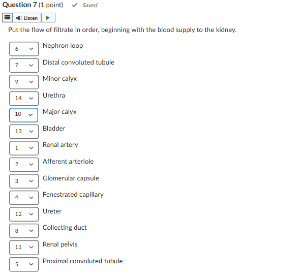
Trace filtrate through the urinary system, starting with blood flow to the kidneys