Neurons and Membrane Potentials
1/31
There's no tags or description
Looks like no tags are added yet.
Name | Mastery | Learn | Test | Matching | Spaced | Call with Kai |
|---|
No analytics yet
Send a link to your students to track their progress
32 Terms
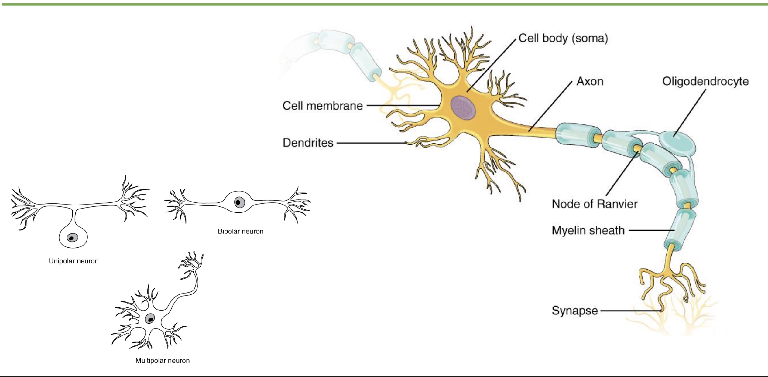
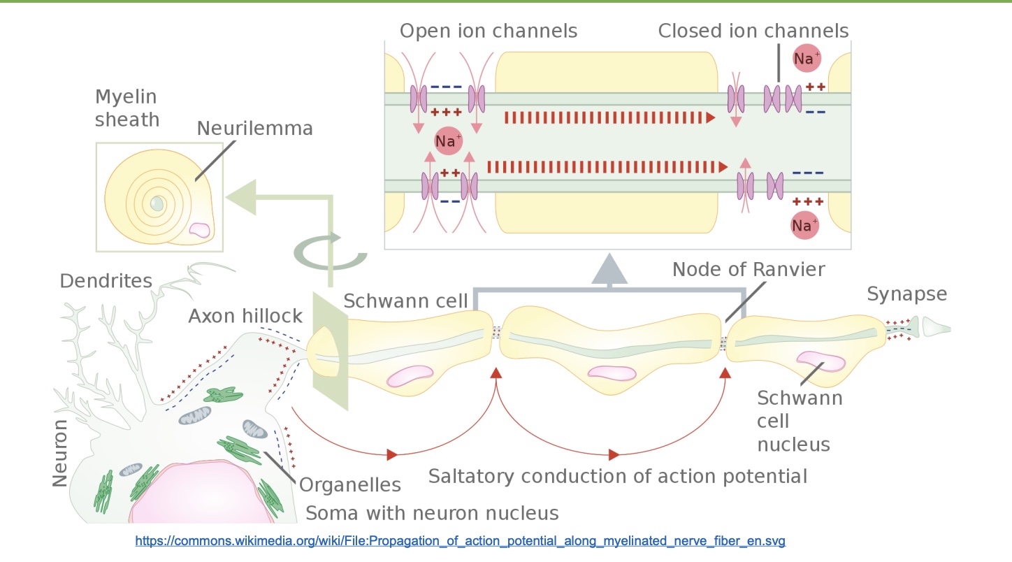
structure of a neuron
-dendrites: receive signals
-cell body: large in photo, realistically smaller?
-axons: sends out signal
-Myelin sheet
-Oligodendrocytes: separate cell that forms myelin, can do multiple segments
-Nodes of Ranvier: AP jumps node to node, so makes it faster
-Synapse
*+ just know there are dif types of neurons
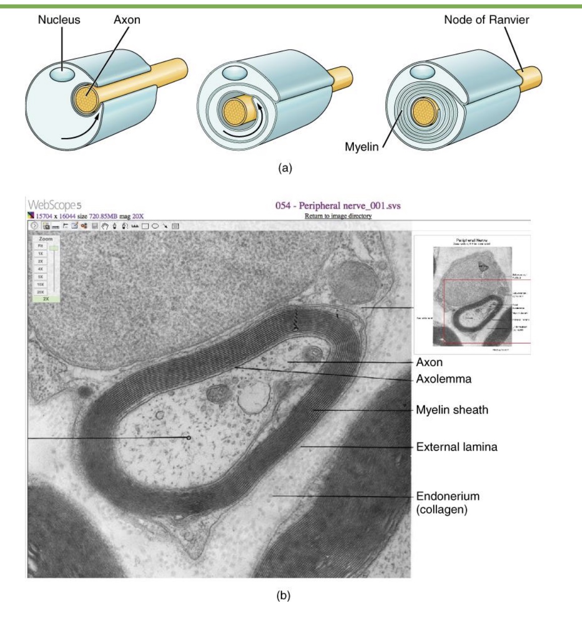
myelination
-electrical insulator, max AP travel faster down axon (increase myelin=increase speed)
-unmyleinated axons=lower priority; bc myelin takes up space, so not everything myleinated
ex: NS
-concentric layers
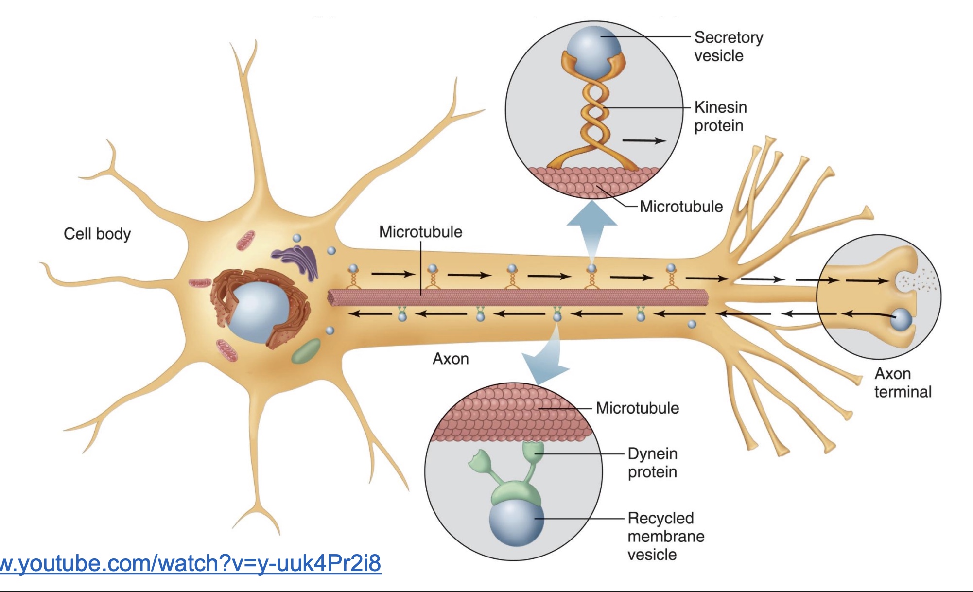
axonal transport
-microtubules: chains of proteins, what motor proteins move on
-Motor proteins: walk/carry things up and down the axon microtubules, uses ATP to move
Kinesin protien: goes towards terminal
Dynein protein: retrograde axonal transport (back to cell body)
functional classification of neurons
-Sensory neurons: Afferent neurons of PNS; Sensory information ascends to the CNS
-Motor neurons: efferent
*ex: motor neurons carry signals to the effector neuron in feedback loops
-Interneurons: communication within CNS and between sensory/motor (*all other types of neurons)
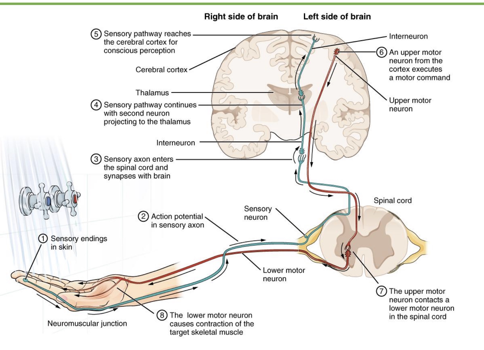
Picture
-1st synapse in spinal cord; reflexes in SC=touch boiling water; reflexes in brain=touch somewhat hot water
-2nd synapse when peripheral sensory neuron synapses w/ interneuron
-Thalamus: process sensory signals and send to right place
-Cerebral cortex: conscious awareness
-Motor neurons originate in the motor cortex
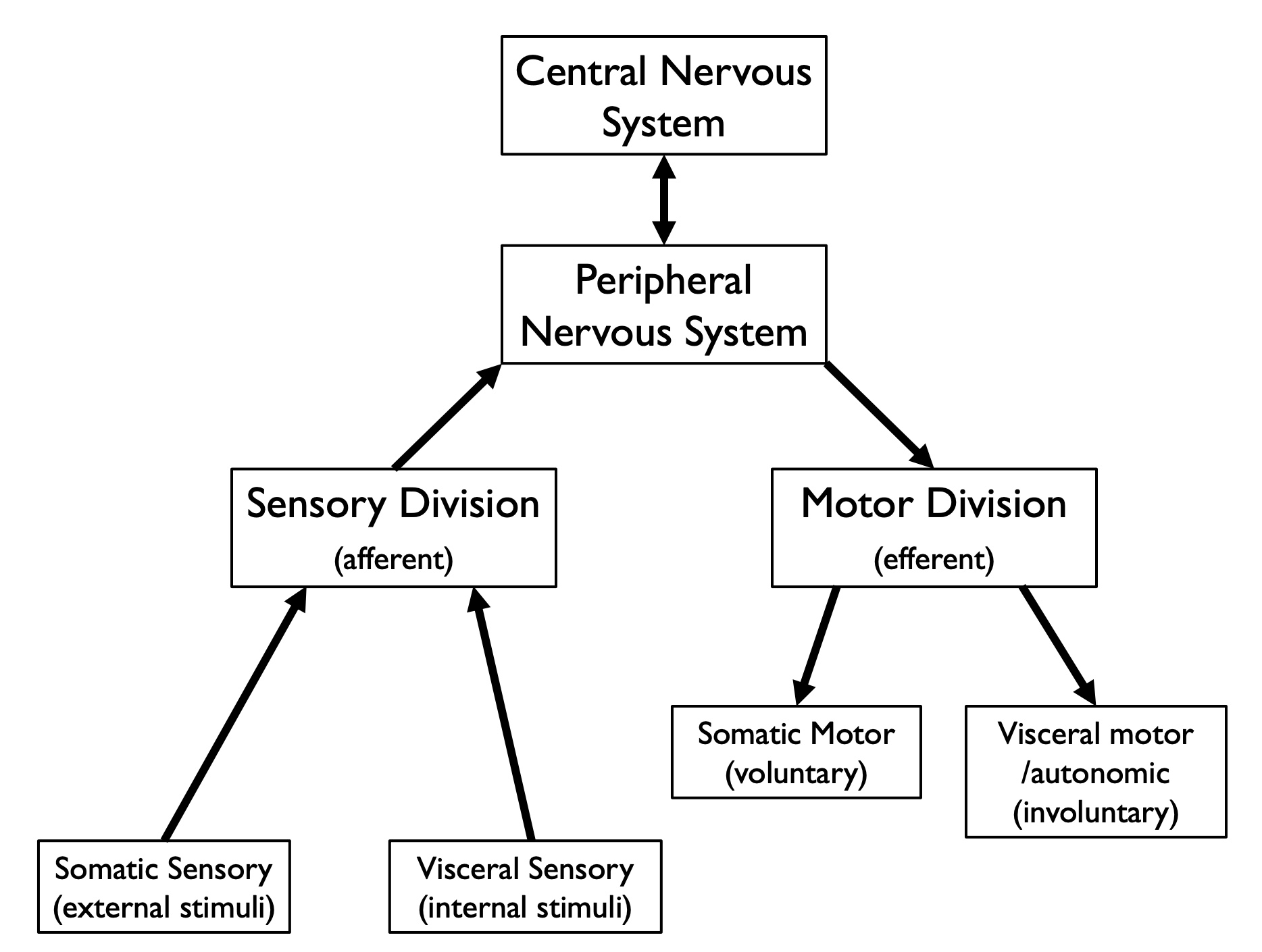
how the CNS and PNS communicate
Sensory
-Somatic: external stimuli, conscious awareness; ex=touch
-Visceral: internal stimuli, not conscious awareness; ex=neurons for BP and HR
Motor
-Somatic: voluntary; ex=skeletal muscle
-Visceral/autonomic: involuntary, not aware of/in the background; ex=CV function (BP, HR), digestive function, etc.
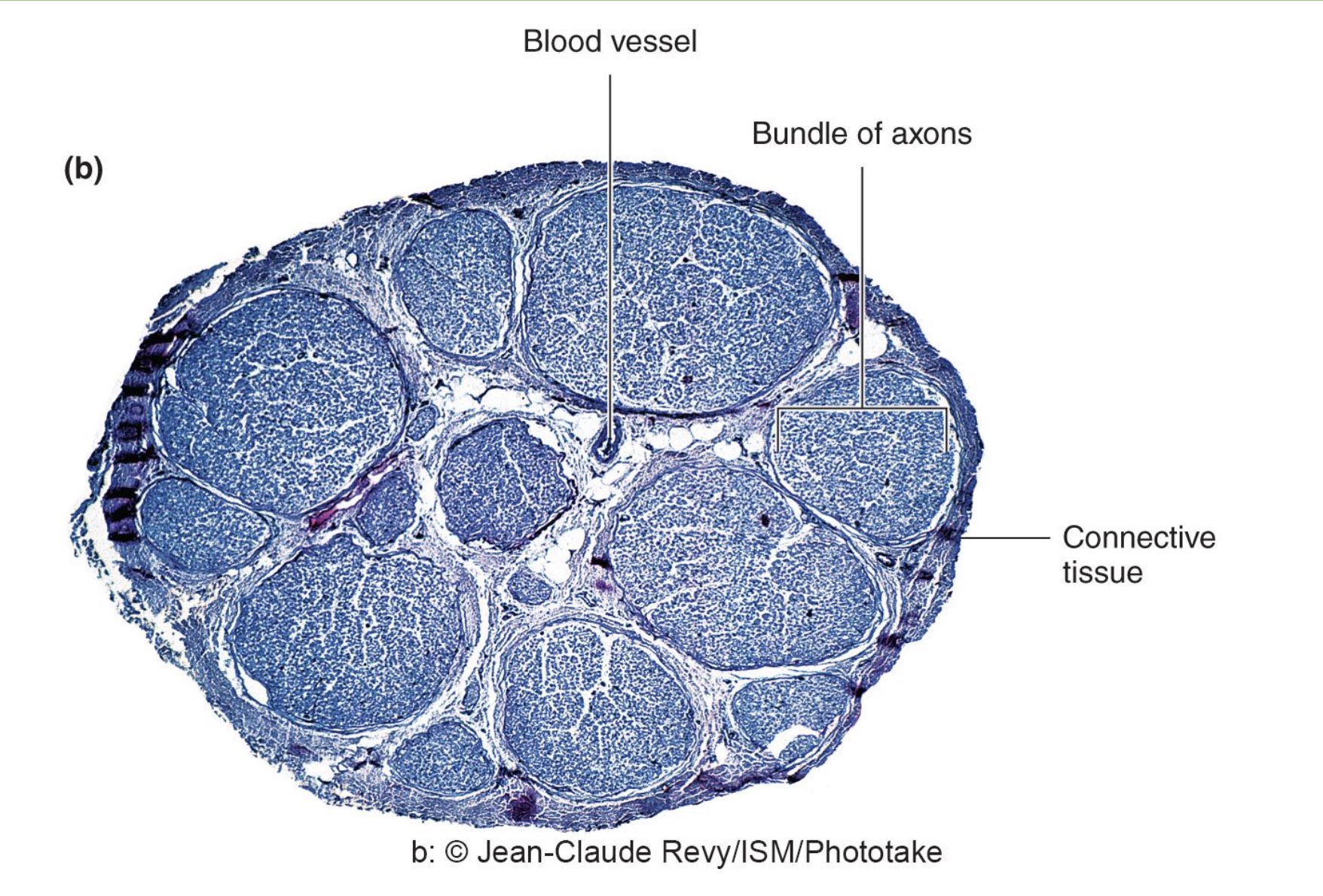
neurons versus nerves
-neuron: single nerve cells
-nerve: bundle of many neuron axons; has all 4 types of tissues; blood vessel provides nutrients; can carry multiple types of info (ex: sensory and motor)
*picture=nerve
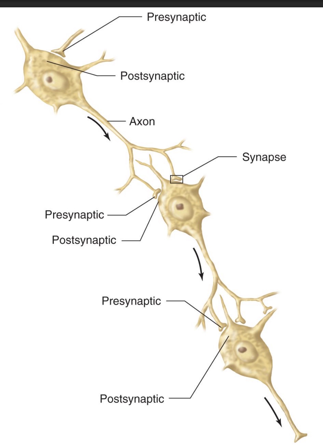
synapses
-At most synapses, a chemical substance (ex: NT) released from the presynaptic terminal causes a response in the postsynaptic neuron (or target organ)
*communication between neurons OR neurons and target organs
-Synapses can be excitatory or inhibitory
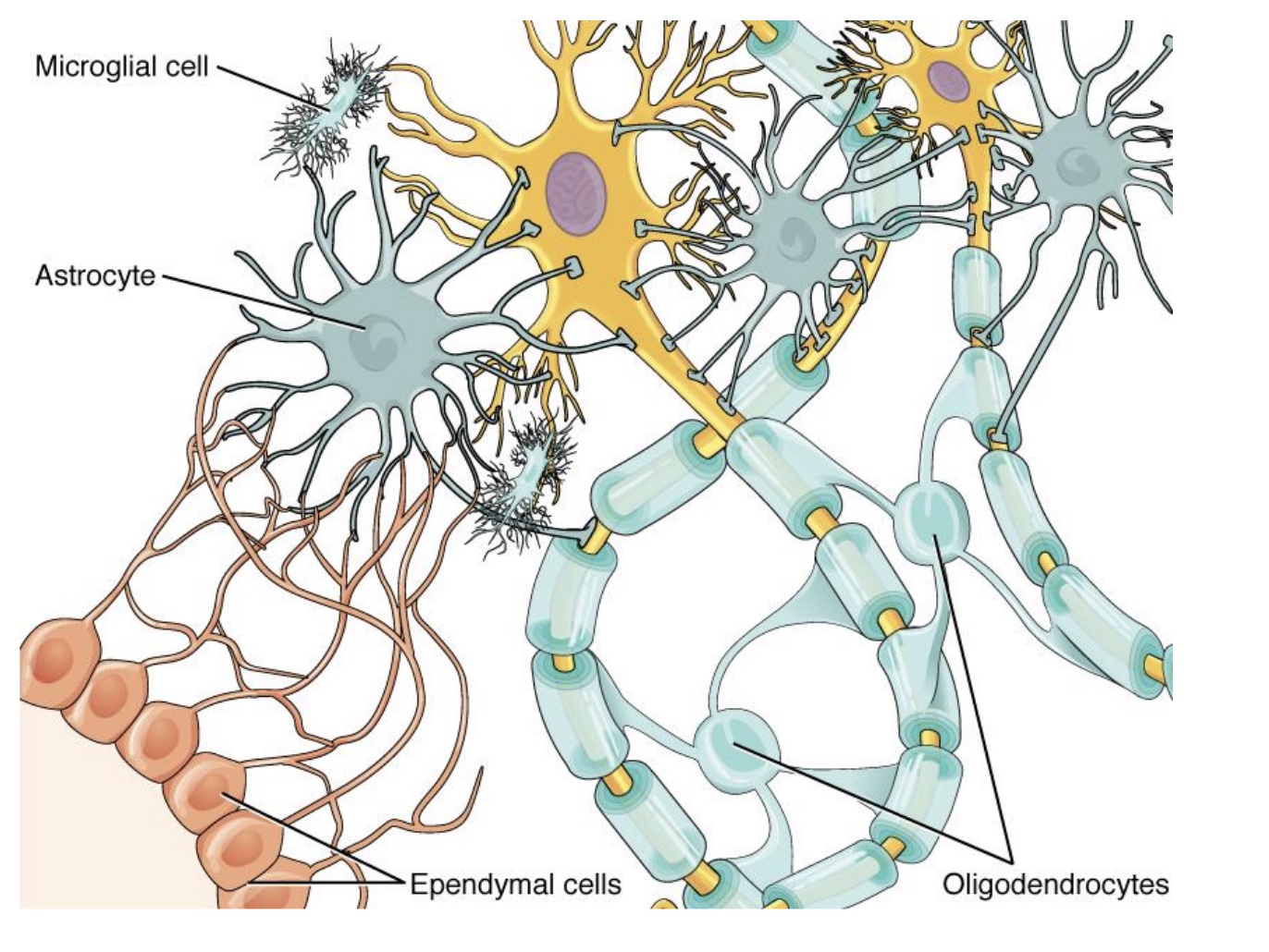
4 types of Glial Cells in the CNS
-Astrocytes: support cells, control extracellular environment of neurons (*lot of CNS structure; ex: regulate NT levels)
-Microglia: “immune system” of the CNS (*usually not much immune in CNS otherwise; looks for pathogen or disease and can clean up damage)
-Ependymal cells: ciliated, involved with production of CSF and CSF movement (*surround ventricles in brain)
-Oligodendrocytes: responsible for myelination of axons
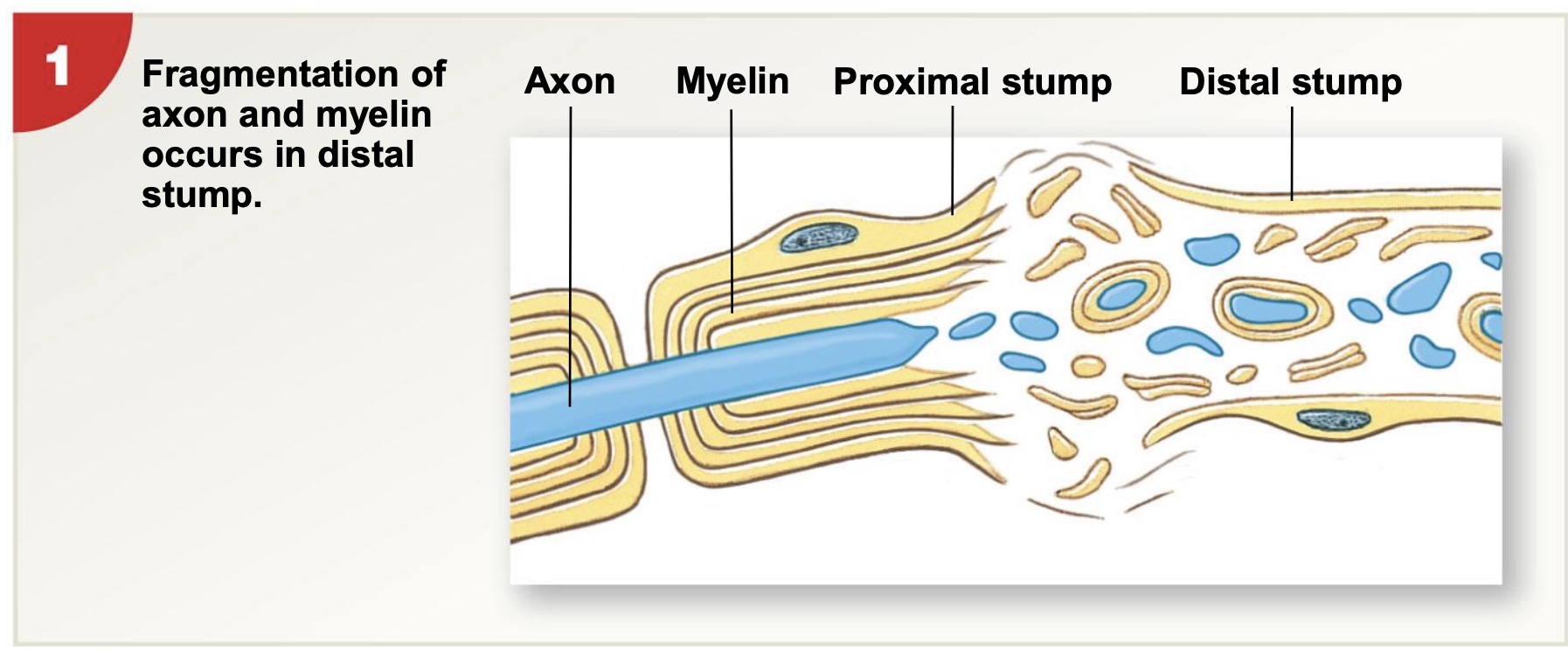
Peripheral nerve regeneration after injury
-PNS regeneration can occur, if the axons finds its target
-nerve and muscle in general hard to heal; epithelium easier (ex: get cut on skin)
1) fragmentation of axon and myelin occurs in distal stump (cuts it off and dies, left with mess; now cut how does it find its target again?)
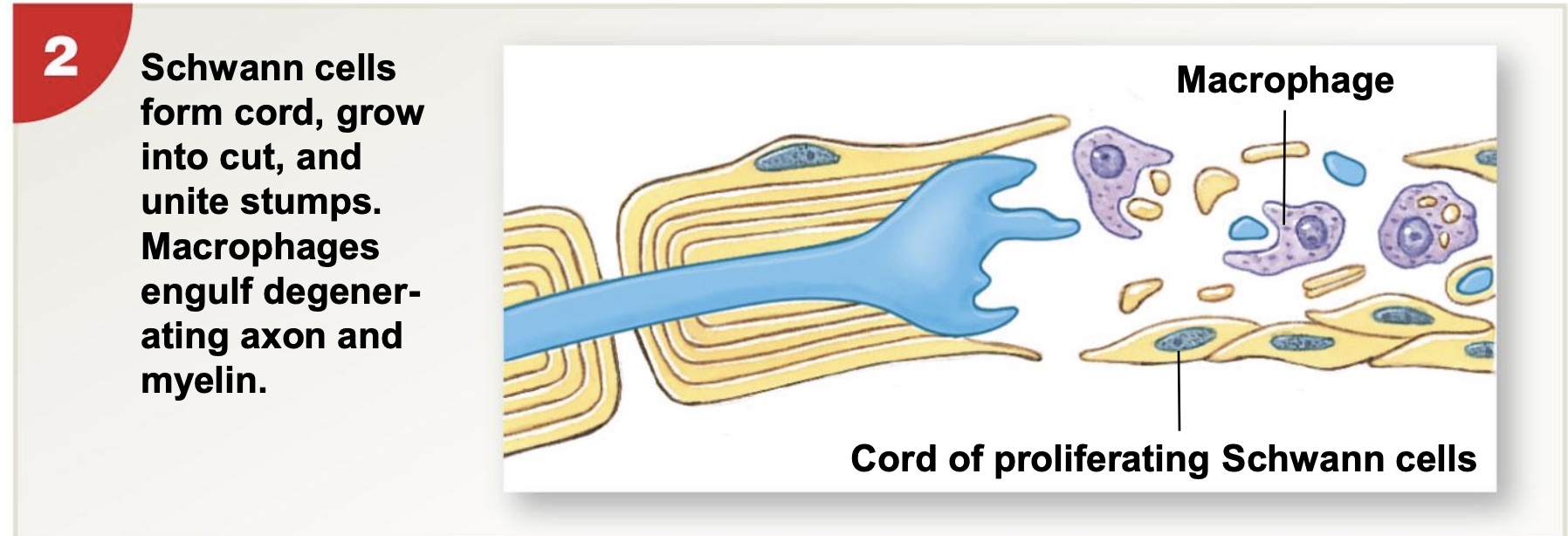
2) Schwann cells form cord, grow into cut, and unite stumps. Macrophages engulf degenerating axons and myelin
3) axon sends buds into network of Schwann cells and then starts growing along cord of Schwann cells
4) axons continues to grow into distal stump and is enclosed by Schwann cells
Schwann cells
supporting cells that form myelin for PNS axons; can form regeneration pathway for cut off distal stump
CNS after injury
-CNS neurons are extremely limited in ability to regenerate
*ex: brain damage, but get some plasticity so other parts may take over
Development of the Nervous System
-Early in development (fetal→child), the brain has much greater potential for remodeling in response to stimulation or injury than in the adult brain, a characteristic known as plasticity.
*can remodel/reshape; better if younger
-The basic shapes and locations of major neuronal circuits in the mature central nervous system do not change once formed
-The creation and removal of synaptic contacts begun during fetal development continue, however at a slow pace throughout life as part of normal growth, learning, and aging
*ex: hippocampus (for learning and memory) overall structure the same, but nerve connections changing all the time and can grow some more
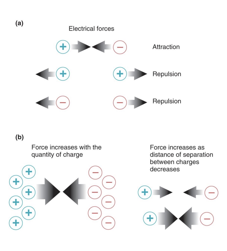
basic principles of electricity
-opposite attract; like repel
-force increases with the quantity of charge
-force increases as get closer together (as distance of seperation between charges decreases)
Resting Membrane Potentail
-overall negative charge inside cell (~ -70), positive charge outside cell
grouping of positive charge outside cell, and grouping of negative charge inside cells; seperated by membrane but attracted to each other
-Na+ higher outside cell (~145); K+ higher inside cell (~15)
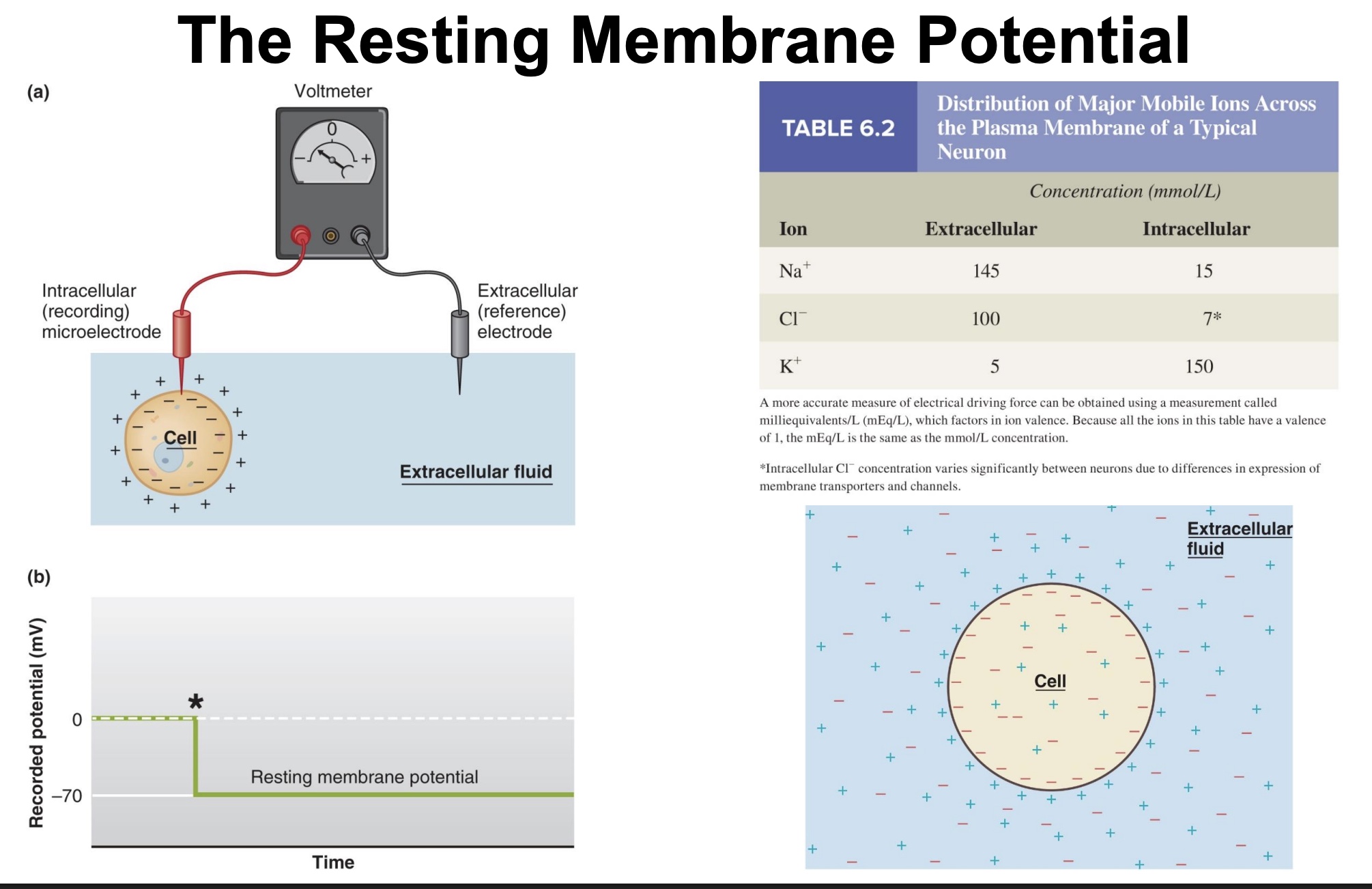
Resting Membrane Potential: major ions concentrations
-Major ions: Na+ and K+
Na+ : 10x higher concentration outside cell compared to inside
K+ : 30x higher concentration inside cell compared to outside
RMP + electrochemical gradient
-Electrochemical gradient: sum of electrical forces AND (chemical/diffusion) concentration gradient
-Na+: chemical gradient in + smaller electrical gradient in= Na+ electrochemical gradient in
-K+: big chemical gradient out + smaller electrical gradient in= K+ electrochemical gradient out

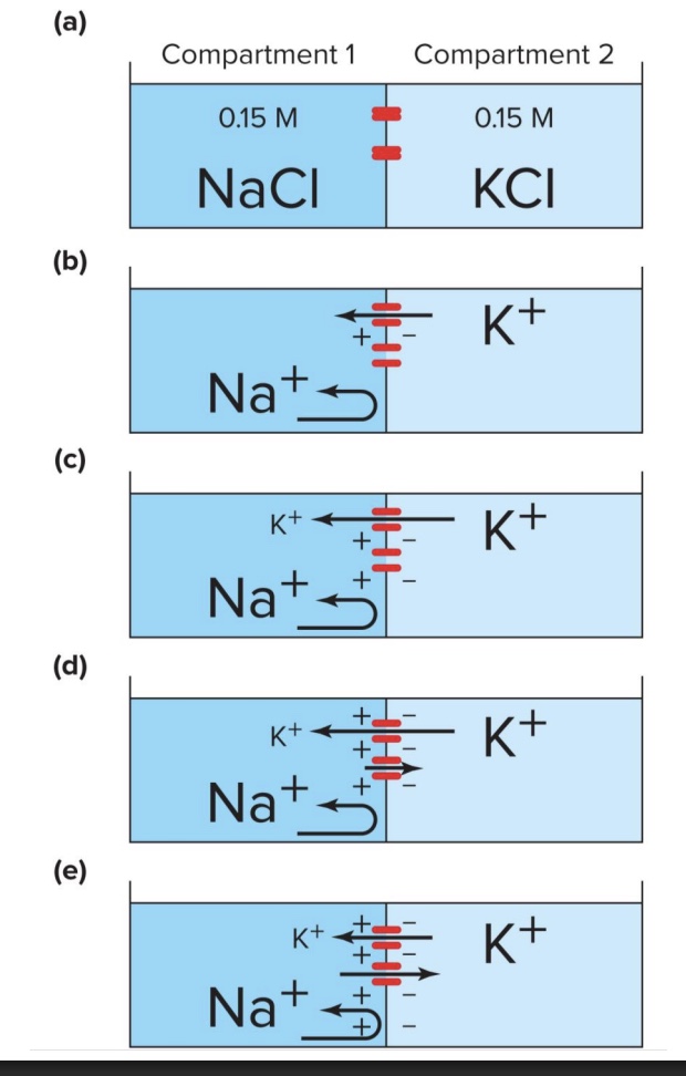
the RMP exists because…
-Concentration gradients intracellular-extracellular
-Selective permeability of membrane (more permeable to K+)
-Sodium-potassium ATPase (establishes gradients and moves more (+) charge out of cell)
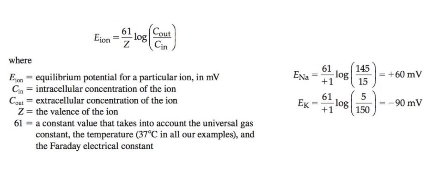
Nernst Equation
*don’t have to calculate, but know exist
-tells us equilibrium potential based on ion concentration inside/outside the cell
-At equilibrium potential, electrical and chemical gradients are balanced so that there is no net movement of an ion across the membrane
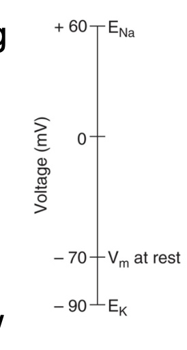
Equilibrium potentials and resting membrane potentials
-K+ is trying to leave the cell and make it more negative (trying to bring cell to its equilibrium potential of -90 mV)
-Na+ is trying to enter the cell and make it more positive (trying to bring cell to its equilibrium potential of +60 mV)
-Actual resting potential of a cell is about -70 mV
-Why not right in the middle? Permeability differences (closer to K+ bc more permeable to K+)
-Because the membrane has many open K+ channels and few for Na+ , the resting potential ends up much closer to the equilibrium potential of K+
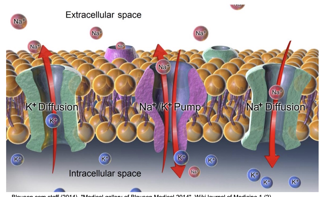
Summary of membrane potential development
-Movement of ions at resting potential:
Leak channels: K+ leaks out, Na+ leaks in (to a much lesser degree)
K+ is pumped in, Na+ pumped out by Na+ /K+ ATPase. This maintains electrical and concentration gradients
*at rest to maintain negative charge
*K+ positive charge constant diffuse out, so inside of cell is negative (also have negative proteins inside the cell)
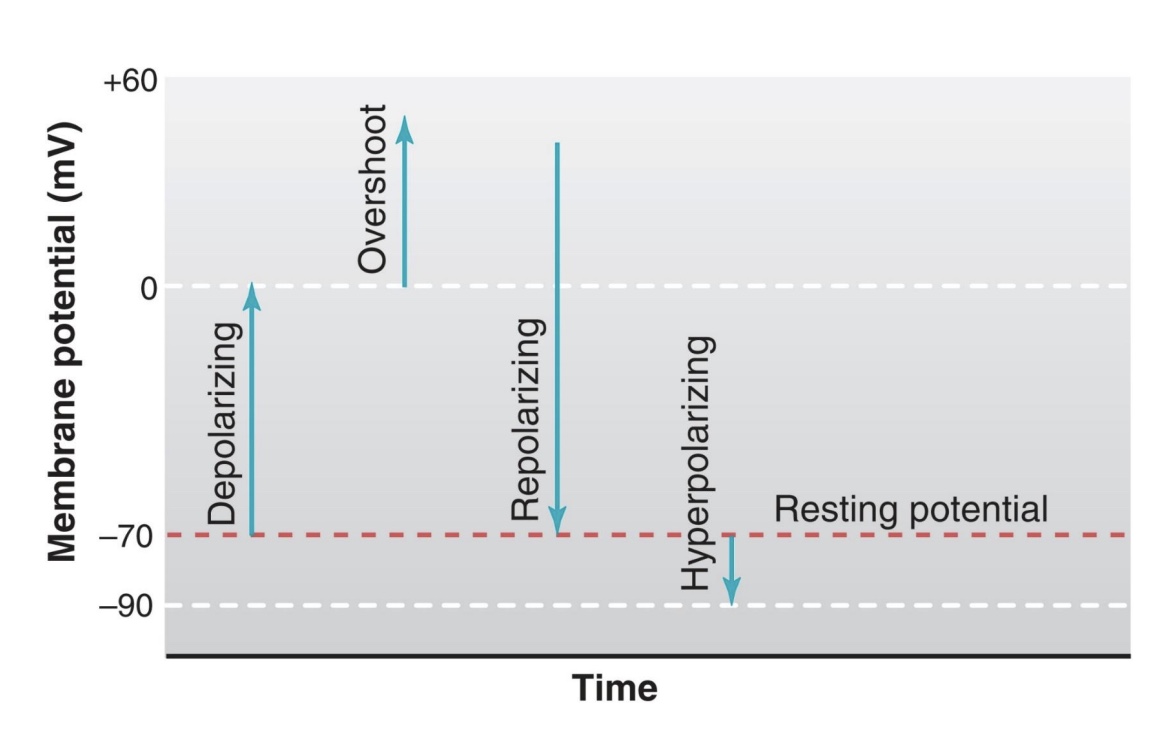
RMP terminology:
-deplorizating
-repolorizing
-hyperpolarizing
-deplorizating: move in positive direction
-repolorizing: move in negative direction and towards RMP
-hyperpolarizing: below RMP
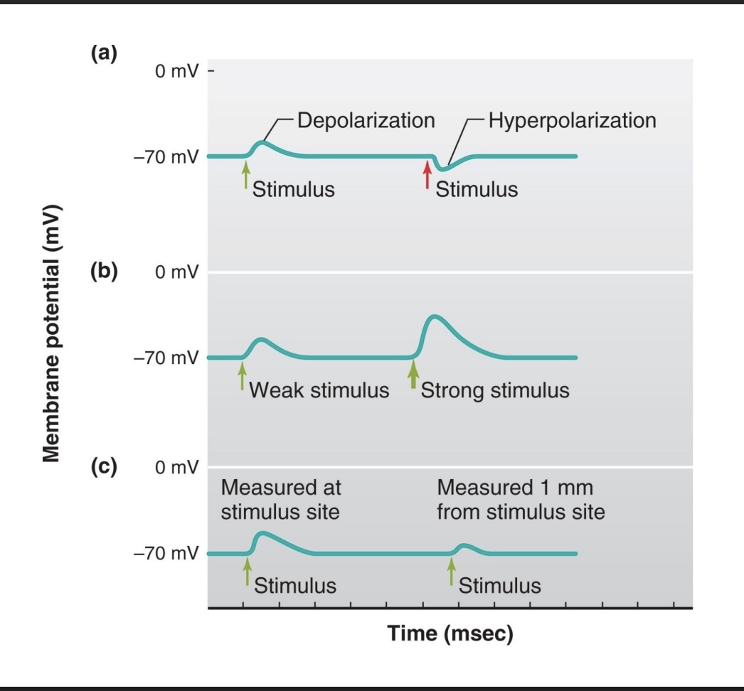
graded potentials
-Changes in membrane potential affecting a local/small area
-Called graded because magnitude can vary
-Signal decreases as distance from stimulus increases
-Can be in depolarizing or hyperpolarizing direction
*graded potentials lead to an AP
factors to change the RMP/size of graded potentials
-depolarization vs hyperpolarization
-weak vs strong stimulus (ex: increase # NT)
-how close the stimulus is to site (stronger at stimulus site)
Action potentials
-Large, all-or-none depolarizations that spread throughout cell
*all look the same throughout the cell
-Very rapid, may repeat at frequencies of several hundred per second
-The ability to generate action potentials is known as excitability. This ability is possessed by neurons, muscle cells and some other types of cells.
Ion channels involved in AP
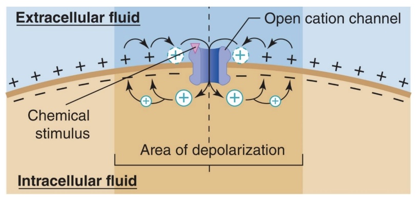
-Ligand-gated channels and mechanically gated channels often serve as the initial stimulus for an action potential
mechanically gated: mechanical force on protein opens it; ex=touch receptors
-Action potential can result from multiple graded depolarizations within a short period of time
*can summate to reach threshold and get all or nothing
-Action potentials spread when voltage-gated channels open
Voltage-gated Na+ channels depolarize the cell
Voltage-gated K+ channels repolarize the cell
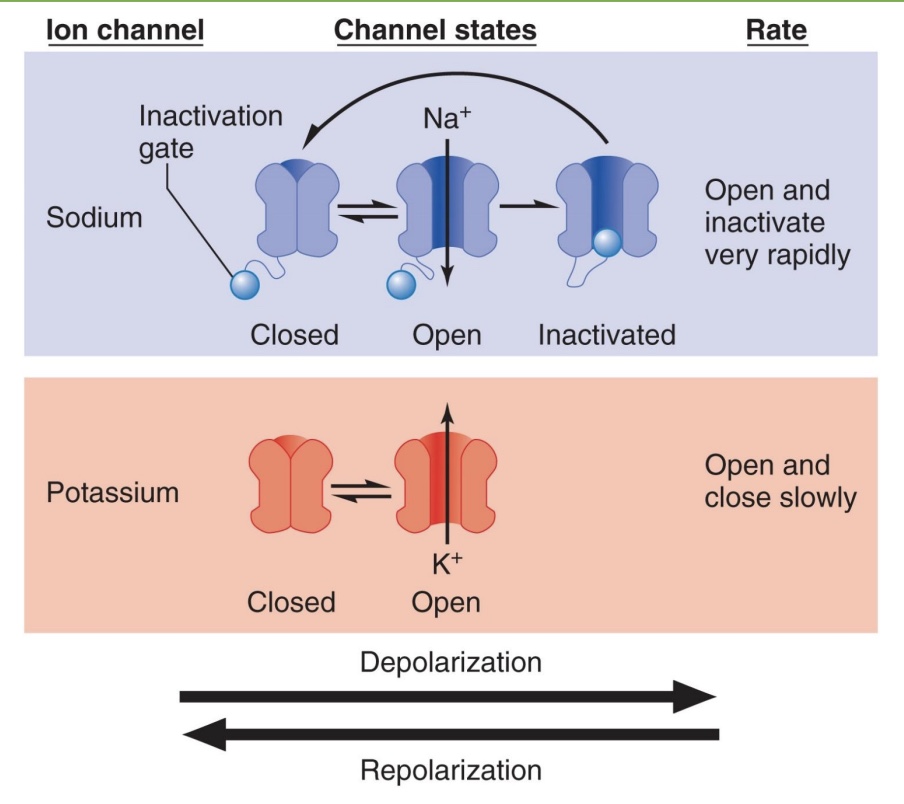
Na+ and K+ channels when closed, open, or inactivated
*voltage causes open or close
Sodium
-closed at resting state (ball out of channel); makes sure AP stay organized
-ball comes out to open channel
-inactivated: ball blocks pore so ions can’t get through; changes bc sensitive to electric current; need to repolarize cell to reset
Potassium
-closed at resting state
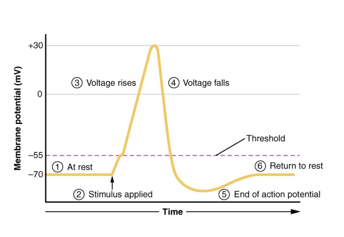
stages of an AP
1. At rest: voltage-gated Na+ and K+ channels closed, leak channels open
2. Stimulus applied: Na+ voltage-gated channels open
3. Voltage rises: depolarize, Na+ in
-at peak/+30: Na+ channels close and voltage-gated K+ channels open
4. Voltage falls: replorize, K+ out
5. Hyper polarize: overshoot below RMP, bc K+ slow to close
6. Return to rest
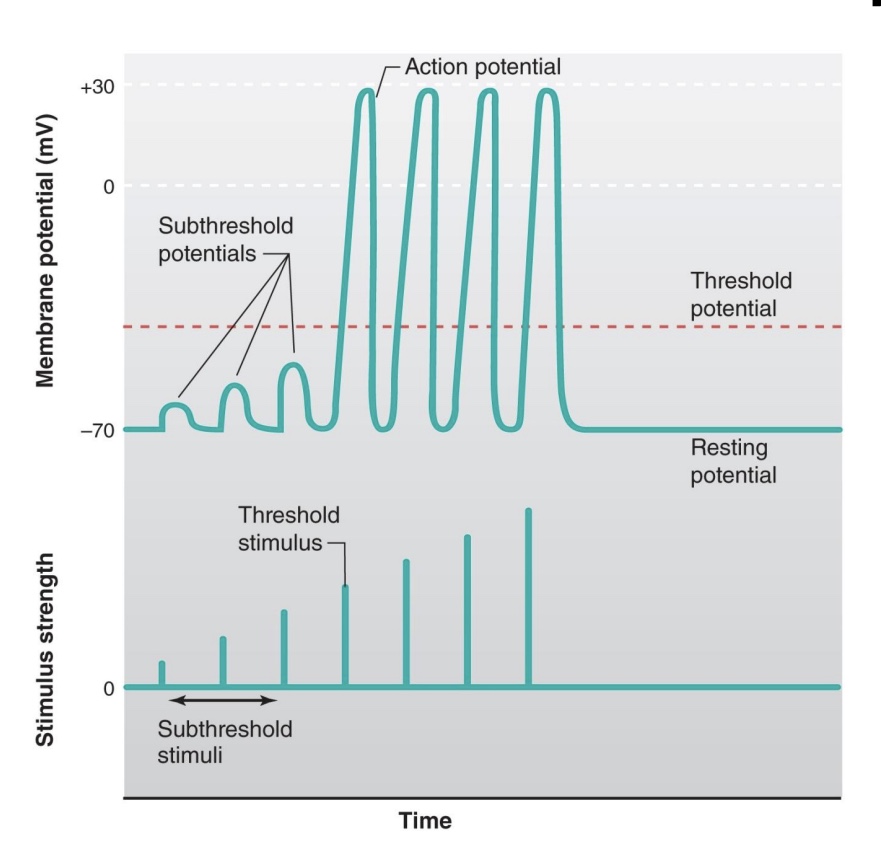
Threshold and the All-or-noe Principle
-increase stimulus=increase graded potentials
-once reach threshold, get identical AP (same heigh and duration)
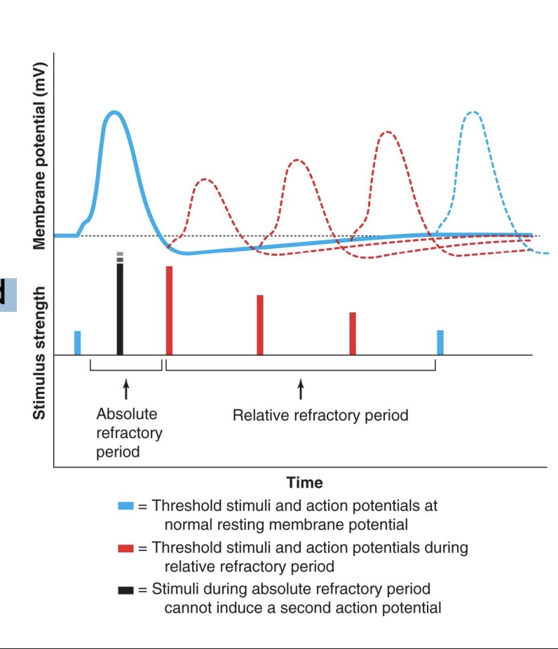
Refractory Period (absolute vs relative)
-Absolute: cell is incapable of depolarizing
*can’t stack on another AP if one AP already going
-Relative: cell can depolarize if supplied with a stronger-than-usual stimulus
*weaker AP, one time AP may look little dif (bc not all Na+ channels avaialbe)
-Helps maintain organization of signals—keeps each action potential as an individual signal
*if not=seizure or heart arrhythmia (heart also has all or nothing so can contract and relax)
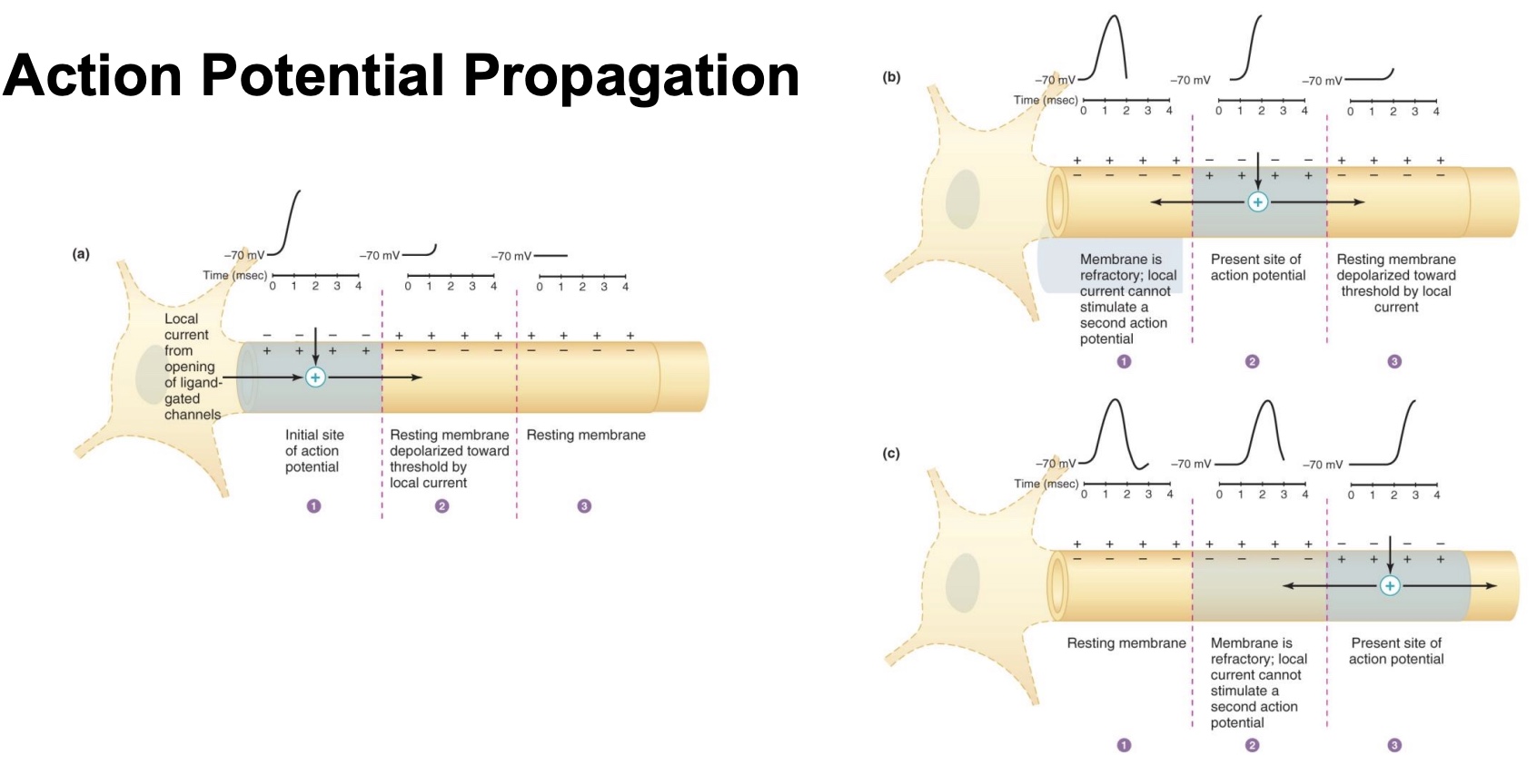
AP propagation
-movement of AP down the axon in one-direction
-starts at axon hillox, spreads to distal end
-refractory period so AP can’t move backwards
Saltatory Conduction
-myelin on most of axon but have nodes so the signal can jump node to node, which makes it faster
*similar to passing vs throwing something
*everything not myelinated, so higher priority things are
Key Points: AP propagation (direction + how faster + velocities)
-Action potentials in neurons are unidirectional (can only go forward down the axon, since the space behind is in its refractory period)
-The velocity with which an action potential propagates along a membrane depends upon fiber diameter and whether or not the fiber is myelinated
Larger diameter: faster
Myelinated: faster
-Conduction velocities range from 0.5 to 100 meters per second (*wide range bc depends on myelination and diameter)
“C fiber” small and unmyeilnated, around the 0.5 m/s, pain signals