Nervous Tissue (Laboratory)
1/82
There's no tags or description
Looks like no tags are added yet.
Name | Mastery | Learn | Test | Matching | Spaced | Call with Kai |
|---|
No analytics yet
Send a link to your students to track their progress
83 Terms
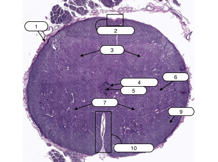
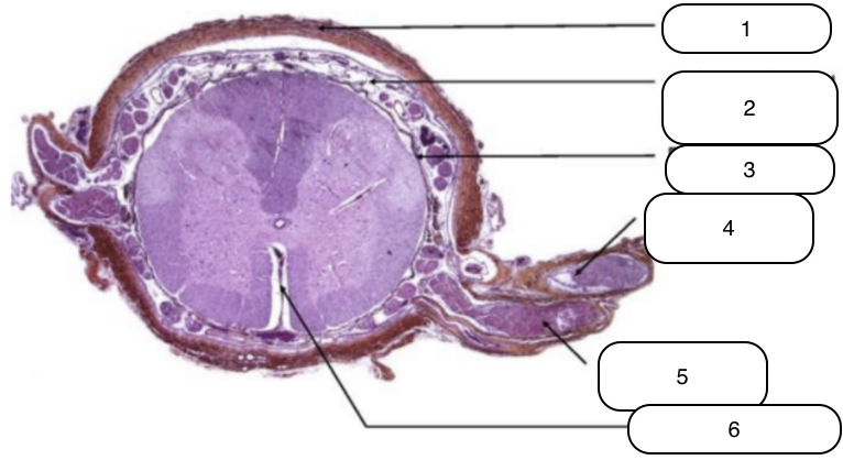
Spinal Cord
Identify the specimen.
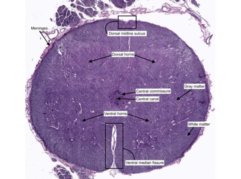
Meninges
Identify the structure labeled #1

Dorsal midline sulcus
Identify the structure labeled #2

Dorsal horns
Identify the structure labeled #3

Central commissure
Identify the structure labeled #4

Central canal
Identify the structure labeled #5

Gray matter
Identify the structure labeled #6
Ventral horns
Identify the structure labeled #7

White matter
Identify the structure labeled #9
Ventral median fissure
Identify the structure labeled #10

Nissl granules
Identify the structure labeled #1

Nucleolus
Identify the structure labeled #2

Nucleus
Identify the structure labeled #3
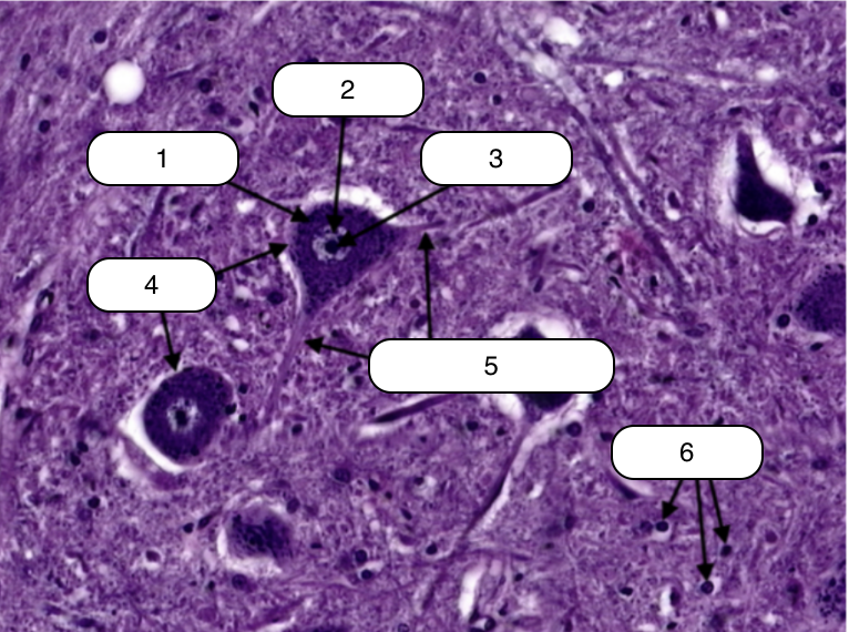
Neurons
Identify the structure labeled #4

Cytoplasmic processes
Identify the structure labeled #5

Glial cells
Identify the structure labeled #6

White matter
Identify the structure labeled #1

Gray matter
Identify the structure labeled #2

Spinal Motor Neuron
Identify the structure labeled #3
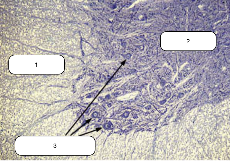
Sensory neuron
Identify the structure labeled #1

Axons
Identify the structure labeled #2
Dura mater
Identify the structure labeled #1
Subarachnoid space
Identify the structure labeled #2

Pia mater
Identify the structure labeled #3

Dorsal nerve root
Identify the structure labeled #4

Ventral nerve root
Identify the structure labeled #5

Linea splendens
Identify the structure labeled #6

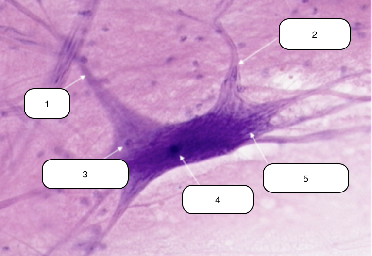
Neuron
Identify the specimen.
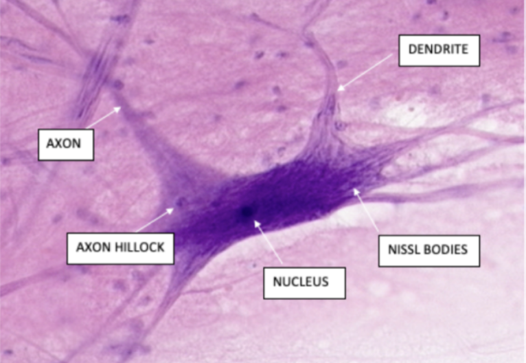
Axon
Identify the structure labeled #1

Dendrite
Identify the structure labeled #2

Axon Hillock
Identify the structure labeled #3

Nucleus
Identify the structure labeled #4

Nissl bodies
Identify the structure labeled #5
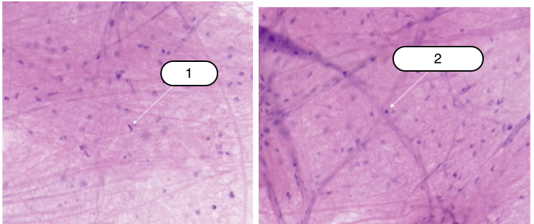
Astrocyte
Identify the structure labeled #1

Oligodendrocyte
Identify the structure labeled #2
Cerebrum
Identify the specimen.

Pyramidal cells
Identify the structure labeled #1

Subdural space
Identify the structure labeled #2

Arachnoid Matter
Identify the structure labeled #3
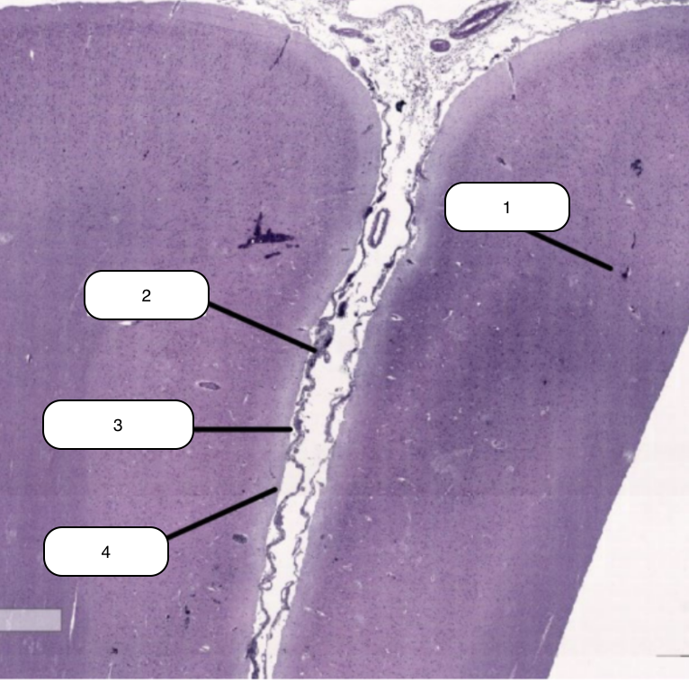
Pia Matter
Identify the structure labeled #4

Cerebrum
Identify the specimen.
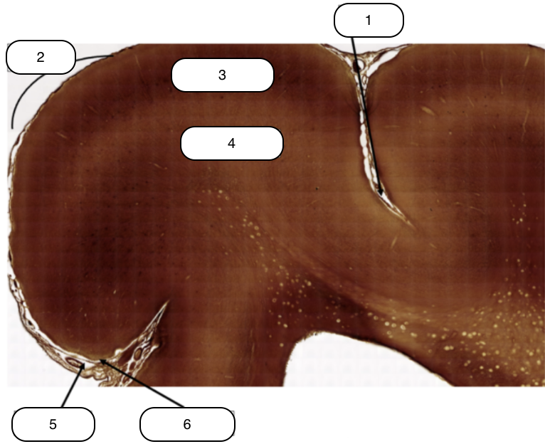
Sulcus
Identify the structure labeled #1

Gyrus
Identify the structure labeled #2

Gray Matter
Identify the structure labeled #3

White Matter
Identify the structure labeled #4

Arachnoid
Identify the structure labeled #5

Pia Mater
Identify the structure labeled #6

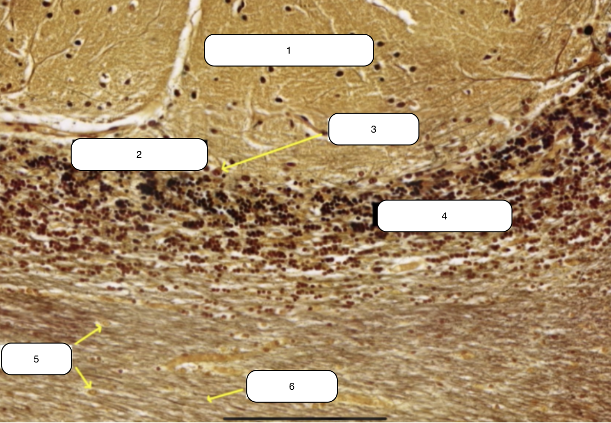
Cerebellum
Identify the specimen.

Transverse Folds (Folia)
Identify the structure labeled #1

White Matter
Identify the structure labeled #2

Gray Matter (Cortex)
Identify the structure labeled #3
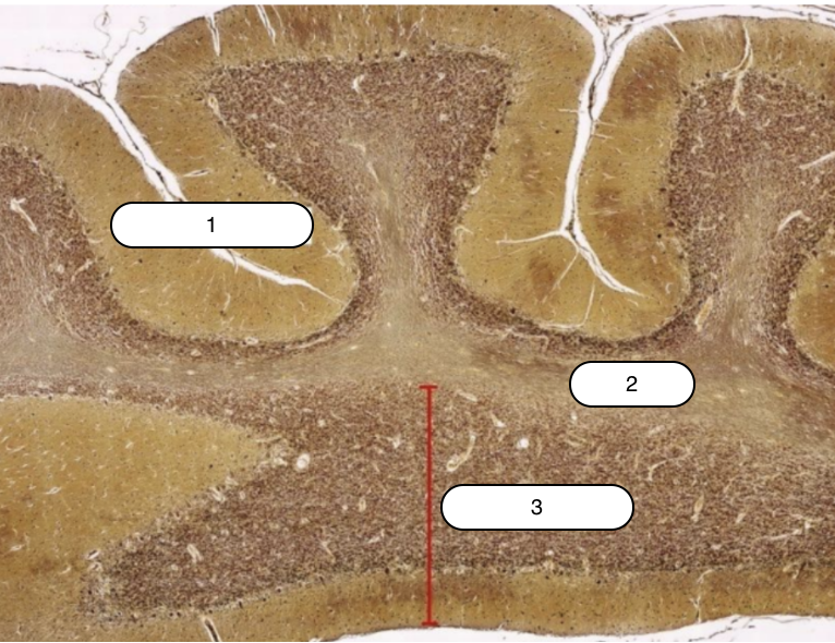
Cerebellar Cortex
Identify the specimen.

Outer Molecular Layer
Identify the structure labeled #1

Layer of Purkinje Cells
Identify the structure labeled #2

Purkinje Cells
Identify the structure labeled #3
Layer of Granule Cells
Identify the structure labeled #4

Glial Cells
Identify the structure labeled #5

Nerve Fiber
Identify the structure labeled #6

Choroid Plexus
Identify the specimen.
Ependymal Cells
Identify the structure pointed by the arrow.

Peripheral Nerve
Identify the specimen.

Nerve bundle/fascicles
Identify the structure labeled #1

Perineurium
Identify the structure labeled #2
Myelin Sheath
Identify the structure labeled #1

Axon
Identify the structure labeled #2

Perineurium
Identify the structure labeled #3
Endoneurium
Identify the structure labeled #4

Schwann Cell
Identify the structure labeled #5

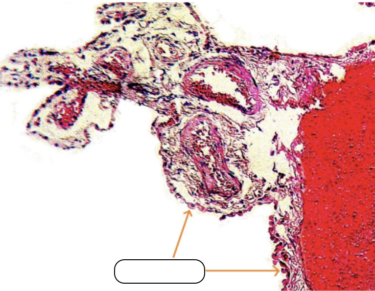
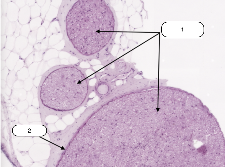
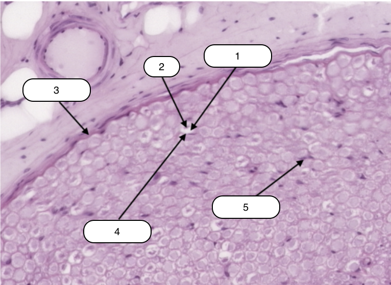
Dorsal Root Ganglion (Spinal Ganglion)
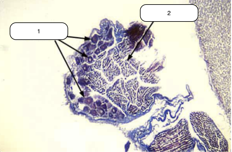
Identify the specimen.
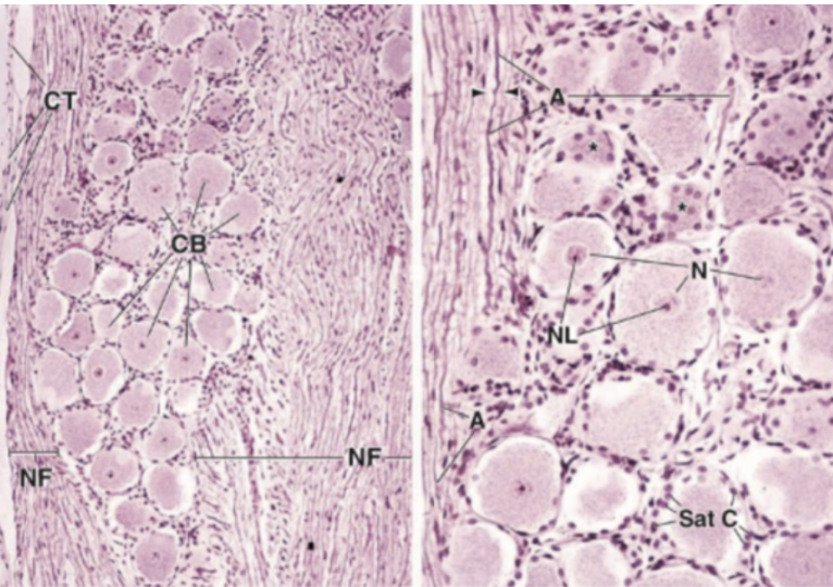
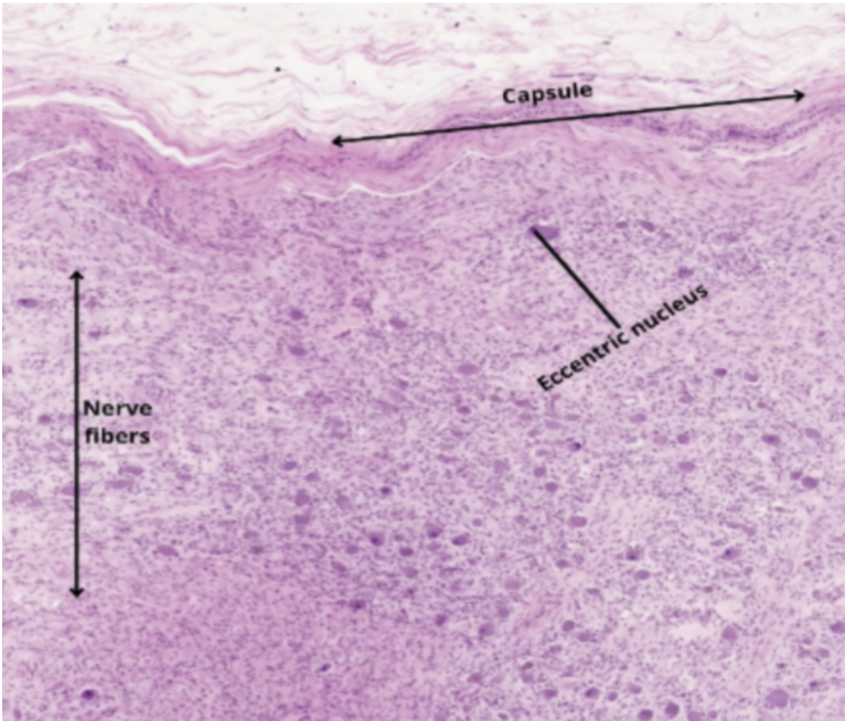
Sympathetic Ganglion
Identify the specimen.
Digestive Tract
Identify the specimen.

Mucosa
Identify the structure labeled #1
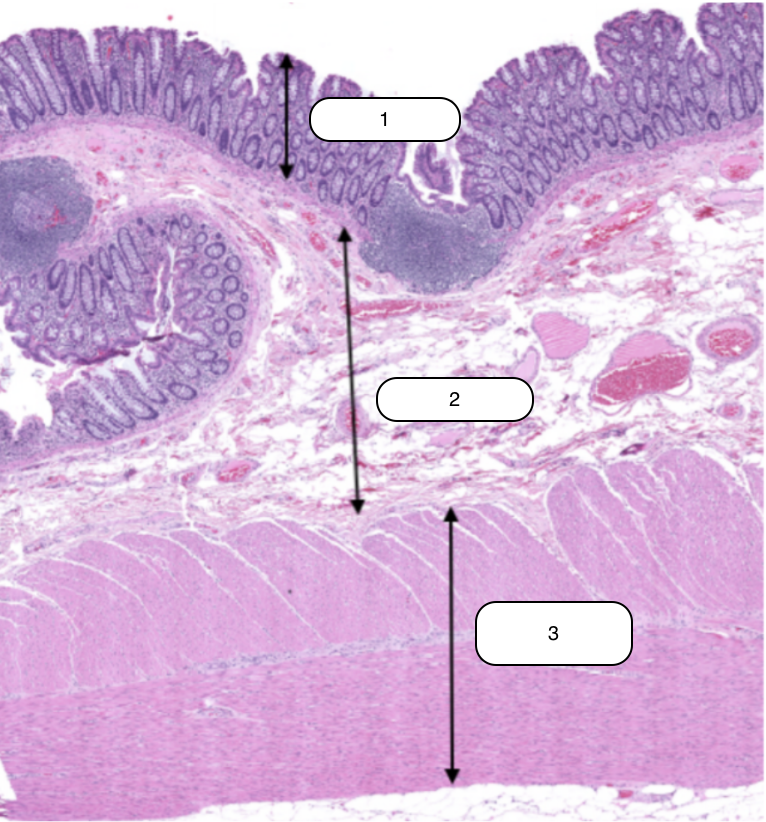
Submucosa
Identify the structure labeled #2

Muscularis Externa
Identify the structure labeled #3

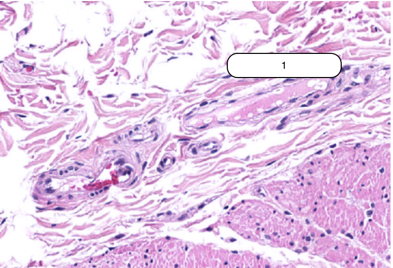
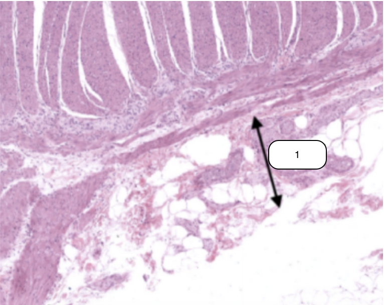
Meissner’s plexus
Identify the structure labeled #1
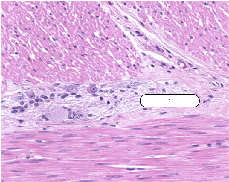
Auerbach’s plexus
Identify the structure labeled #1
Serosa
Identify the structure labeled #1
Lamellar Cells
Identify the structure labeled #1

Meissner’s Corpuscle
Identify the structure labeled #2
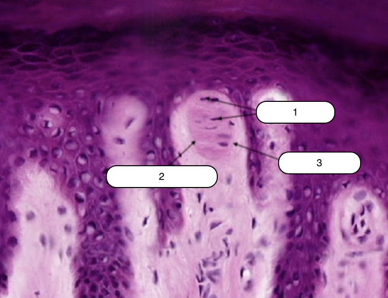
Capsule
Identify the structure labeled #3

Pacinian Corpuscle
Identify the structure labeled #1

Supporting Cells
Identify the structure labeled #2
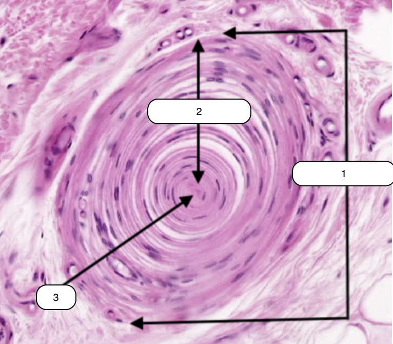
Axon
Identify the structure labeled #3
