Lab Assessment 7
1/55
There's no tags or description
Looks like no tags are added yet.
Name | Mastery | Learn | Test | Matching | Spaced | Call with Kai |
|---|
No analytics yet
Send a link to your students to track their progress
56 Terms
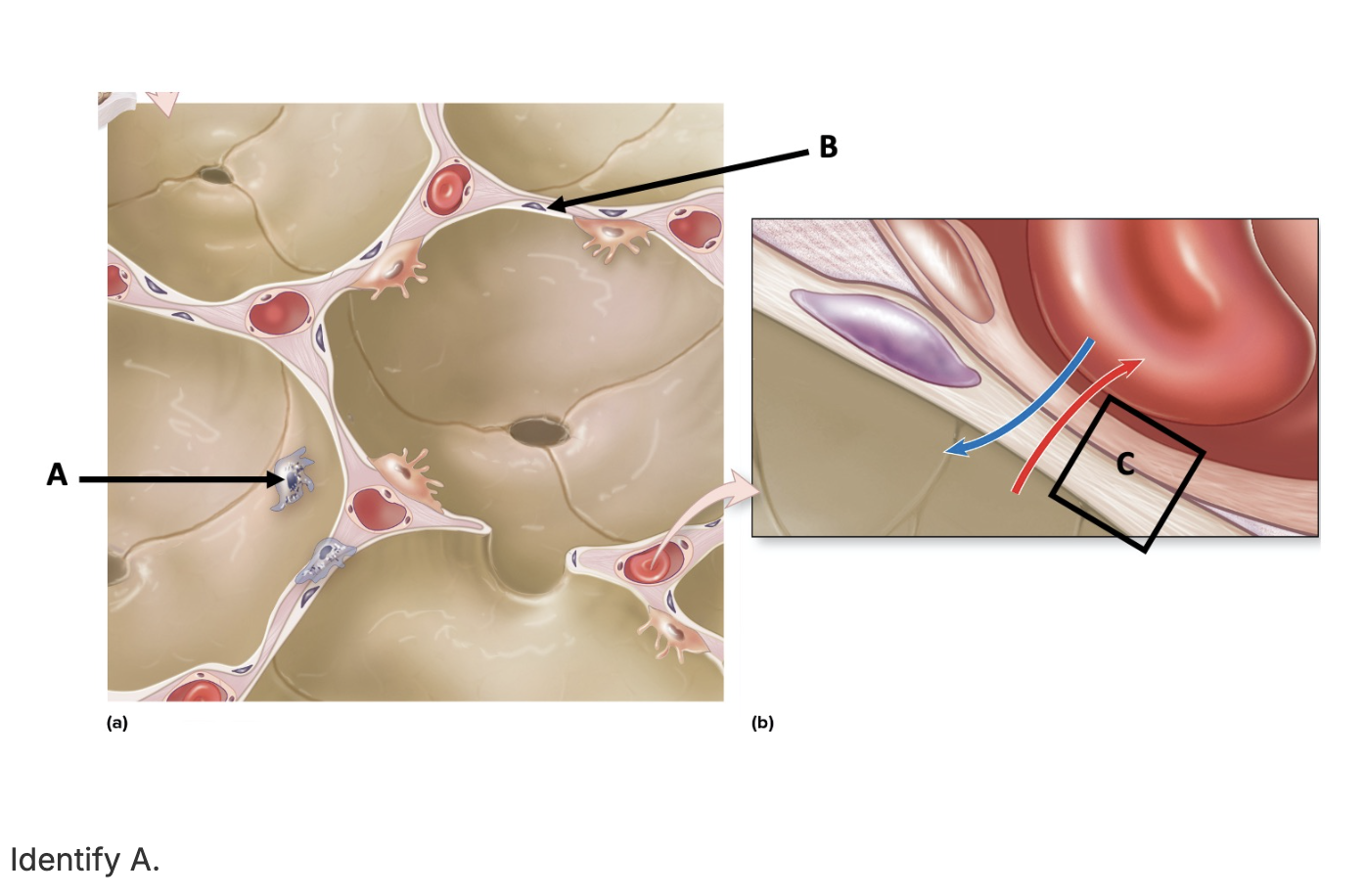
alveolar macrophage
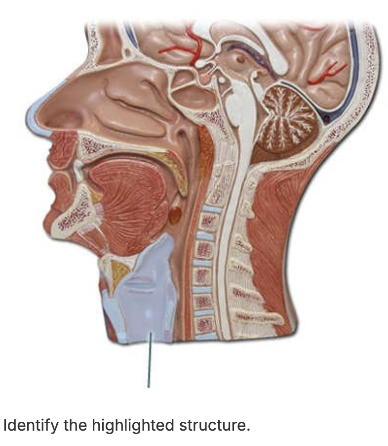
larynx
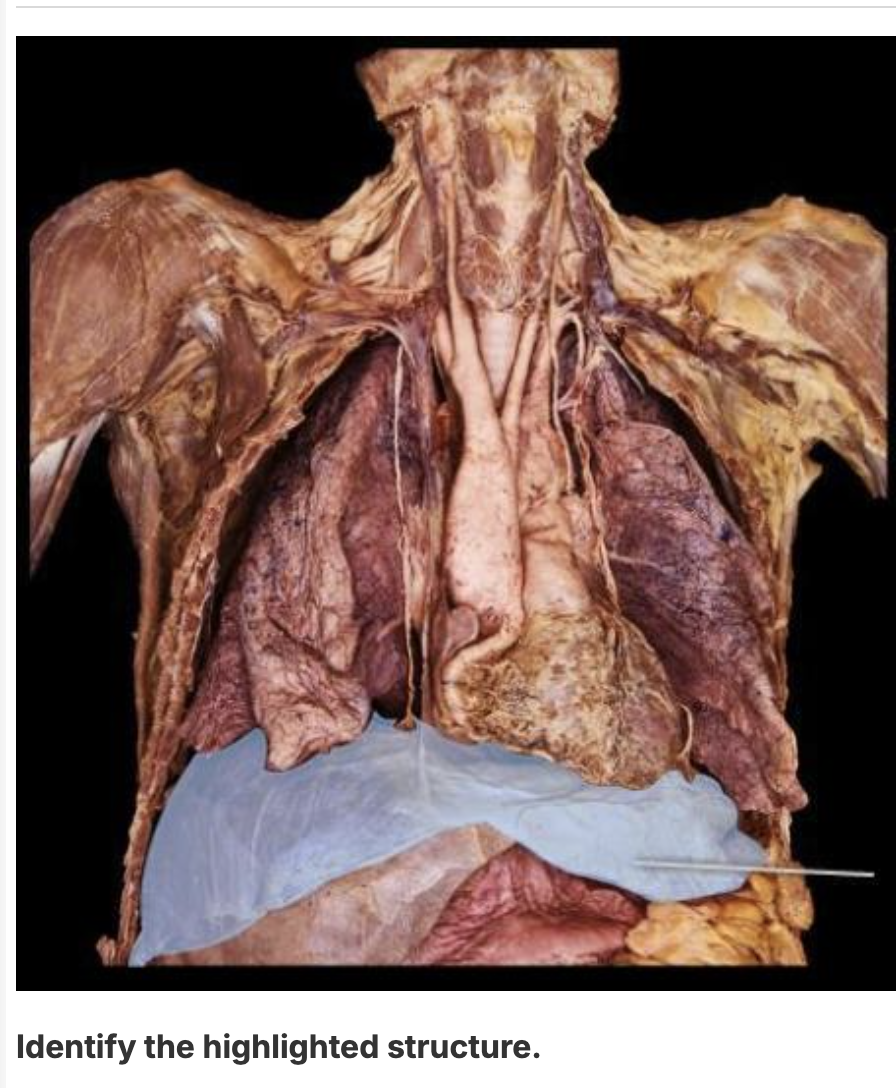
diaphragm
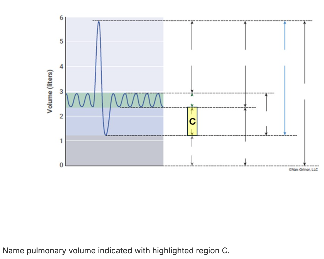
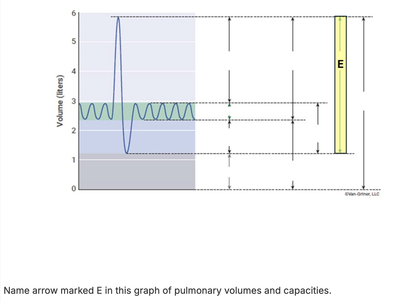
ERV
Air moves out of the lungs when the pressure inside the lungs is:
less than atmospheric pressure.
equal to atmospheric pressure.
greater than atmospheric pressure.
less than intrapleural pressure.
greater than atmospheric pressure.
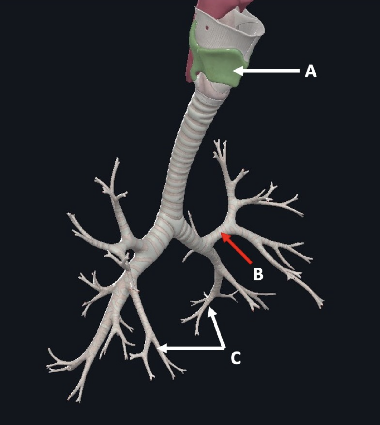
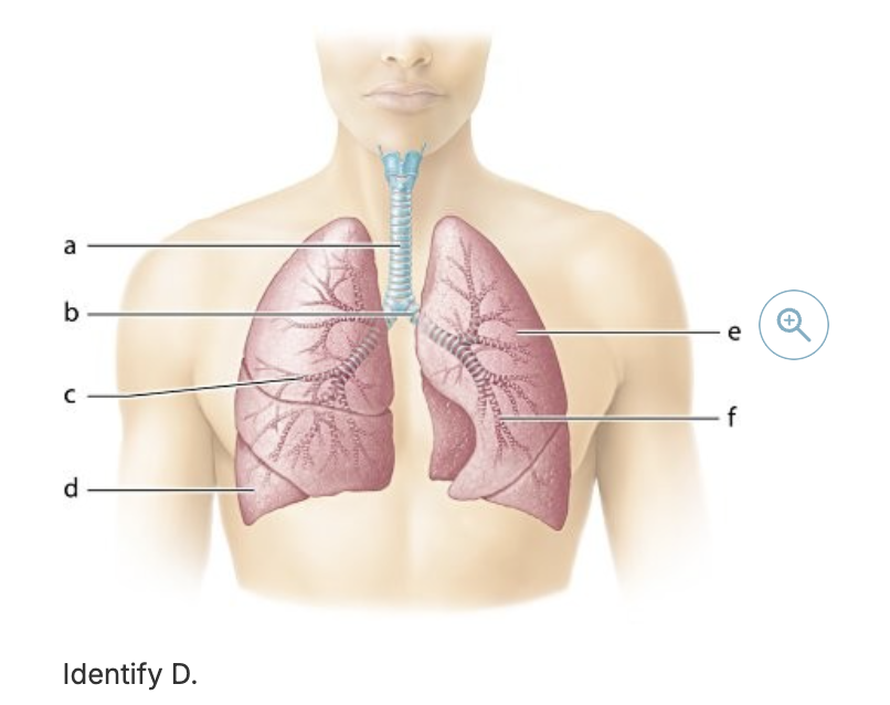
Identify C
Tertiary bronchi
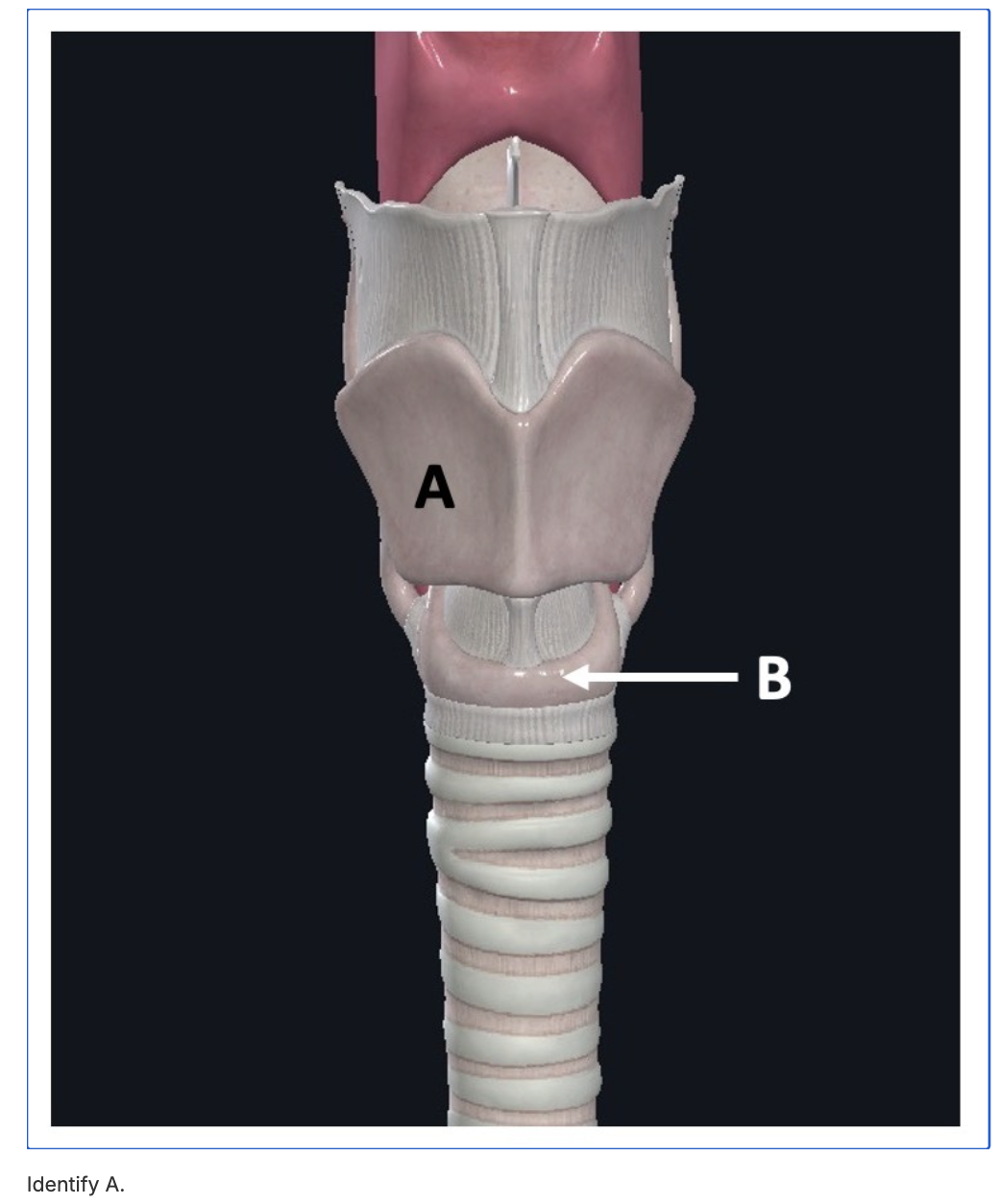
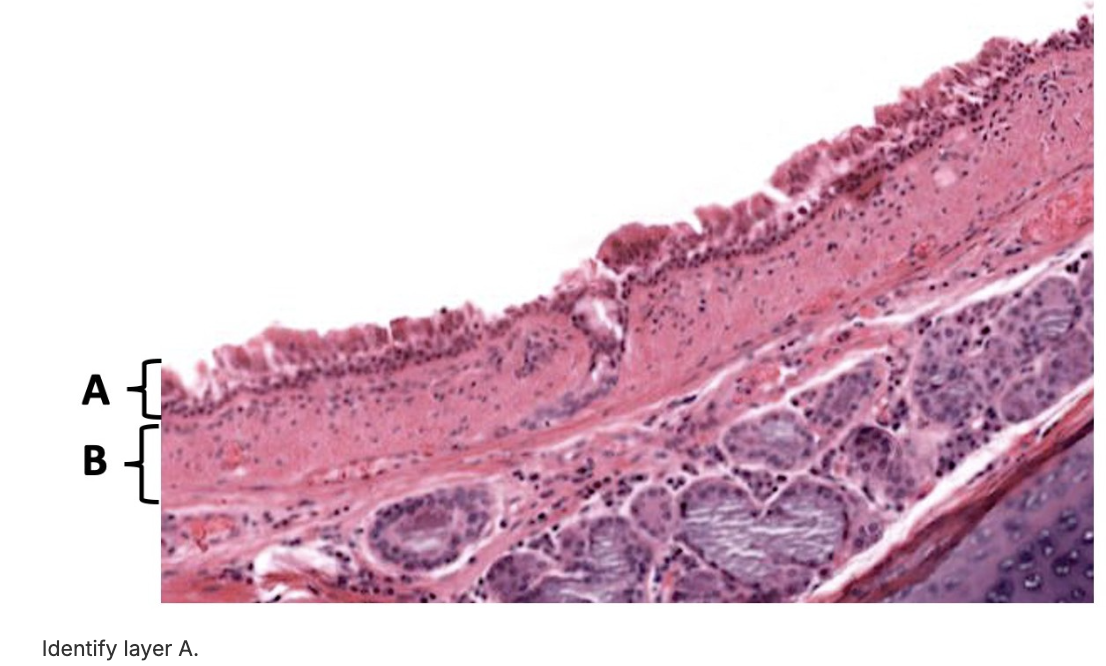
Identify A
thyroid cartilage
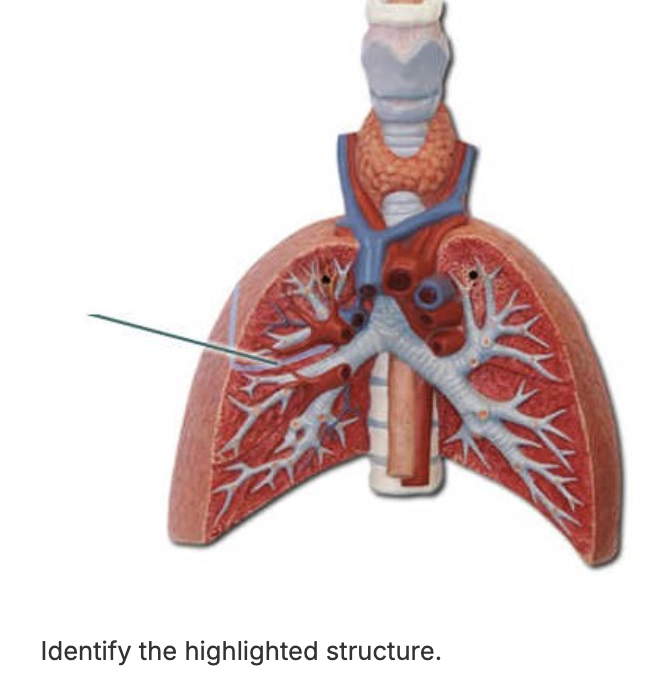
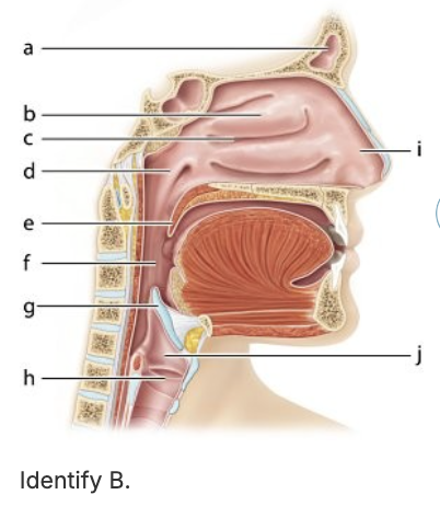
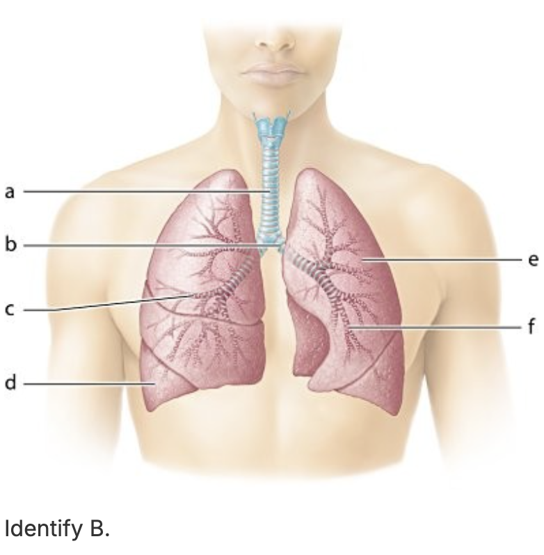
Identify B
left secondary bronchus
alveoli
submucosa
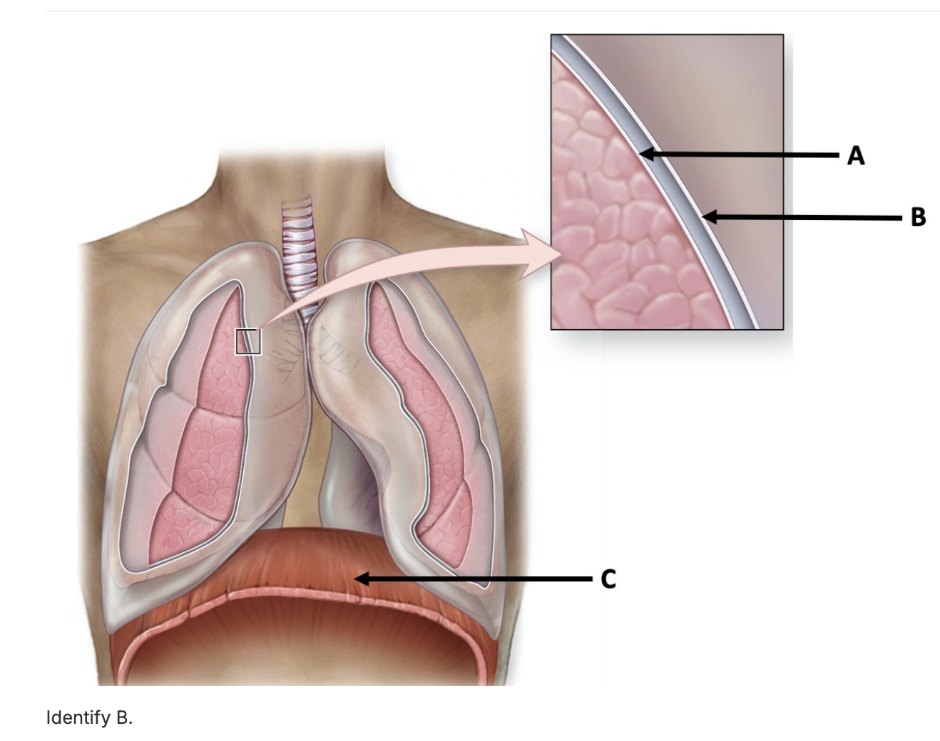
parietal pleura
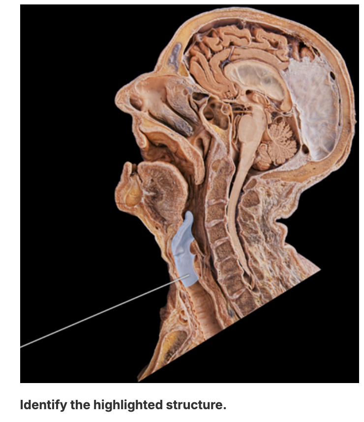
larynx
trachea
superior nasal concha
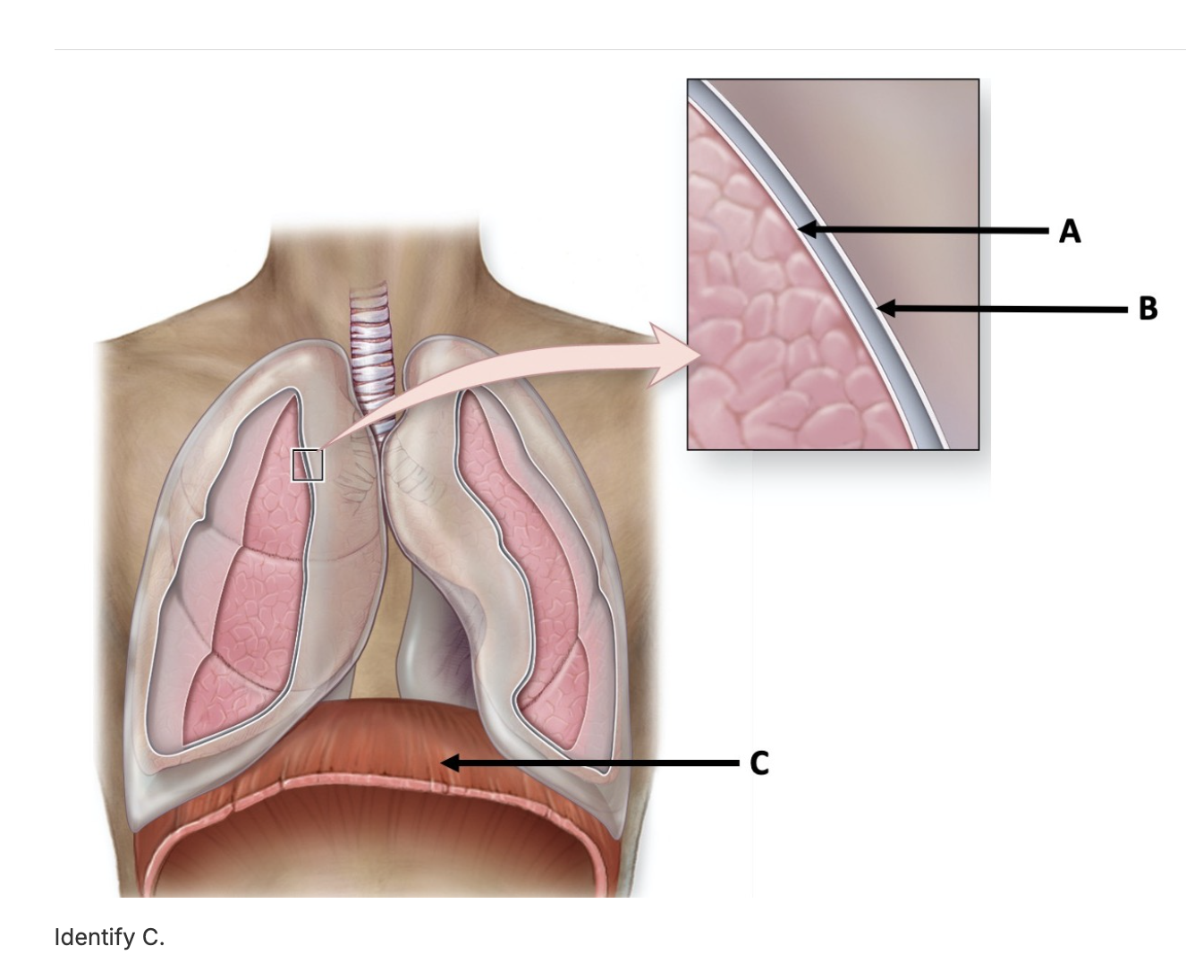
diaphragm
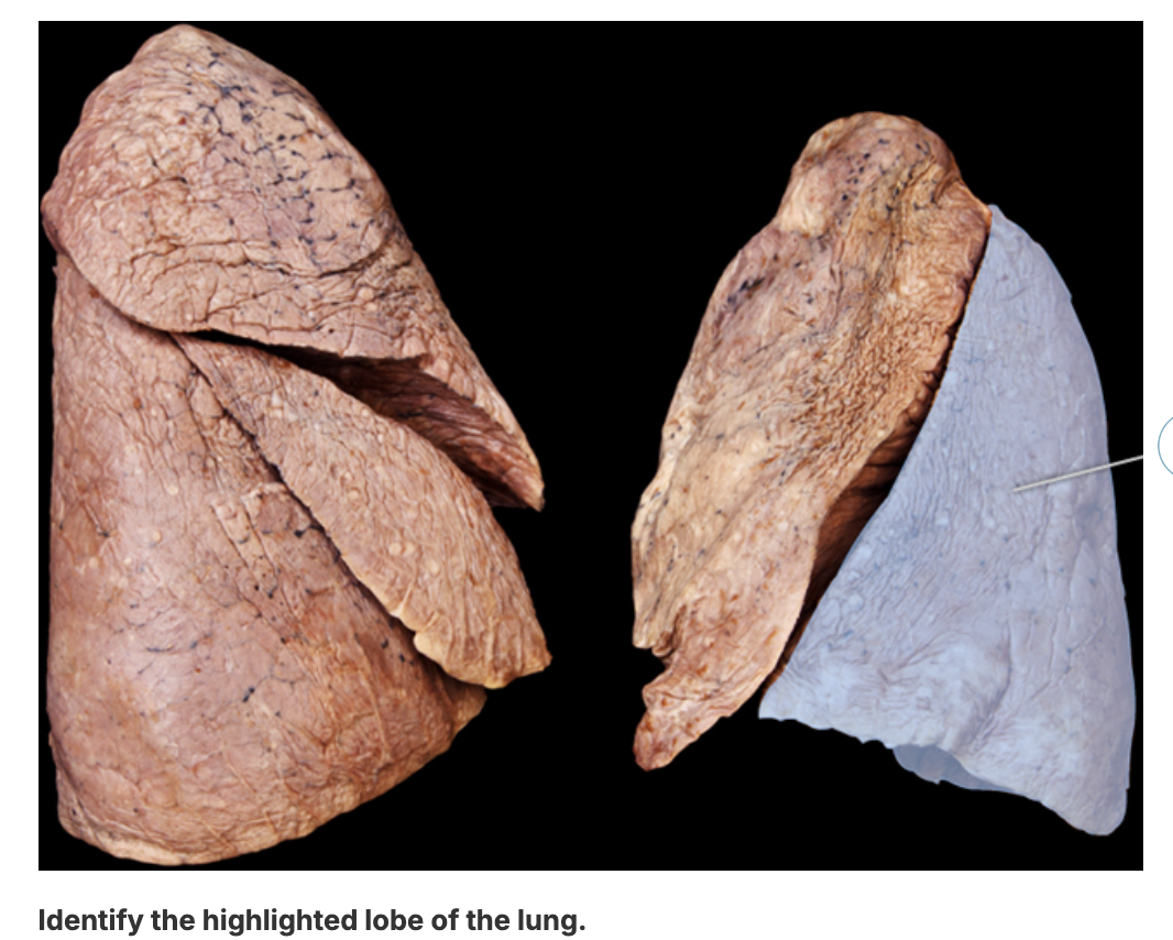
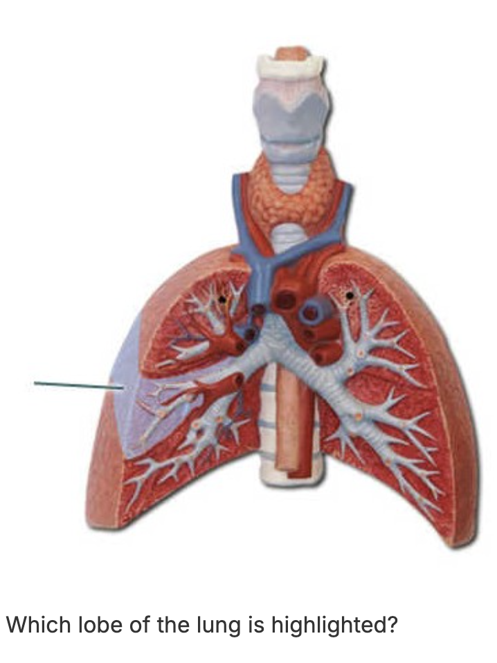
left inferior lobe
ethmoid
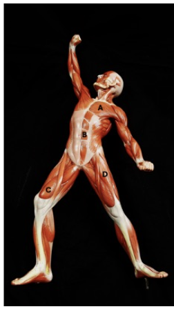
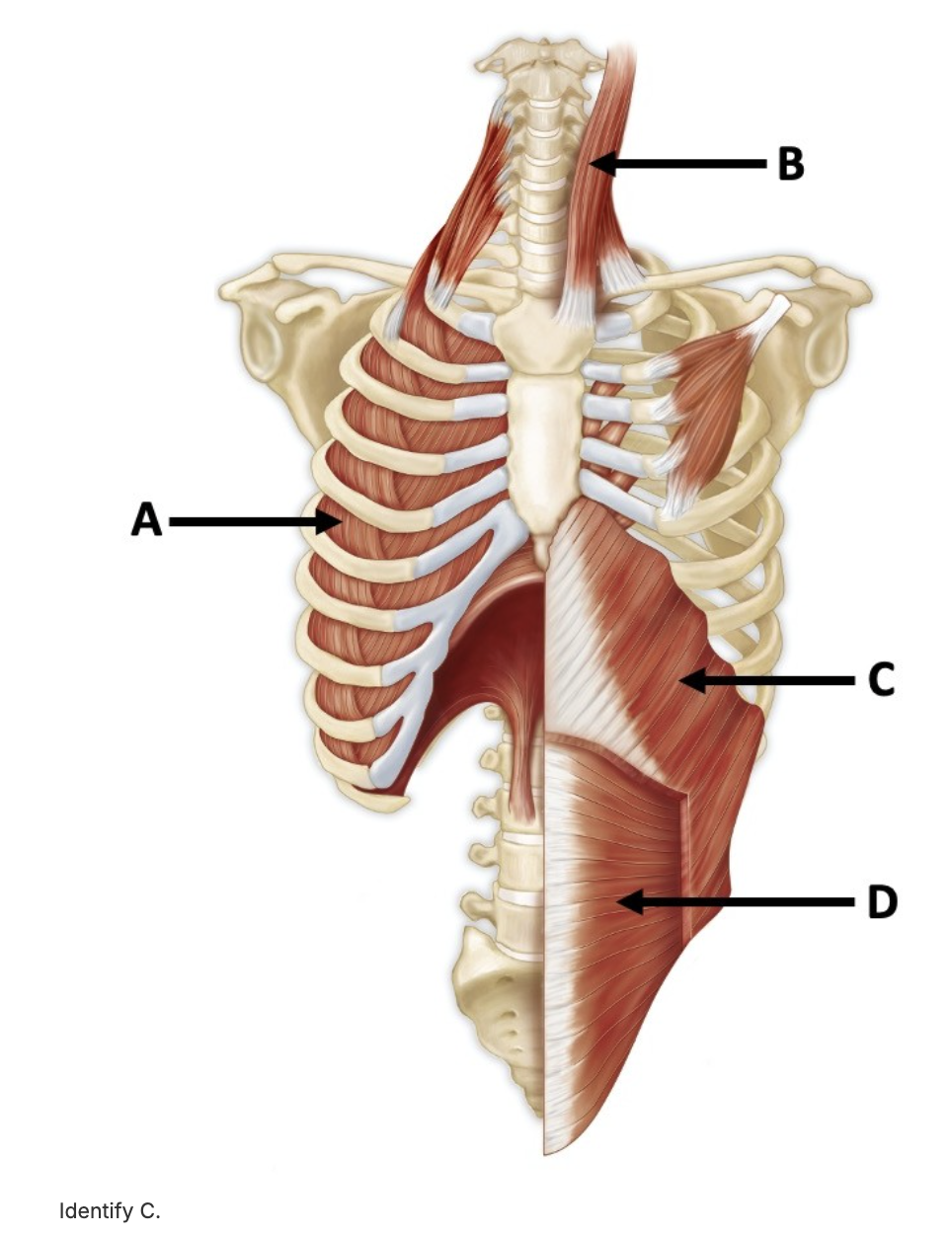
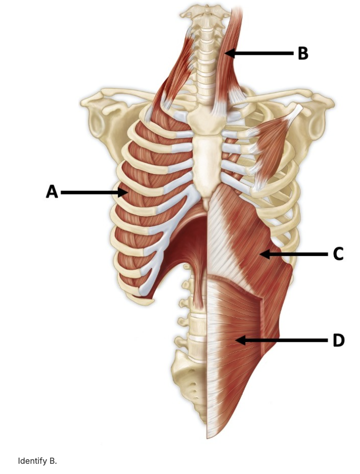
identify the muscle labeled B
rectus abdominis
horizontal fissure
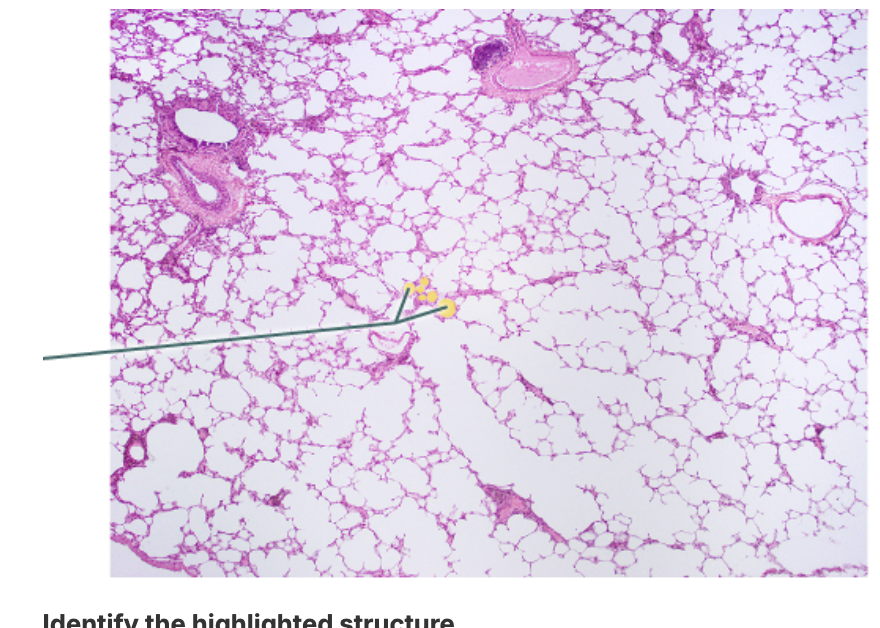
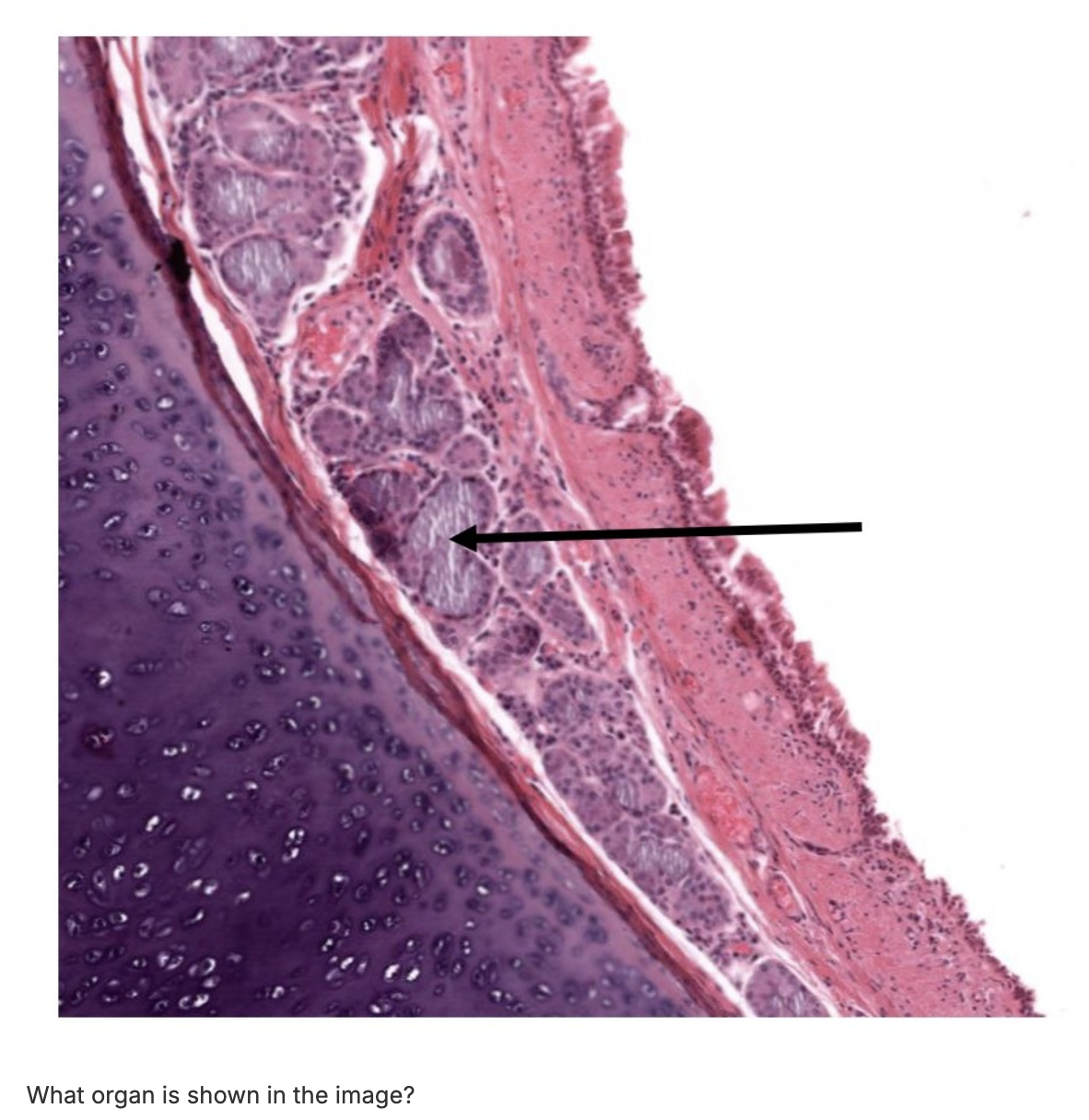
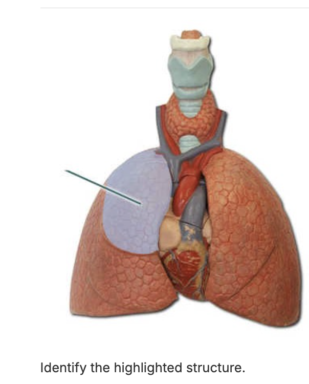
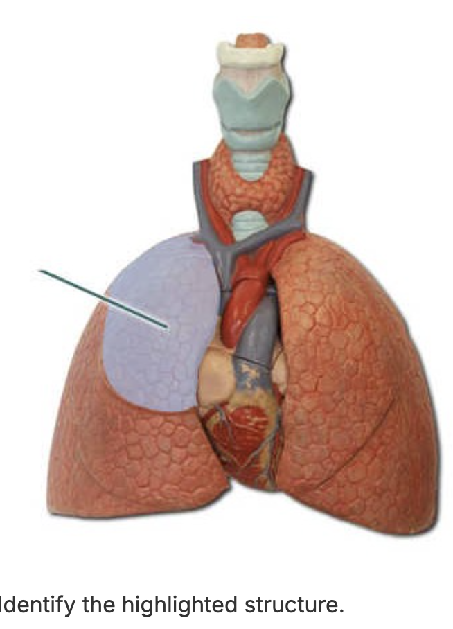
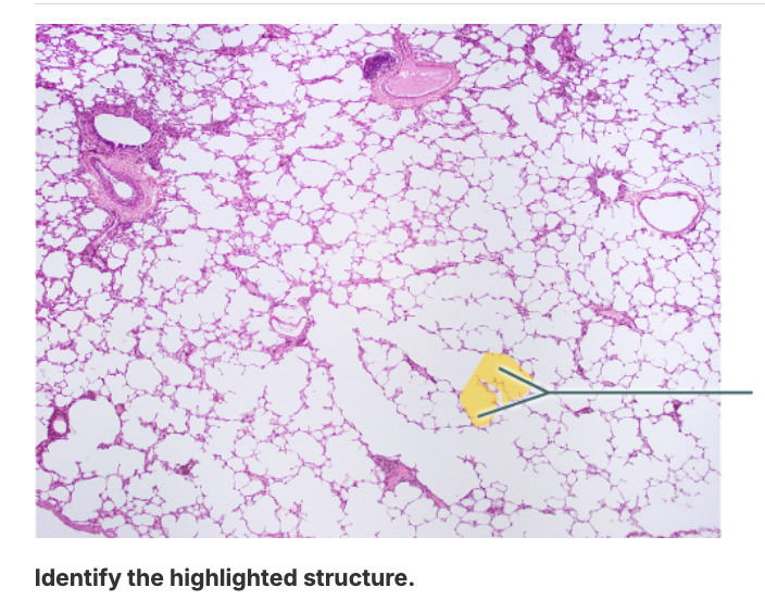
name the organ shown in this picture
lung
What statement best describes tidal volume?
Tidal volume is the air exhaled after normal inspiration.
Tidal volume is the air exchanged during normal breathing.
Tidal volume is the air remaining in the lungs after forced expiration.
Tidal volume is the air forcibly expelled after normal expiration.
Tidal volume is the air exchanged during normal breathing.
right superior lobe
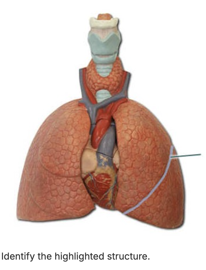
left oblique fissure
right superior lobe
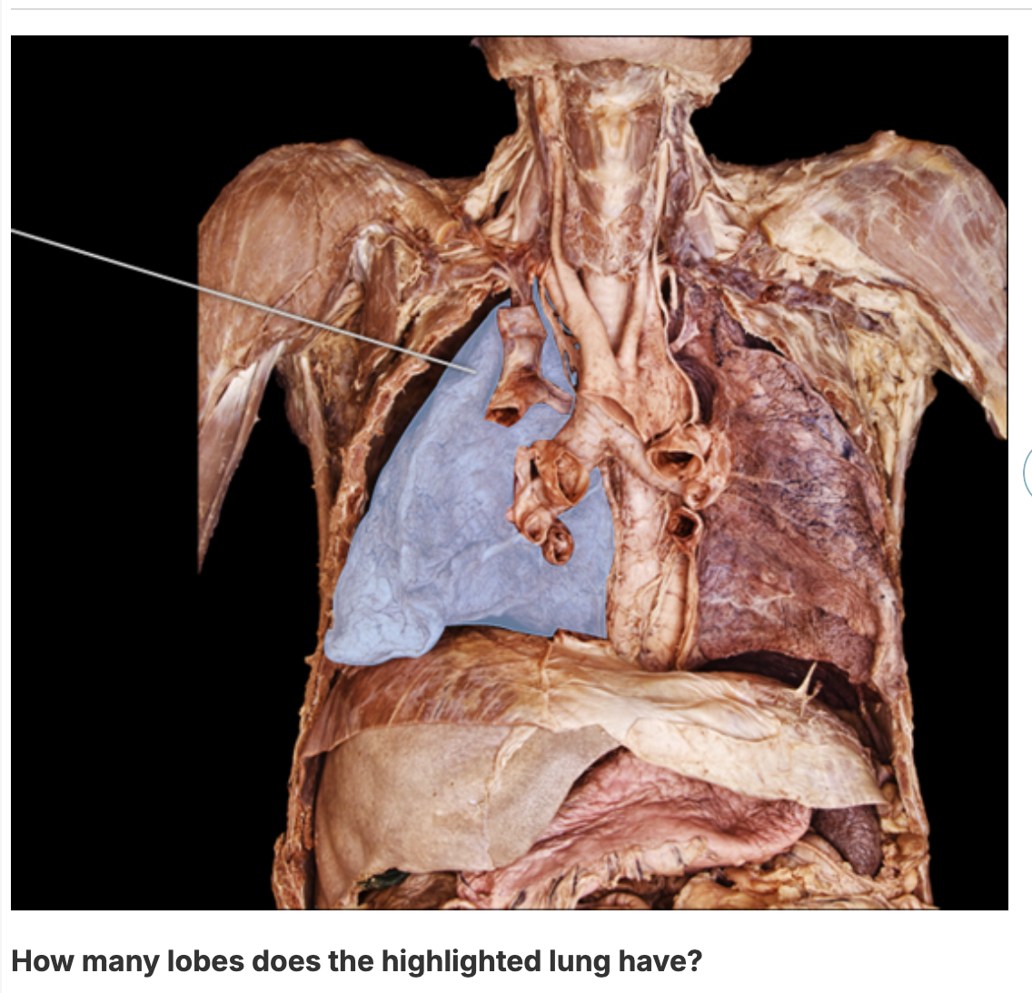
3
Thyroid cartilage
external obliques

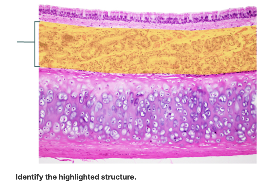
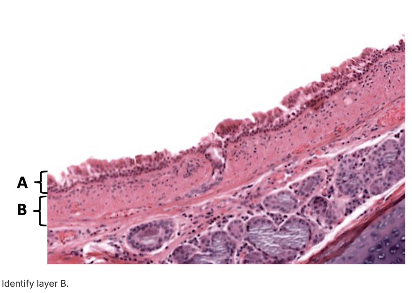
Identify the tissue shown in layer B.
PCCE
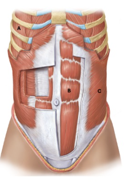
Identify the muscle labeled B.
rectus abdominis
The volume that remains in the lungs after a forced expiration is the:
functional residual capacity.
residual volume.
dead space volume.
vital capacity.
residual volume.
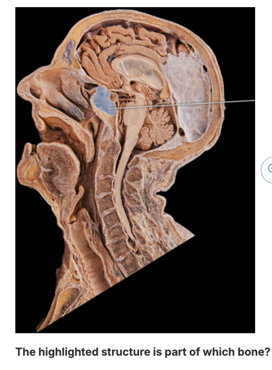
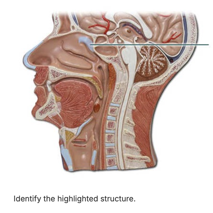
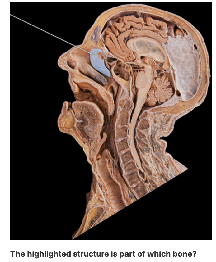
sphenoid bone
TV
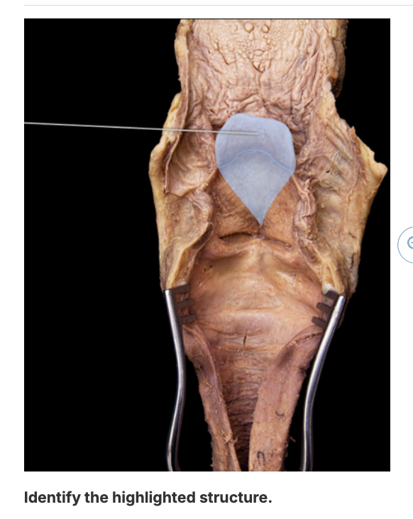
epiglottis
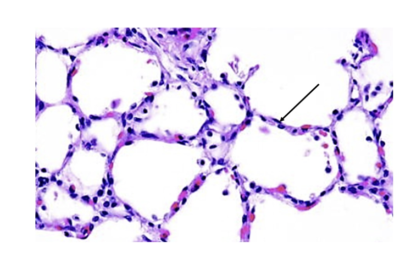
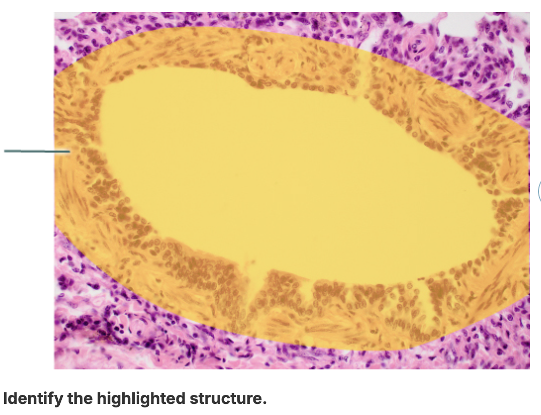
Name the circular structures shown in this picture.
alveoli
mucosa
middle nasal concha
alveolar sacs
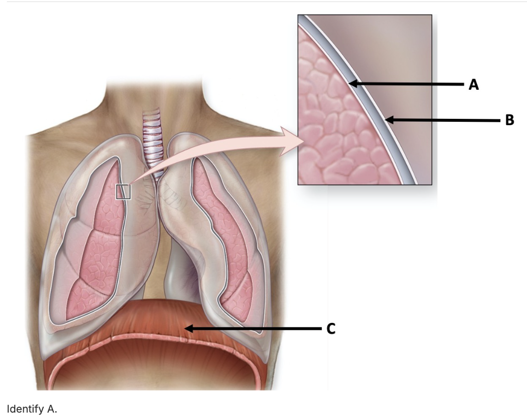
visceral pleura
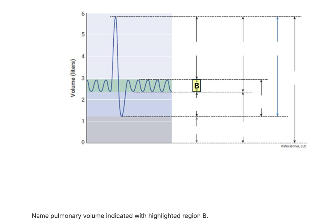
The amount of air that can be forcibly expired after a normal tidal expiration is known as the:
tidal volume.
vital capacity.
expiratory reserve volume.
inspiratory reserve volume.
expiratory reserve volume.
vital capacity
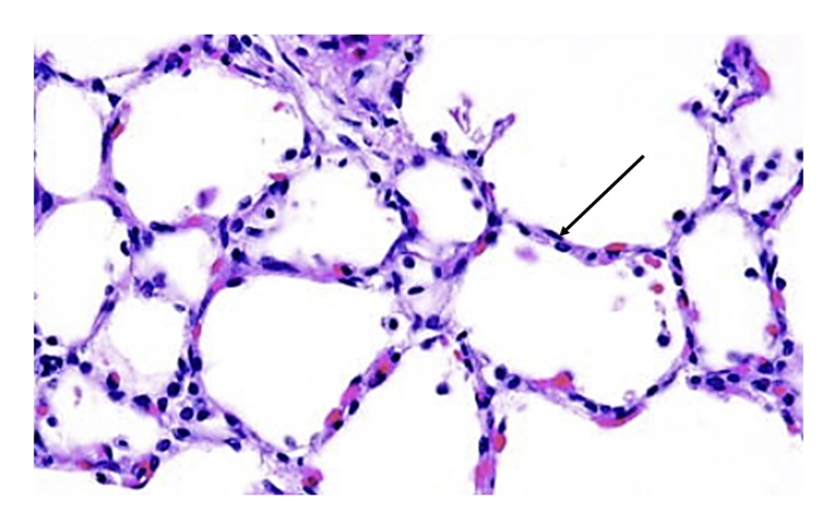
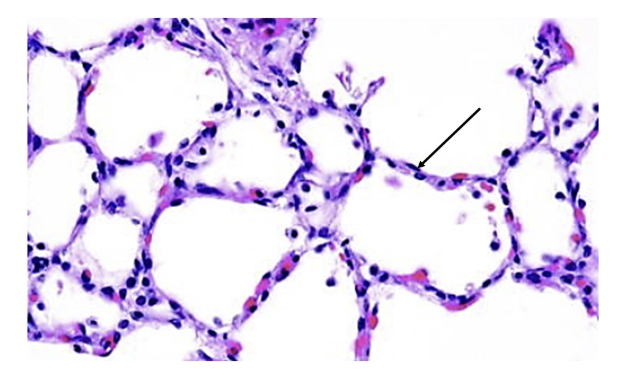
Name the cells that make up the thin walls seen in this picture.
type I alveolar cells
Which of the following reduces alveolar surface tension?
water
surfactant
gas
mucus
surfactant
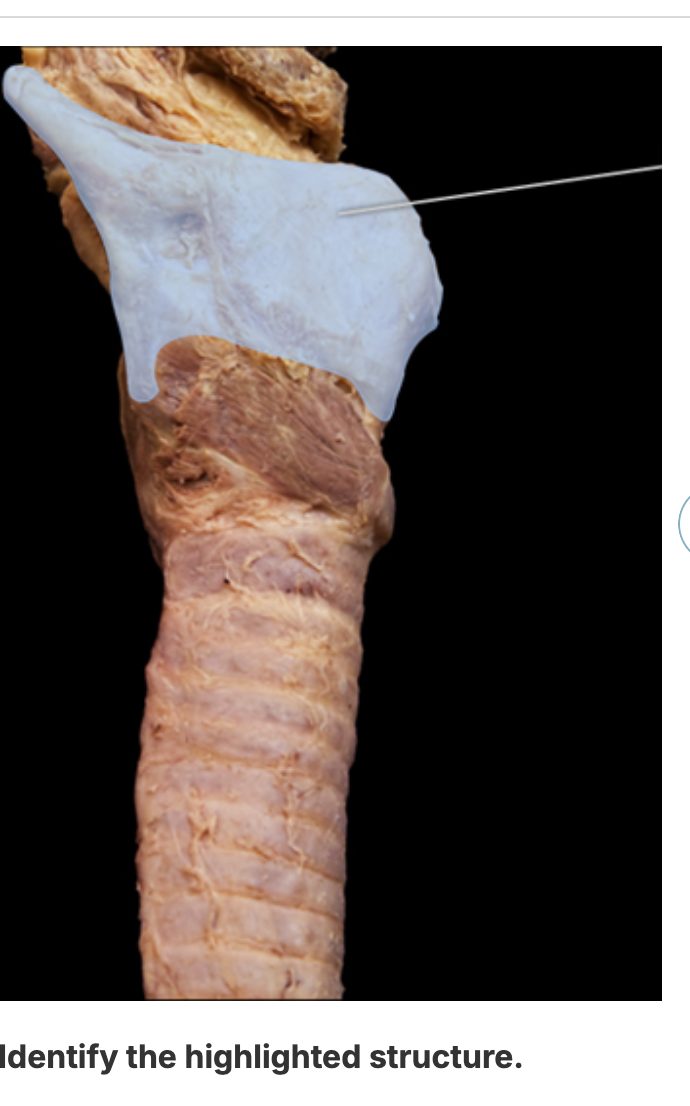
thyroid cartilage
right middle lobe
submucosa
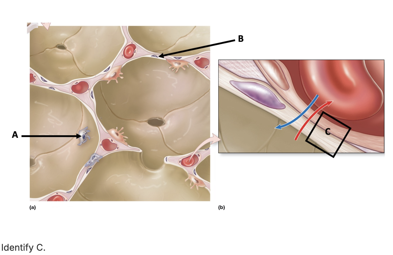
respiratory membrane

Identify the LAYER marked B.
mucosa
bronchiole
left primary bronchus
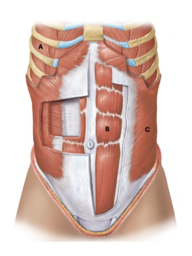
Identify the muscle labeled A.
external intercostals
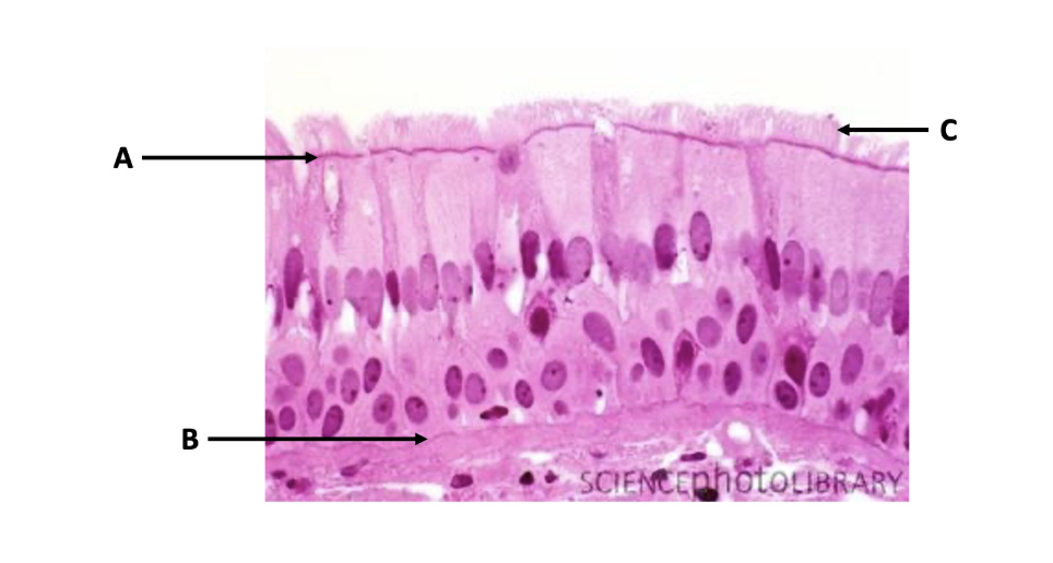
Identify the structure labeled C.
cilia
right inferior lobe
sternocleidomastoid
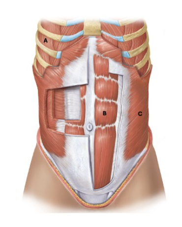
Identify the muscle labeled C.
external oblique
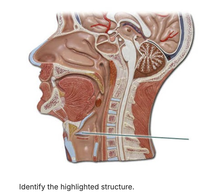
vestibular fold
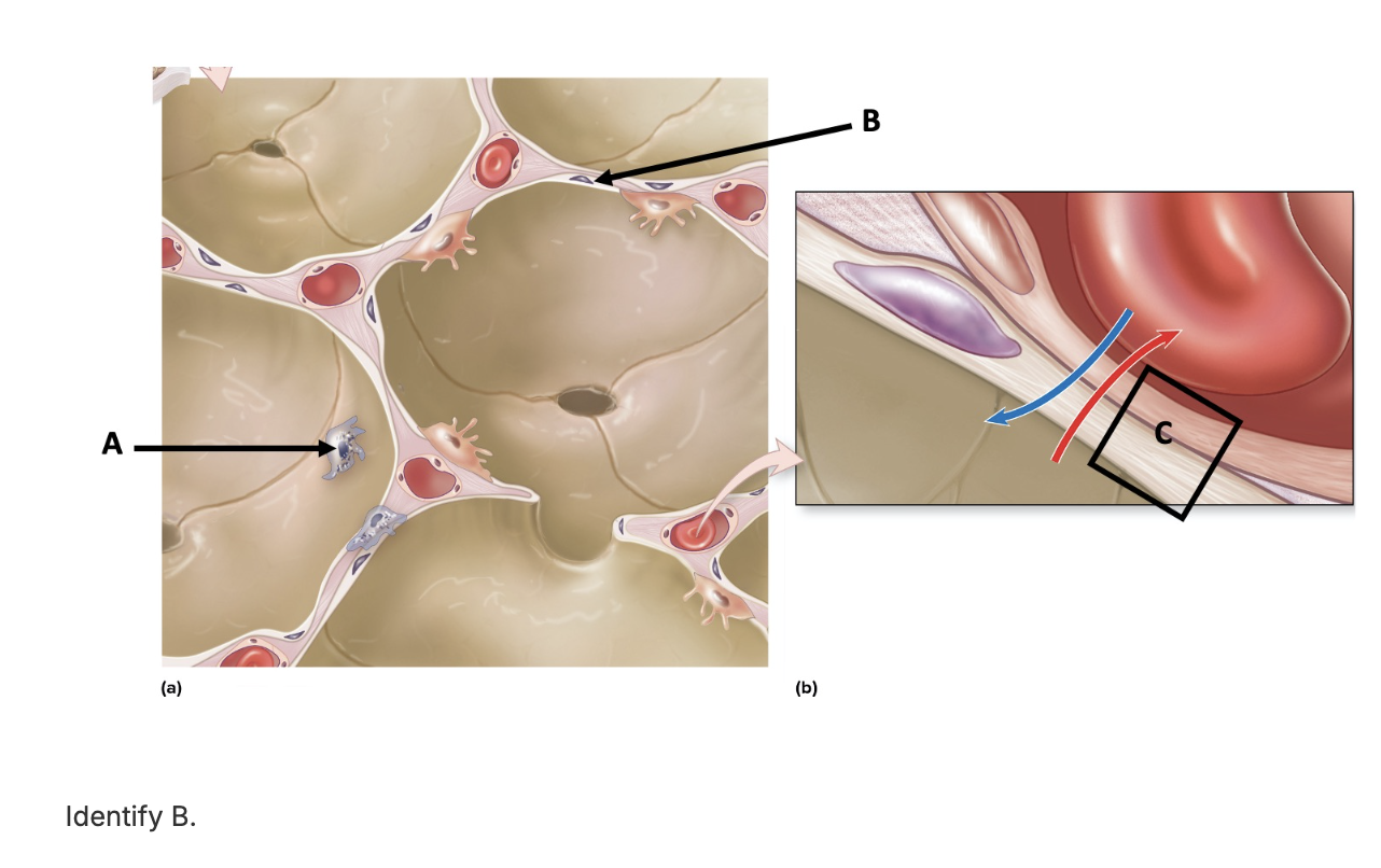
type I alveolar cell