IB Bio Unit 5
1/109
There's no tags or description
Looks like no tags are added yet.
Name | Mastery | Learn | Test | Matching | Spaced | Call with Kai |
|---|
No analytics yet
Send a link to your students to track their progress
110 Terms
Neuron
Are specialized cells that carry electrical impulses
3 Major types of neurons
Sensory neuron: transmit nerve impluses from sense receptros to the central nervous system
Interneuron: are located within the central nervous system. Interneurons transmit nerve impulses between neurons.
Motor neuron: transmit nerve impulses from the central nervous system to an effector.
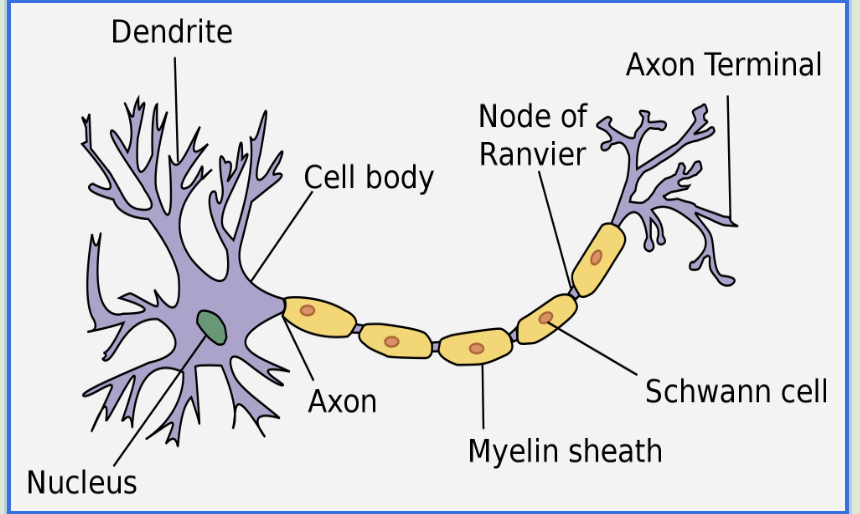
Parts of a neuron
Dendrite
Cell body
Axon
Dendrite
Are short nerve fibers which receive nerve impulses from receptors or other neurons
Cell body
Contains the cell’s nucleus and most of its cytoplasm. The cell body determines if a signal coming from the dendrites is to be passed down.
Axon
Nerve signals in the form of an action potential travel down the axon to axon terminals
Resting potential
The electrical potential difference (voltage) of the cytoplasm of a neuron, relative to its surroundings, when not stimulated or involved in the passage of a nerve impulse
Approximately -70 mV
It is negative due to the distribution of ions inside and outside the plasma membrane
Resting potential in regards to Potassium/Sodium pump
The sodium-potassium pump actively transports sodium(Na+) ions out of a cell and potassium(K+) ions into a cell
Uses ATP energy to maintain the resting potential of a neuron
3 Na+ goes out of the cell, 2 K+ goes into the cell, resulting in a net negative charge on the inside (AKA more positive on the inside)
The sodium potassium pump transports Na+ and K+ against their concentration gradients, and is an example of active transport.
Is an exchange transporter, as Na+ and K+ travel in opposite directions
Resting Potential and the Sodium Potassium Pump
Three Na+ attach to the sodium ion binding sites on the sodium potassium pump protein.
ATP attaches to the sodium potassium pump.
ATP is hydrolyzed, with a phosphate remaining attached to the protein pump. ADP is released.
The phosphate causes the pump to change shape, moving the sodium across the axon membrane, releasing Na+ outside the axon.
Two K+ attach to the potassium ion binding sites on the sodium potassium pump protein.
The phosphate is released from the pump.
The pump returns to its original shape moving the K+ into the axon.
The process can be repeated.
Summary of why the resting potential is negative
Three Na+ ions are pumped out of the neuron for every two K+ ions that are pumped in.
Some K+ ions leave the neuron through simple diffusion, but no Na+ ions enter.
Nerve impulses
Are action potential that are propagated along nerve fibers
Action potential
Is electrical as it involves the flow of ions along nerve fibers such as axons
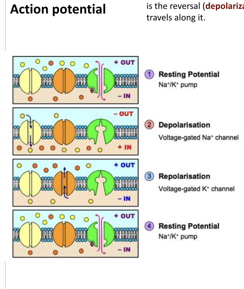
*Steps of a nerve impulse from resting potential to restoration of resting potential
Resting potential: The Na+/K+ Pump maintains the resting potential of -70mV
Depolarization: In response to a stimulus, voltage-gated Na+ channels open to generate an action potential, and sodium enters the neuron by diffusion. The entry of Na+ causes the membrane potential to become positive (depolarisation)
Repolarisation: The depolarisation of the membrane potential causes the voltage gated Na+ channels to close and the voltage gated K+ channels to open. K+ diffuses out of the neuron rapidly, and the membrane potential becomes negative again (repolarisation) to end the action potential
Resting potential: The Na+/K+ pump restores resting potential of -70mV.
*Look at video from slides*
Coefficient of determination (R²)
It can be used to determine the strength of a relationship between two variables
Animal size and speed of nerve impulses
As animal size increases, the speed of nerve impulses decreases (negative correlation)
Adaptations of large animals to increase speed of nerve impulses
Wider diameter of axons
Myelination of axons
Relationship between axon diameter and speed of nerve impulses
As axon diameter increases, the speed of nerve impulses increases as well (positive correlation)
Myelinated neurons
Myelin is a multilayer of phospholipids and proteins, that surrounds axons.
it acts as an insulating layer, and increases the speed of nerve impulses
Parts of Myelin
Myelin is wrapped around the axon of the neuron by Schwann cells to form myelin sheath
The spaces between myelin sheaths are nodes of Ranvier.
Myelinated neurons are capable of saltatory conduction
Speed of nerve impulses in squid : myelinated VS non-myelinated
speed of Myelinated: 25 ms-1
speed of non-myelinated: 0.5ms-1
Saltatory Conduction
describes the way an action potential jumps between nodes of Ranvier as it moves down an axon.
Action potential jumps from node to node when traveling down an axon
*It is much faster than non-saltatory conduction in non-myelinated neurons and requires less energy (as the sodium-potassium pumps are only active at the nodes of Ranvier)
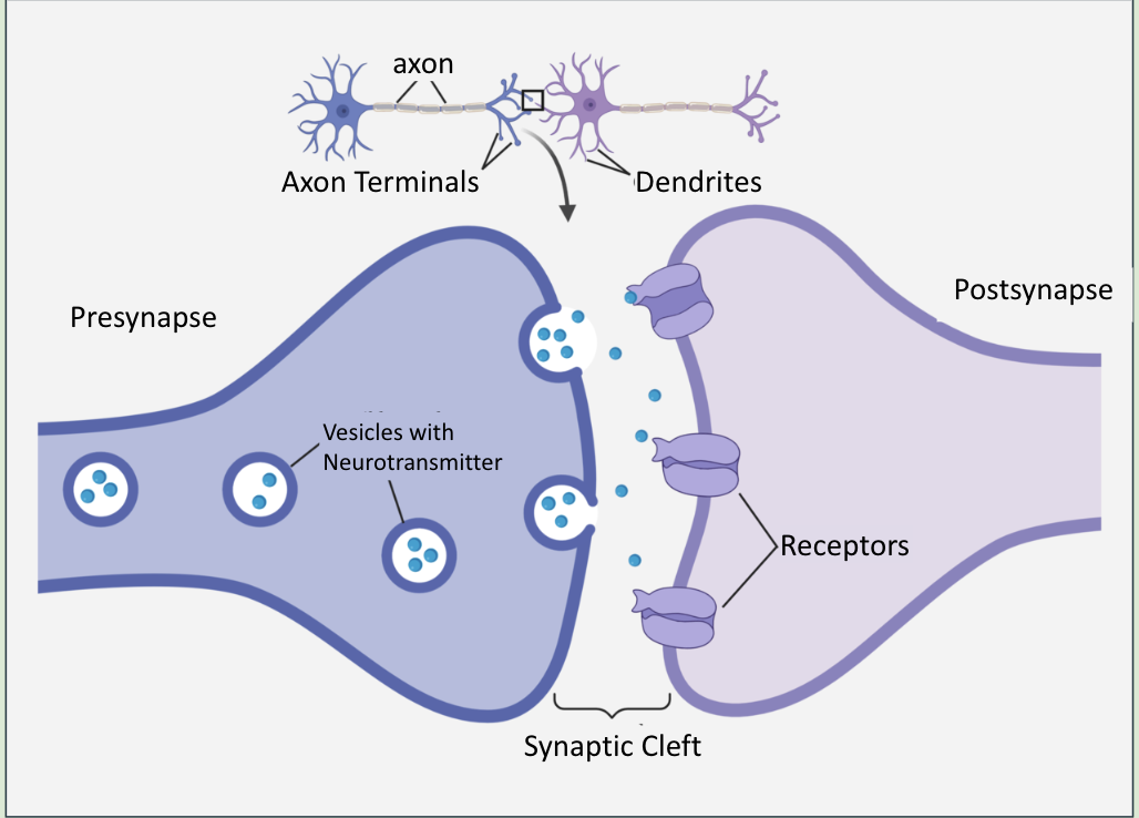
Synapse
Are junctions between two neurons or a neuron and an effector (such as muscles or glands)
Direction the nerve impulses can travel at a synapse?
Nerve impulses can only pass from the presynaptic membrane to the postsynaptic membrane.
Electrical → Chemical → Electrical
Synaptic transmission
Nerve impulses are transmitted across synapse using chemical signaling chemicals known as neurotransmitters
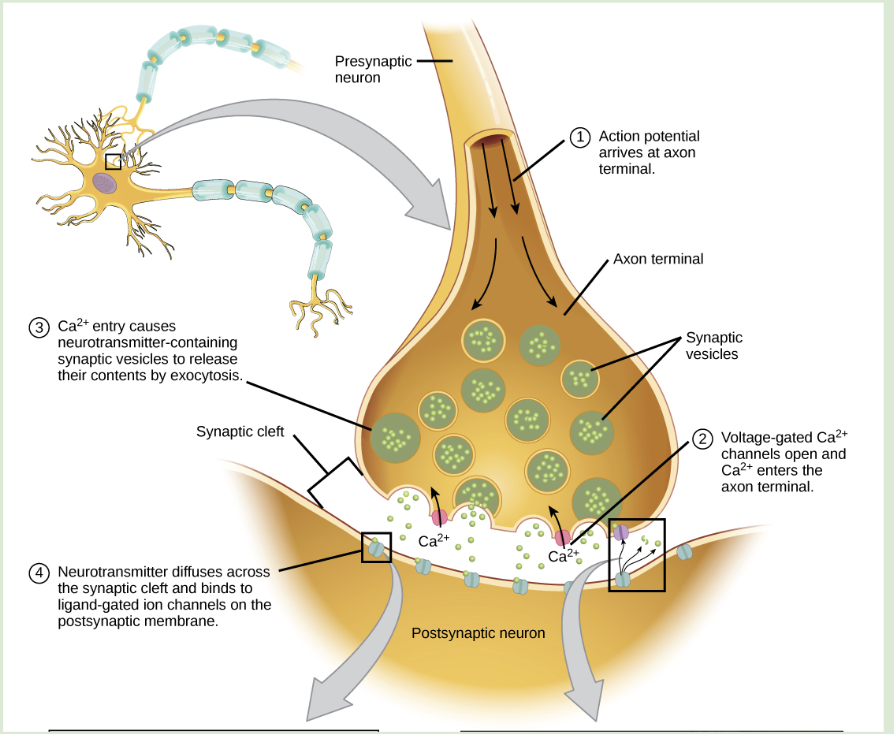
Steps involved in synaptic transmission
Action potential arrives at the axon terminal
Voltage-gated calcium channels open: Calcium ions diffuse into the axon terminal
Movement of vesicles: Calcium ions trigger the movement of vesicles containing neurotransmitters to the presynaptic membrane for release into the synaptic cleft through exocytosis
Neurotransmitters diffuse across the synaptic cleft and bind to ligand-gated ion channels on the post synaptic membrane
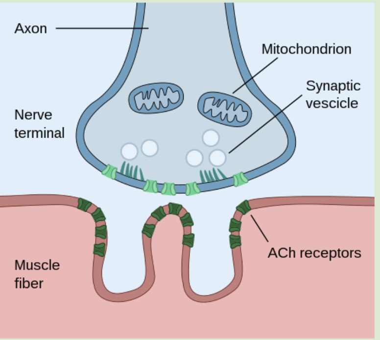
Acetylcholine
Is an example of a neurotransmitter that exists in many types of synapses, including neuromuscular junctions.
Neuromuscular junctions are synapses between axon terminals of motor neurons and muscle fibres.
Release of acetylcholine into the synaptic cleft generating an action potential in the postsynaptic membrane
The arrival of an action potential at an axon terminal results in the release of ACh into the synaptic cleft
ACh diffuses across the synaptic cleft
ACh binds to transmembrane ACh receptors on the postsynaptic membrane, which opens ligand-gated sodium channels
Sodium ions (Na+) flow into the neuron, generating an excitatory postsynaptic action potential
Protein receptors
Play a key role in cell-to-cell communication
Protein receptors have binding sites with specific shapes and chemistry that allows ligands to bind with them
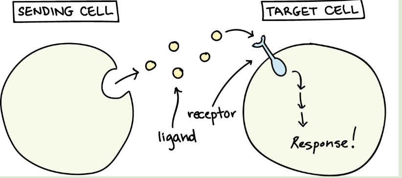
Ligand
Are signaling chemicals that bind to protein receptors, and cause a change in metabolism within the cell
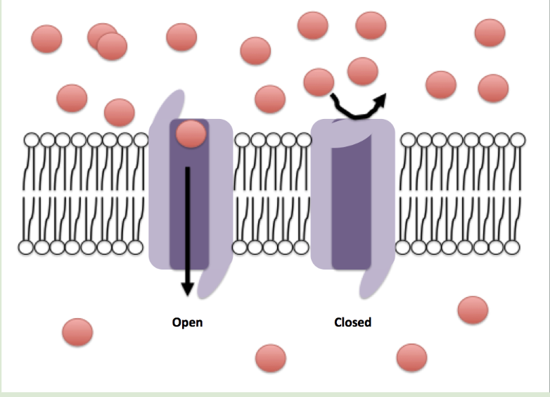
Ion channels and their function
Are integral proteins that allow specific ions to pass through by facilitated diffusion. The pore in the ion channels is hydrophilic, allowing specific ions to enter and pass through.
Gated ion channels
Channels allowing the movement of ions under controlled conditions
Ex) Voltage gated channels, Ligand gated channels.
Voltage gated channels
which respond to changes in membrane potential difference. (voltage) -They open and close in response to voltage
Ex)
Na+/K+ voltage gated channels : are involved in the movements of action potential along neurons
Ca+ voltage gated channels: are involved in synaptic transmission, the transfer of a nerve impulse from one neuron to another.
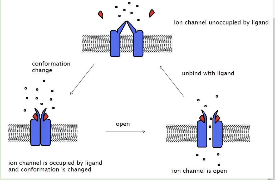
Ligand (neurotransmitter) gated channels
which respond to a ligand attaching to the channel
Ex)
aCH
Acetylcholine is a ligand that attaches to a sodium ion channel.
When acetylcholine is attached to the channel, the channel opens, allowing sodium ions to enter a neuron through the postsynaptic membrane.
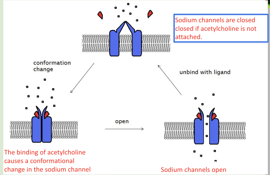
Acetylcholine receptors
Are transmembrane protein receptors found in the postsynaptic membrane of axons
What occurs when ACh binds to the receptor?
ACh binds to ligand-gated sodium channels
Acetylcholine is a ligand which attaches to a sodium ion channel.
When acetylcholine is attached to the channel, the channel opens, allowing sodium ions to enter a neuron through the postsynaptic membrane.
Sodium ions entering the cell changes the voltage across the plasma membrane and may lead to an action potential.
Exogenous chemicals
Come from sources outside of living things
may interfere with synaptic transmission.
Ex)
Neonicotinoids, which bind to cholinergic protein receptors and prevent the binding of acetylcholine.
Cocaine, which prevents the reuptake of dopamine.
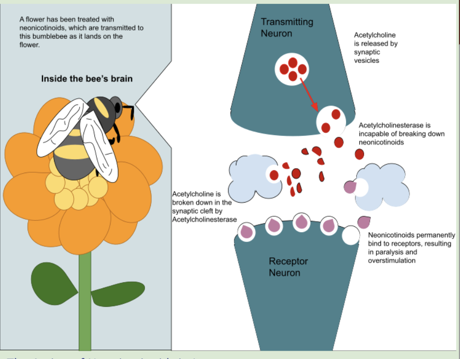
Neonicotinoids
Are a group of chemicals that are used in pesticides around the world
However, evidence shows that it kills bees (non-targeted insects)
The neurotransmitter acetylcholine binds to nicotinic cholinergic receptors in the postsynaptic membrane.
Neonicotinoids are a group of chemicals that also bind to these receptors.
The neonicotinoids bind irreversibly to the acetylcholine receptors of insects.
This blocks synaptic transmission, resulting in paralysis and death of the insect.
Neonicotinoids doesn’t have a great effect on humans because….
The shape of the acetylcholine receptors in humans is different than insects.
Neonicotinoids do not bind to human receptors as strongly as they do to insect receptors.
Neonicotinoids are less toxic to humans.
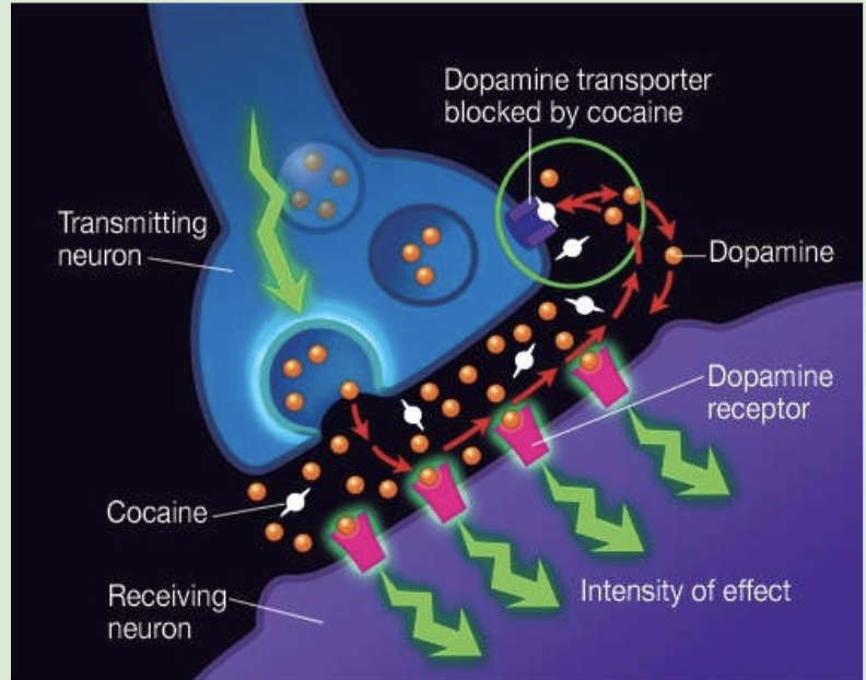
Relationship between cocaine vs dopamine
Dopamine is a neurotransmitter that is responsible for feelings of pleasure and motivation.
Dopamine transporters remove dopamine from synaptic clefts(synaptic gap) between neurons in the brain.
Cocaine binds to and blocks the dopamine transporters, preventing the reuptake of dopamine.
Stays in the synaptic cleft longer and continues binding to the dopamine receptors in the postsynaptic membrane.
Excess dopamine= Euphoria
*Cocaine blocks dopamine from being reuptaken!
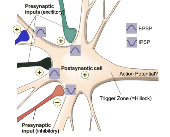
Excitatory neurotransmitters
increase the likelihood of an action potential being generated at the postsynaptic neuron.
They open sodium channels, allowing sodium ions to enter the neuron.
Inhibitory neurotransmitters
decrease the likelihood of an action potential being generated at the postsynaptic neuron.
Inhibitory neurotransmitters bind to protein receptors that allow negative ions to enter the neuron, resulting in the neuron being hyperpolarized.(voltage/potential difference is lower than resting potential)
Summation of effects of excitatory and inhibitory neurotransmitters in a postsynaptic neuron
Neurons have many synapses with other neurons.
Inhibitory and excitatory neurotransmitters may be released at different synapses, sending inhibitory and excitatory signals to the cell body.
The combination of signals is called summation. The summative effects determine whether neurons are activated or not.
Perception of Pain
Nociceptors
are a group of pain receptors found in the skin, which respond to a range of stimuli, including high temperature, acid, or certain chemicals such as capsaicin in chilli peppers.
Respond to:
High temp
Acid
Certain chemicals
When a nociceptor is stimulated…..
Sodium channels open
Sodium ions flow into the nociceptor, causing the threshold potential to be reached.
An action potential is generated, which travels to the brain, where pain is perceived.
Emergent properties
arise from the interaction of component parts.
The whole is greater than the sum of its parts.
Consciousness
is the state of being aware of self and surroundings.
*is an emergent property of neurons in the brain.
Consciousness is an emergent property
Consciousness is an example of an emergent property due to interaction of neurons in the brain.
Consciousness is an emergent property of the interaction of many neurons in the brain, as no single neuron is conscious.
Quorum sensing
is a form of cell to cell communication in bacteria, allowing the bacteria to regulate their behaviour according to population density.
Bacteria produce and release autoinducers, which are chemical messengers that allow cell to cell communication in bacteria.
As a bacterial population grows, the concentration of autoinducers increases.
Once the concentration of the autoinducers reaches a critical concentration, they bind to specific protein receptors.
The binding of the autoinducer to its protein receptor triggers a cascade of metabolic reactions within the bacterial cells.
In many cases, the cascade of reactions leads to activation or repression of specific genes.
leads to a common response from all of the bacteria present.
Example of Quorum sensing: Bioluminescence in Vibrio fischeri
Vibrio fischeri bacteria use quorum sensing to express genes for bioluminescence.
Vibrio fischeri release autoinducers into the environment.
The concentration of the autoinducers increases as the population of Vibrio fischeri bacteria increases.
High concentrations of autoinducer binds to LuxR receptor proteins, and triggers a cascade of metabolic reactions,
which activates genes responsible for bioluminescence in the Vibrio fischeri bacteria.
The enzyme luciferase is synthesised, and catalyzes a chemical reaction, resulting in the emission of light.
*The same process happens in all of the bacterial cells in the population.
Chemical signaling in animals
Hormones
Neurotransmitters
Cytokines
Calcium ions
Hormones
are chemical messengers that are secreted by endocrine glands into the bloodstream.
Hormones travel through the bloodstream to their target tissues. (can travel long distances)
Hormones may bind to receptors on the surface of plasma membranes, or to receptors in the cytoplasm of cells.
Ex) epinephrine, insulin, oestradiol, progesterone and testosterone
Neurotransmitters
are chemical messengers that are released by neurons into synapses.
allow for cell to cell communication between neurons, and between neurons and effectors such as muscles and glands.
Ex) ACh (Acetylcholine )
Cytokines
are chemical messengers involved in the immune response through cell to cell communication within the immune system.
regulate the development, activation and behaviour of cells within the immune system.
Calcium ions
act as secondary messengers within cells in a wide variety of processes.
can be stored intracellularly, or enter cells through gated calcium channels in response to stimuli
act as secondary messengers in muscle contractions and the release of neurotransmitters.
Chemical Diversity of Hormones
There are a wide variety of chemicals used as hormones in animals.
Hormones:
Amine Hormones
Protein Hormones
Steroid hormones
Amine Hormones
are derived from amino acids. The amino acid is modified, including the removal of the carboxyl group of the amino acid.
Ex) Epinephrine
Protein Hormones
are large polypeptides that act as hormones.
Ex) insulin, FSH and LH.
Steroid hormones
are steroids that act as chemical messengers.
Ex) oestradiol, progesterone and testosterone.
Chemical Diversity of Neurotransmitters
Amino acids
Peptides
Amines
Nitrous oxides
Amino acids
Individual amino acids can act as neurotransmitters
Peptides
Chains of amino acids can act as neurotransmitters
Amines
Modified amino acids can act as neurotransmitters
Nitrous oxides
The gas nitrous oxide (NO) is a neurotransmitter
Why is there such diversity in the types of signaling compounds that we have?
Natural selection: The process of natural selection would have selected for genes which produced any signalling molecule that was advantageous.
Diversity of roles: There are many different functions of chemical signals, resulting in a variety of different chemicals being used as chemical signals.
Hormones
Produced by: Endocrine glands
Released into: The bloodstream
How do they travel? Travel long distances around the body
Where do they bind? Attaches to protein receptors on target organs
Where do they act? On many cells
Neurotransmitters
Produced by: Neurons
Released into: Synapses
How do they travel? Quickly diffuse across the synapse
Where do they bind? Protein receptors on the postsynaptic membrane
Where do they act? only act locally
Transmembrane Receptor Proteins
are integral proteins embedded within the plasma membrane of cells.
The signalling chemicals, ligands, are unable to pass through plasma membranes, and they bind to a specific binding site on the receptor protein outside the cell.
The section of the transmembrane protein within the fatty acid tails is hydrophobic.
The sections of the transmembrane protein at the inner and outer surfaces of the plasma membrane are hydrophilic
Ligands and their binding sites on transmembrane proteins are able to interact because they are both hydrophilic.
Intracellular Receptor Proteins
are located within the cytoplasm or the nucleus of the cell.
The signalling chemicals, ligands, diffuse through the plasma membranes, and bind to a specific binding site on the receptor protein inside the cell.
Chemical signals that can diffuse through the phospholipid bilayer are hydrophobic
The binding site of the intracellular receptor proteins will also be hydrophobic.
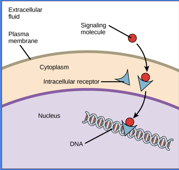
Initiation of Transduction Pathways
The binding of a signalling chemical to a protein receptor initiates a sequence of responses, a cascade of reactions, within the cell.
The binding of a ligand to an intracellular protein receptor initiates a change in gene expression.
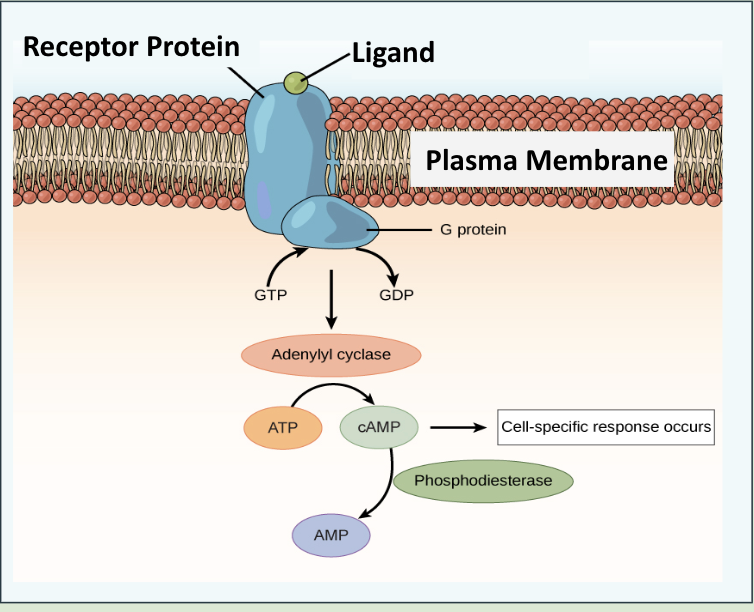
G Proteins***
are found in the cytoplasm of cells coupled with a transmembrane receptor protein, known as a GPCR protein.
composed of three polypeptide subunits: alpha (α), beta (β) and gamma (γ).
In the G proteins’ inactive state, GDP (guanosine diphosphate) is attached to the alpha subunit of the G protein.
Activation of g proteins ***
When an extracellular ligand (hormone) binds to a GPCR protein, the GPCR protein changes shape.
The change in shape of the GPCR protein causes the G protein to release GDP, which is replaced by GTP (Guanosine triphosphate).
This activates the G protein, causing the alpha subunit to dissociate from the other subunits.
The alpha subunit interacts with a secondary messenger, which initiates a cascade of reactions within the cell.
*G protein coupled-receptors are the largest class of cell surface receptors in humans.
Epinephrine = Adrenaline
adrenaline” and “epinephrine” were coined by researchers and are based on the production of the hormone by the adrenal gland; “adrenaline” comes from Latin ad = at and ren = kidney and “epinephrine” comes from old Greek epi = above and nephros = kidney, respectively.
Epinephrine***
is a peptide hormone, and cannot pass through the plasma membrane.
binds to a binding site on a GPCR protein, which changes shape to activate a G protein.
The alpha subunit dissociates from the other two G protein subunits.
The alpha subunit activates an enzyme which converts ATP to cyclic AMP (cAMP).
cAMP is a secondary messenger which triggers a cascade of reactions within the cell, resulting in a change of metabolism.
Tyrosine Kinase Receptors***
are a group of transmembrane receptor proteins that activate kinase enzymes.
have an extracellular ligand binding site, with tyrosine kinase sections within the cytoplasm of a cell
The intracellular tyrosine kinases of the protein receptor are enzymes that catalyse the phosphorylation of itself.
*Phosphorylation is the addition of a phosphate to a molecule.
The hormone insulin binds to a tyrosine kinase receptor.
This initiates a sequence of reactions leading to vesicles containing glucose transporters moving to the plasma membrane.
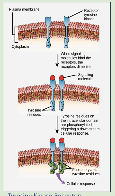
Action of insulin
The hormone insulin binds to transmembrane receptors with kinase activity, which leads to the following events:
Two proteins combine and form a dimer which activates the tyrosine kinase.
The activation of the protein causes the phosphorylation of the tyrosine section of the protein.
Phosphorylated tyrosines attract and bind to other proteins.
This initiates a cascade of reactions, which leads to the movement of glucose transporter vesicles to the plasma membrane.
Glucose is removed from the bloodstream.
Mucles and motility
Movement in sessile species
do not perform locomotion – they remain in a fixed position but move individual body parts.
Ex) Barnacles move appendages known as corral fans, which they use to filter food from water.
Movement in Motile organism
move around while feeding within their territory – some further distances than others.
Ex) Mammals, like cheetahs, have muscles attached to bones which allow them to move.
Muscles
are composed of muscle fibres, which are atypical cells.
Muscle fibres are formed by the fusion of many cells and are multinucleate.
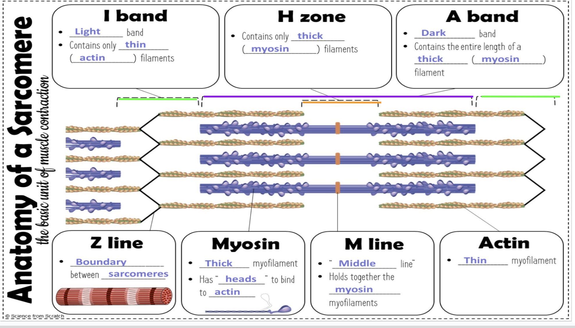
Myofibrils and Sarcomeres
Muscle fibres contain many myofibrils.
Myofibrils are composed of many sarcomeres/basic unit of muscle
The sarcomere contains the protein filaments actin(thin) and myosin(thick), which allow muscles to contract.
Muscle Contraction
Muscles contract as sarcomeres shorten.
Actin and myosin filaments slide over each other.
Sarcomeres
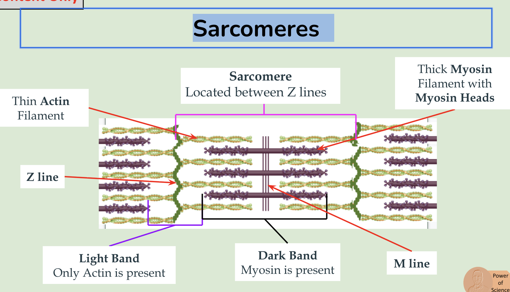
Sarcomere
Steps of muscle contraction***
Contraction of sarcomeres in myofibrils causes muscles to contract.
An action potential arrives at a muscle fibre.
The action potential stimulates the sarcoplasmic reticulum to release calcium ions (Ca2+).
Calcium ions bind to troponin on actin filaments, causing troponin to change shape…
… which causes tropomyosin to move and expose the myosin binding sites on the actin filaments.
Myosin heads form cross-bridges by attaching to the exposed binding sites on actin.
ATP is hydrolyzed to ADP and a phosphate, which causes the myosin head to cock (The myosin head moves towards the Z lines in the sarcomere).
ADP is released from the myosin head, causing a powerstroke, which shortens the sarcomere by the sliding action of actin and myosin.
ATP attaches to the myosin head, breaking the cross-bridge.
This process repeats, resulting in the contraction of the muscle.
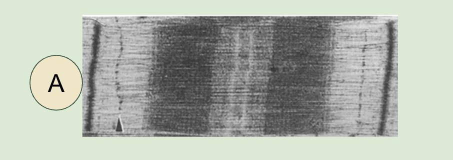
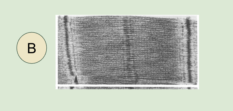
Micrograph of Relaxed Sacromere
Micrograph of contracted Sacromere
as the light band is much shorter, and the Z lines are closer to the center of the sarcomere.
Antagonistic Muscles
Muscles contract to transfer force to bones, but muscles are not able to relax by themselves.
Muscles work in antagonistic pairs.
As one muscle contracts, the other muscle is stretched, and relaxed.
Titin
is a long fibrous elastic protein that stretches from the Z line to the M line (in the middle of a sarcomere).
acts as a molecular spring.
helps sarcomeres to recoil after stretching, and prevents over-extension of the muscle.
Muscle Fibres
are long multinucleate cells which contract when stimulated by an action potential from a motor neuron.
Motor neurons release acetylcholine, a neurotransmitter, which binds to protein receptors on the sarcolemma of muscle fibres.
Neuromuscular Junctions
is the synapse between an axon terminal of a motor neuron and muscle fibre.
The arrival of an action potential at an axon terminal stimulates an action potential in a muscle fibre.
Bones Act as Levers
Vertebrate animals have an endoskeleton.
An endoskeleton is an internal skeleton.
Joints are junctions between two or more bones.
Muscles are anchored to bones by tendons, and the bones act as a lever as muscles contract.
Exoskeletons
Arthropods, such as insects, have a chitinous exoskeleton.
In arthropods, muscles are attached to the exoskeleton.
Synovial Joints
connect bones with a fibrous capsule which contains synovial fluid.
Ball and socket joint
Ex) Hip, shoulder
It allows movement in all axes and planes
Allow the greatest range of movement
Structures found in Synovial joints
Muscles: Muscles contract to generate force, and move muscles.
Bones: Muscles are anchored to bones, which act as levers.
Cartilage: Reduces friction between bones, and absorbs shock.
Synovial Fluid: Lubricates cartilage. Provides cartilage with nutrients and oxygen
Synovial Capsule: Seals synovial fluid, and promotes stability by limiting movement of the joint.
Tendons: Tendons are composed of fibrous tissue, which anchors muscles to bones.
Ligaments: Ligaments are composed of fibrous tissue, which connect bones to bones. The ligament stabilizes the bones in a joint.
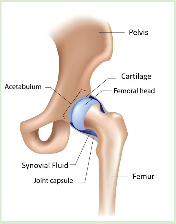
The Hip Joint
is a synovial, ball and socket joint.
The ball on the femur fits into the (acetabulum) socket on the pelvis.
The acetabulum and the femoral head are covered in cartilage.
The joint is stabilized by the joint capsule.
Synovial fluid within the joint capsule lubricates the joint.
Hinge Joints
allow movement in one direction.
allow flexion (bending) and extension of limbs and other body parts.
Ex) Elbows and Knees
Ball and Socket Joints
allow the widest range of motion.
allow flexion, extension, and rotational motion.
Measuring the Range of Joint Movement
Goniometry is the measurement of the range of joint movements.
Students should use a goniometer to measure the range of motion in a number of joints.
Internal and external intercostal muscles
The internal and external intercostal muscles are an example of antagonistic muscles.
The intercostal muscles are involved in ventilation.
The different orientations cause different directions of movement. When one contracts, the other stretches/relaxes, storing potential energy in the titin protein.