Parasitic Platyhelminths - Trematodes
1/20
There's no tags or description
Looks like no tags are added yet.
Name | Mastery | Learn | Test | Matching | Spaced | Call with Kai |
|---|
No study sessions yet.
21 Terms
Trematodes general description
Small dorso-ventrally flattened, non-segmented, worms with simple anatomy.
All digenea (trematode subclass) are parasitic with 2 suckers that they use to attach Whitin the host
→ Oral sucker contains the mouth whose muscular pharynx allows it to pump food into its blind-ending gut
All hermaphrodites except schistosomes
Many have actin spines that help the worms anchor themselves
General treatment of Platyhelminths
Praziquantel → Paralysis of musculature
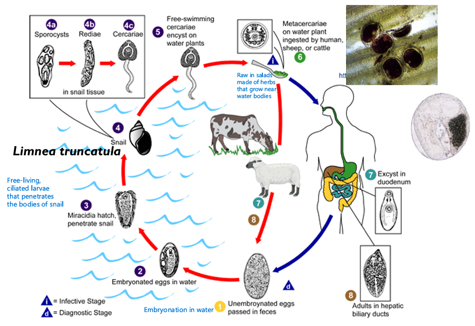
Describe liver flukes
Fasciola hepatica: agent of fascioliasis
Found in bile ducts, gallbladder, and pancreas
Large (3-5 cm)
-Multibranched uterus is situated under the abdominal sucking disk
-Testis are branched too and located in the middle part of the body
Liver fluke life cycle
Mainly:
We acquire the Encysted Metacercaria on aquatic plants/ plants near water bodies
It excysts in the duodenum
Adults are the form found in the hepatic ducts
We (and mammals) shed unembryonated eggs in feces
F. hepatica clinical presentation
Migration through the liver causes symptoms proportionate to worm burden
General symptoms: Fever, abdominal pain, diarrhea, eosinophilia
Heavy infection → Epithelial hyperplasia and fibrosis
Biliary obstruction due to worms can lead to cirrhosis
Fascioliasis diagnosis
Unembryonated eggs in feces
Fascioliasis prevention
Avoiding aquatic vegetables
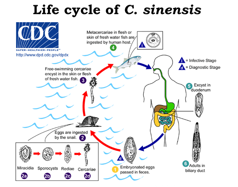
Chinese or Oriental liver fluke
Clonorchis sinensis
Parasite of man, dogs, and cats, and EXTREMELY common southeast Asia
C. sinensis life cycle
Mainly:
We ingest the Metacercariae form in the flesh or skin of freshwater fish
Excystation occurs in the duodenum
Adults are found in biliary ducts
Embryonated (unlike fascioliasis) eggs are passed in feces
Clonorchiasis clinical presentation
Irritation and inflammation of the bile ducts leading to their dilation and the formation of Pigmented Gallstones
Liver: Enlarged, necrotic, tender and high ALT/AST
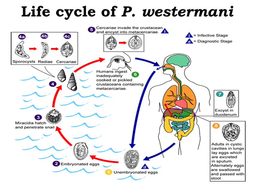
Lung flukes general description
Paragonimus westermani agent of paragonimiasis
Asia, Africa, South America
Plump reddish-brown, oval
Paragonimus westermani life cycle
Mainly:
We ingest crustaceans bearing the encysted form: Metacercariae
They excyst in the duodenum
Adults are found in lungs, where they lay eggs which are then excreted in the sputum or re-swallowed and passed in stools
The form in stools is embryonated eggs
Lung fluke disease clinical presentation
Adult worms in the lungs get encapsulated in granulomas (often 2 at a time), Rupture can result in:
Dry cough
Blood-stained rusty-brown sputum
Inflammation of the pleural membrane
*Note: X-ray looks like X-rays for T
Chronic high worm burden can result in:
Chronic bronchitis
Dyspnea
Fibrosis
Cerebral paragonimiasis symptoms
Granulomatous abscesses → Epilepsy, headaches, fever, nausea, vision impairment, seizures
Paragonimiasis diagnosis
Eggs in sputum or stool samples
May take biopsy to look for eggs in tissues
Serology
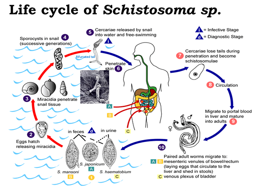
Blood flukes description
Schistosoma spp - agents of Schistosomiasis (Bilharziasis)
Female is long and slender, male is thick and shorter, and forms a characteristic groove in which the female reposes
Schistosoma life cycle
Mainly:
Eggs released from humans
Eggs mature in the marine environment (Snail involvement)
Snails eventually release the Cercariae form, which has a bifurcated tail (like fish) and is capable of free-swimming
Cercariae penetrates the human skin and loses its tail becoming a schistosomulae that can travel hematogenously to the liver
Reach adulthood in liver
Paired male and female adults will migrate to different sites depending on the specie:
S. japonicum and S. mansoni worms will migrate to the mesenteric venules of the bowel/rectum
S. haematobium
Schistosoma virulence factors
Collagenases
Eggs have spines that facilitate their retention
Necrosis-causing enzyme releases leads to the release of the eggs into the intestines or bladder
Schistosomiasis clinical presentation
Type 1/4 Hypersensitivities
Schistosoma dermatitis
Acute schistosomiasis:
Cough
Hepatosplenomegaly (Hyperplasia to make up for lack of blood flow)
Lymphadenopathy (due to obstructions)
Eosinophilia
Chronic schistosomiasis
S. japonicum / mansoni:
Hepatosplenomegaly
Portal hypertension
Esophageal varices
S. haematobium:
Inflammation & fibrosis
Obstruction
Uremia
Hydronephrosis
Pulmonary hypertension
Risk factor for Squamous Cell Carcinoma of the Bladder
Diagnosis of schistosomiasis
Diagnose different species depending on spine position
S. haematobium: Apical spine
S. mansoni: Lateral spine
S. japonicum: Vestigial spine
H→M→J (Straight→Diagonal→Useless)
Schistosomiasis prevention
Avoid sewage water