BAM Week 1
1/24
There's no tags or description
Looks like no tags are added yet.
Name | Mastery | Learn | Test | Matching | Spaced |
|---|
No study sessions yet.
25 Terms
Neurons
Cells arranged in circuits that underlie simplest and most complex behaviors; use electrical, chemical, neurotransmitters, and hormones.
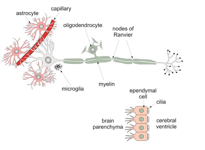
Glial cells + functions
Cells that provide various forms of support and contribute to information processing
Functions:
surround neurons and hold them in place
supply nutrients and oxygen
synaptic transmission
clearance of neurotransmitters
release gliotransmitters (Astrocytes release chemical messengers (e.g. glutamate, ATP) to modulate neuronal activity and synaptic communication.)
Insulation
Destroy pathogens and remove dead neurons
Features of a glial cells
Astrocyte: Supportive glial cells that maintain the blood-brain barrier, provide nutrients to neurons, and maintain ion balance.
Capillary: Small blood vessels that allow exchange of oxygen, nutrients, and waste products between blood and brain tissue.
Microglia: Immune cells in the brain that act as the first line of defense, clearing debris and pathogens.
Oligodendrocyte: Glial cells that create the myelin sheath in the central nervous system (CNS), aiding in faster nerve signal transmission.
Myelin: Fatty substance that insulates axons, speeding up electrical signal conduction.
Nodes of Ranvier: Gaps in the myelin sheath where action potentials are regenerated, speeding up signal transmission via saltatory conduction.
Brain parenchyma: The functional tissue of the brain, including neurons and glial cells.
Ependymal cell: Cells lining the ventricles of the brain and spinal cord, producing and circulating cerebrospinal fluid (CSF).
Cilia: Hair-like structures on ependymal cells that help circulate cerebrospinal fluid.
Cerebral ventricle: Cavities in the brain that contain cerebrospinal fluid (CSF), cushioning and protecting the brain.
Afferent vs Efferent
Afferent: Incoming sensory information coming into the CNS
Efferent: Outgoing information leaving the CNS
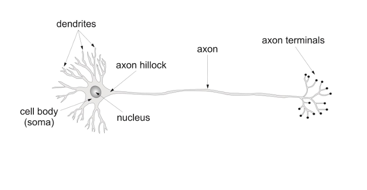
Neuron Structure
Cell Body (Soma): Integrates incoming signals and maintains neuron life; contains the nucleus and organelles.
Dendrites: Receive electrical signals from other neurons and send them to the cell body.
Nucleus: Contains DNA and controls cell functions like protein synthesis and response to stimuli.
Axon Hillock: Initiates the action potential and decides if a signal should be sent down the axon.
Axon: Transmits electrical signals away from the cell body to other neurons, muscles, or glands.
Axon Terminals: Release neurotransmitters to communicate with other neurons, muscles, or glands at the synapse.
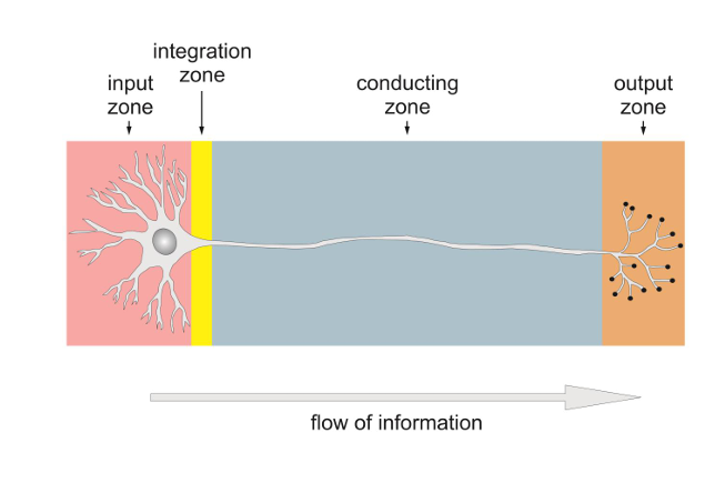
Flow of information in neurons
Input Zone: Composed of dendrites and cell body; receives signals from other neurons.
Integration Zone: Located at the axon hillock; integrates incoming signals and decides whether to initiate an action potential.
Conducting Zone: The axon; transmits electrical signals (action potentials) over long distances.
Output Zone: The axon terminals; release neurotransmitters to communicate with other neurons, muscles, or glands.
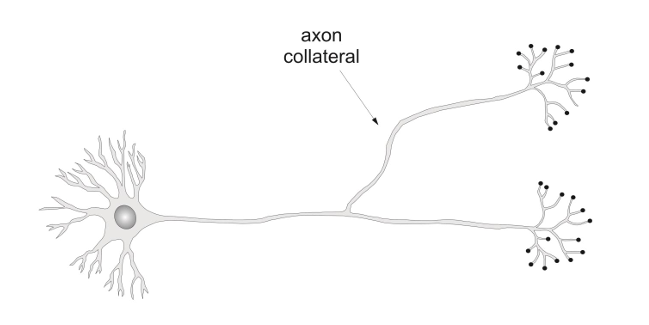
Axon collateral
An axon collateral is a branch of an axon that allows a neuron to send its signal to multiple target cells or regions. It helps in transmitting the action potential to different places, increasing the reach of the neuron’s output.
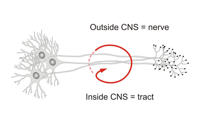
Bundle of neurons
Outside CNS = nerve
Inside CNS = tract
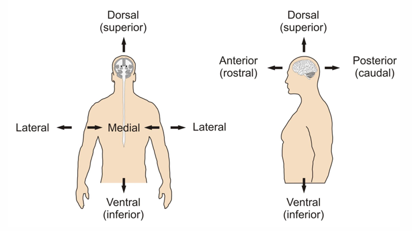
Anatomy Language
Dorsal (Superior): Toward the top or back (e.g. top of the brain).
Ventral (Inferior): Toward the bottom or belly side.
Medial: Toward the midline of the body or brain.
Lateral: Away from the midline, toward the sides.
Anterior (Rostral): Toward the front or nose end.
Posterior (Caudal): Toward the back or tail end.
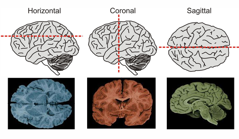
Anatomical orientation
Horizontal Plane: Divides the brain into top and bottom halves.
Coronal Plane: Divides the brain into front and back.
Sagittal Plane: Divides the brain into left and right sides.
Three parts of the nervous system
Central nervous system (mediated behavior)
brain and spinal cord
Somatic nervous system (transmits sensation, produces movement)
cranial nerves and spinal nerves
Automatic nervous system (balances internal functions)
sympathetic division (arousing, fight/flight)
Parasympathetic division (calming, rest/digest)
Central Nervous System (CNS)
The part of the nervous system that consists of the brain and spinal cord, responsible for processing information and coordinating actions.
Somatic Nervous System (SNS)
The part of the nervous system responsible for voluntary motor control and transmitting sensory information to the central nervous system, consisting of cranial and spinal nerves.
Cranial Nerves
Part of the SNS
Cranial Nerves:
Afferent sensory input to the brain from eyes, ears, nose and mouth
Efferent motor output to facial muscles, tongue and eyes
Both afferent and efferent functions like sensation and motor control in the face
Spinal Nerves
Part of the SNS
Spinal Nerves:
Functionally equivalent to the cranial nerves
31 pairs of nerves (8 cervical, 12 thoracic, 5 lumbar, 5 sacral, 1 coccygeal)
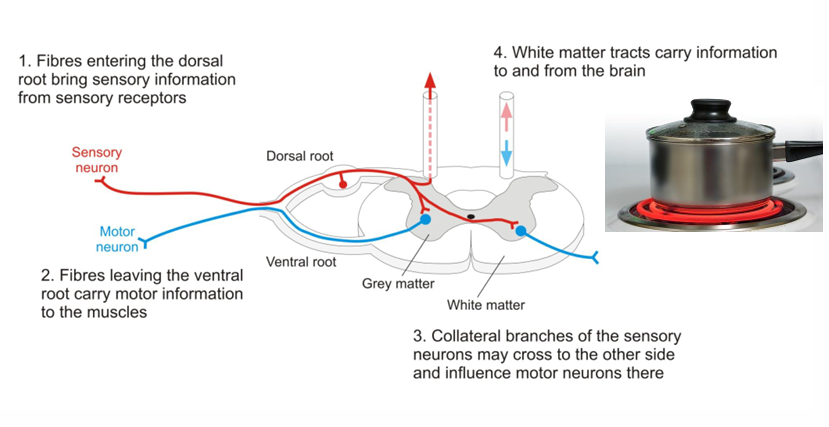
Connections of the spinal nerves:
Emerges from the spinal cord via dorsal (sensory) and ventral (motor) roots.
Dorsal root carries sensory input to the spinal cord.
Ventral root carries motor output to muscles.
Roots merge to form a mixed spinal nerve (both sensory & motor).
Then branch into dorsal and ventral rami to innervate different body regions.
Automatic Nervous System (ANS)
The part of the nervous system that regulates involuntary bodily functions, including heart rate and digestion, and is divided into the sympathetic and parasympathetic divisions.
Sympathetic vs Parasympathetic nervous system
Both part of the ANS
Sympathetic nervous system
Neurons in the spinal cord that innervate the sympathetic chain, which runs along each side of the spinal column
Sympathetic activation prepares the body for action.
Parasympathetic nervous system
Neurons that arise in the cranial nerves and the sacral spinal cord
Parasympathetic activation is often in opposition to sympathetic activity
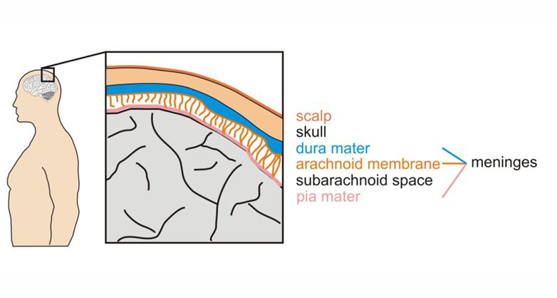
Surface features of the brain
Scalp: Outer skin covering the head.
Skull: Bone that encases the brain.
Meninges (3 protective layers):
Dura Mater: Tough outer layer
Arachnoid Membrane: Web-like middle layer
Pia Mater: Thin inner layer on brain surface
Subarachnoid Space: Between arachnoid and pia; contains cerebrospinal fluid (CSF)
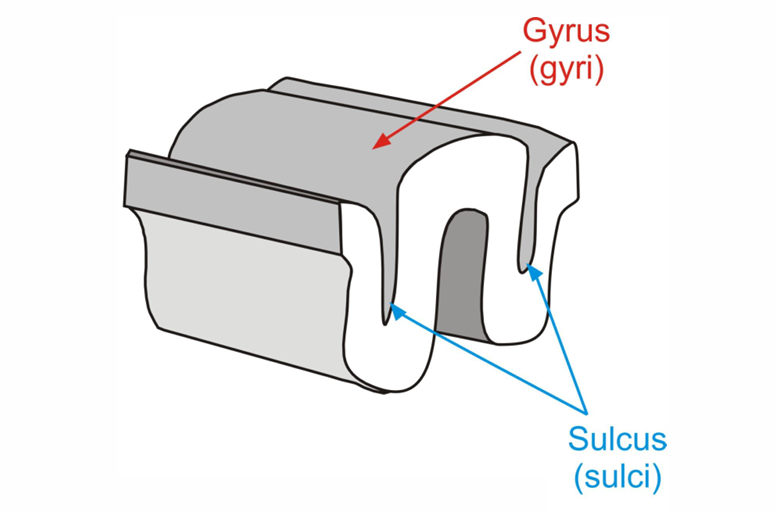
Gyrus: Raised ridge or fold on the brain's surface.
Sulcus: Groove or valley between gyri; increases surface area for neurons.
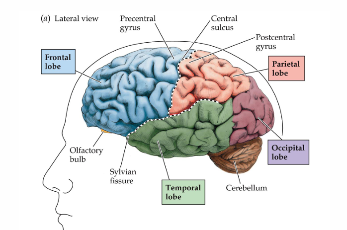
Lobes of the brain
Frontal Lobe: Involved in decision-making, movement, and planning.
Parietal Lobe: Processes sensory information like touch and spatial awareness.
Occipital Lobe: Primary center for visual processing.
Temporal Lobe: Involved in hearing, language comprehension, and memory.
Internal Features of the brain
Cerebral Ventricles: Fluid-filled cavities within the brain that contain cerebrospinal fluid (CSF), which cushions and nourishes the brain.
Choroid Plexus: A network of cells in the ventricles that produces cerebrospinal fluid.
Cerebrospinal Fluid (CSF): Fluid that circulates around the brain and spinal cord, providing protection, nutrients, and waste removal.
Gray Matter: Areas of the brain rich in neuron cell bodies, responsible for processing information.
White Matter: Consists of myelinated axons that transmit signals between different parts of the brain.
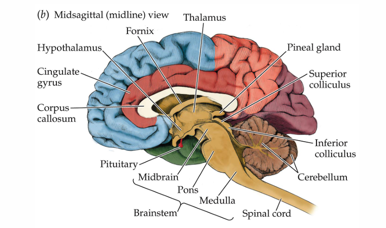
Parts of the CNS
Spinal Cord: Transmits signals between the brain and body; involved in reflexes.
Brainstem and Cerebellum: Controls basic functions (e.g., heart rate, breathing) and motor coordination (balance, posture).
Hindbrain: Includes the medulla, pons, and cerebellum; involved in vital functions and motor control.
Midbrain: Controls sensory processing and reflexes, especially vision and hearing.
Diencephalon ("Between Brain"): Includes the thalamus (sensory relay) and hypothalamus (controls homeostasis and hormone release).
Forebrain: Largest part of the brain; involved in complex functions like thinking, emotion, and motor control.
Cortex: Outer layer of the brain; responsible for higher functions like thinking, memory, and perception.
Basal Ganglia: Involved in motor control and habit formation.
Limbic System: Regulates emotions, memory, and arousal; includes structures like the hippocampus and amygdala.
Olfactory Bulbs: Responsible for processing odors and sending signals to the brain related to smell.
Cortical communication tracts
Cortical Communication: Refers to the way different regions of the brain communicate with each other, allowing for integration of sensory, motor, and cognitive functions.
Projection Tracts: Connect higher cortical areas with lower spinal cord centers (e.g., Corticospinal tract).
Association Tracts: Link different regions within the same hemisphere (e.g., Superior longitudinal fasciculus).
Commissural Tracts: Connect regions between the two hemispheres (e.g., Corpus callosum)
Brain Imaging
Computerized Tomography (CT): Uses X-rays to pass through tissues at different rates. Useful for diagnosing tumors or brain atrophy.
Positron Emission Tomography (PET): Involves a mildly radioactive tracer to track brain activity. While lacking precise detail, it's useful for studying neurotransmitter receptor locations.
Magnetic Resonance Imaging (MRI): Generates a strong magnetic field to create detailed brain images.
Functional MRI (fMRI): Tracks changes in brain activity over time by monitoring blood flow and oxygen levels.
Endocrine System + Hormones
The endocrine system is a network of glands that produce and release hormones to regulate various body functions such as metabolism, growth, and mood.
Hormones are chemical messengers that travel through the bloodstream to target organs, influencing processes like metabolism, growth, and homeostasis.
Major endocrine glands
Pituitary gland – serves as the master gland, controlling the secretions of all other glands.
Thyroid – secretes thyroxine which regulates growth, metabolism and appetite
Adrenal gland - secretes hormones involved in the stress response.
Gonad- secretes sex hormones, which are important for successful reproduction, and regulate sexual motivation and behavior.
Pancreas- secretes hormones that regulate blood sugar