Neuroanatomy NSCI320 w/pics

0.0(0)
learnLearn
examPractice Test
spaced repetitionSpaced Repetition
heart puzzleMatch
flashcardsFlashcards
Card Sorting

1/96

encourage image

There's no tags or description

Looks like no tags are added yet.

Study Analytics
Name
Mastery
Learn
Test
Matching
Spaced

No study sessions yet.

97 Terms

1
New cards

Dura Mater

Meninge closest to the skull. Thickest of the three and is an added layer of protection for the brain and spinal cord
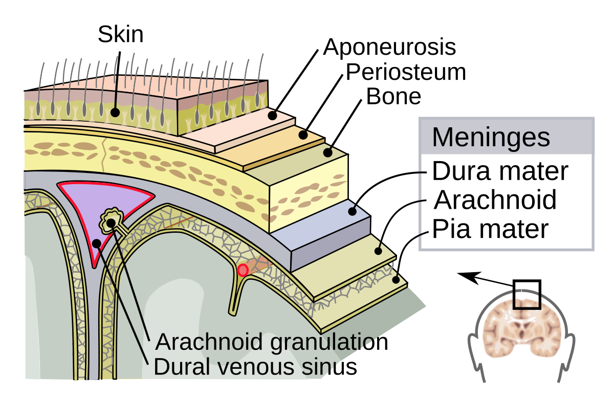
<p>Meninge closest to the skull. Thickest of the three and is an added layer of protection for the brain and spinal cord</p>
2
New cards

Arachnoid Membrane

Middle meninge. Important for protection, blood-brain barrier, and CSF
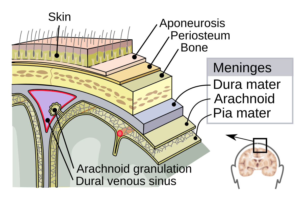
<p>Middle meninge. Important for protection, blood-brain barrier, and CSF</p>
3
New cards

Pia Mater

Meninge closest to the brain. Important for protection, supporting nervous blood vessels, and seperating the brain from CSF
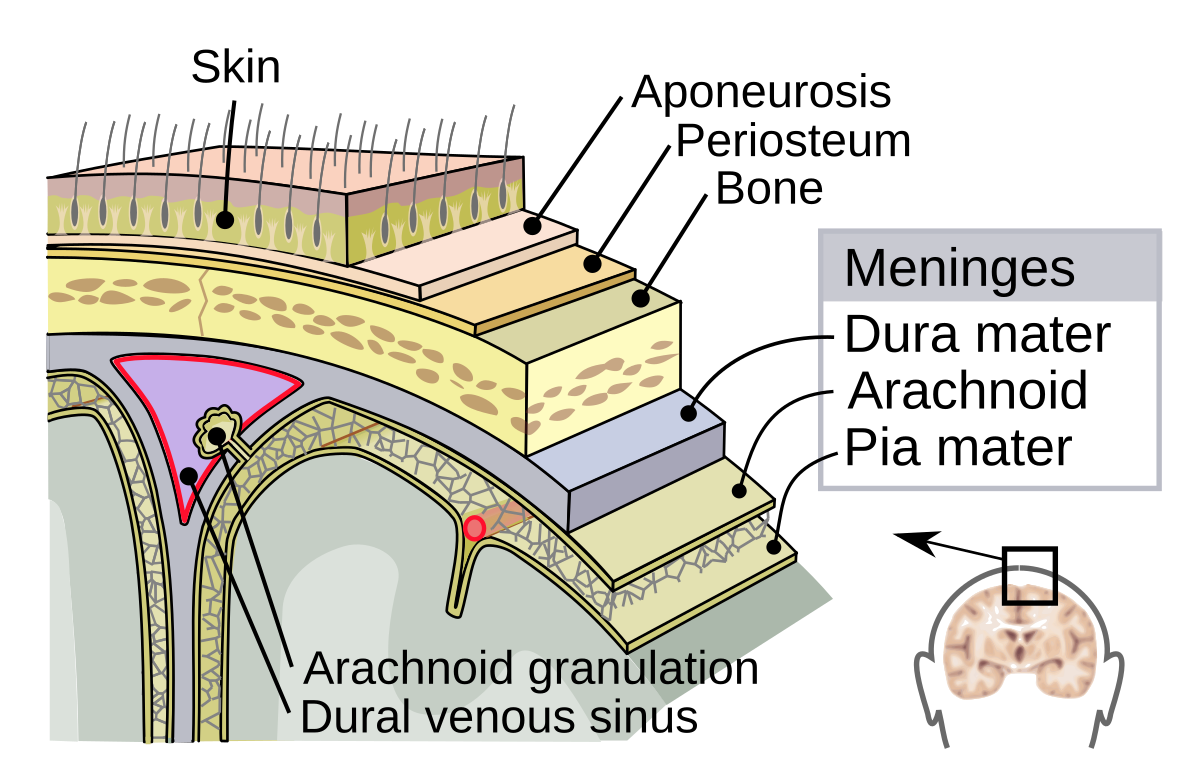
<p>Meninge closest to the brain. Important for protection, supporting nervous blood vessels, and seperating the brain from CSF</p>
4
New cards

Cerebral Cortex / Cerebrum / Telencephalon

Outermost area of the brain. The pink stuff we see. Contains four lobes
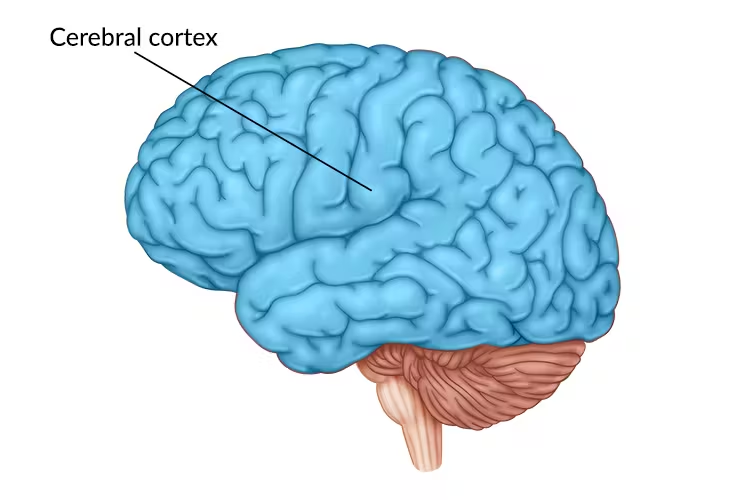
<p>Outermost area of the brain. The pink stuff we see. Contains four lobes</p>
5
New cards

Longitudinal Fissure

Deep grove that runs from the back of the brain to the front. Creates the two hemispheres of the brain
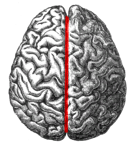
<p>Deep grove that runs from the back of the brain to the front. Creates the two hemispheres of the brain</p>
6
New cards

Cerebellum

portion of the brain located in the back of the head. Important for polishing motor controls and maintaining balance and posture
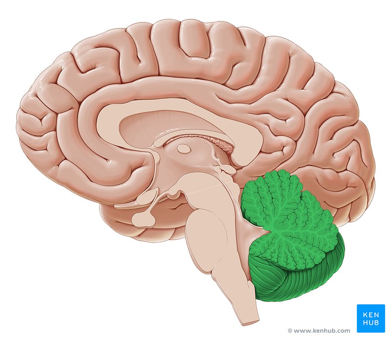
<p>portion of the brain located in the back of the head. Important for polishing motor controls and maintaining balance and posture</p>
7
New cards

Medulla

bottommost region of the brain stem that controls vital functions like breathing, heart rate, and blood pressure
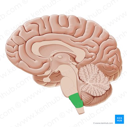
<p>bottommost region of the brain stem that controls vital functions like breathing, heart rate, and blood pressure</p>
8
New cards

Pons

middle region of the brainstem important for motor control and sensory processing
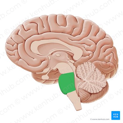
<p>middle region of the brainstem important for motor control and sensory processing</p>
9
New cards

Midbrain

topmost area of brainstem important for motor control and sensory processing
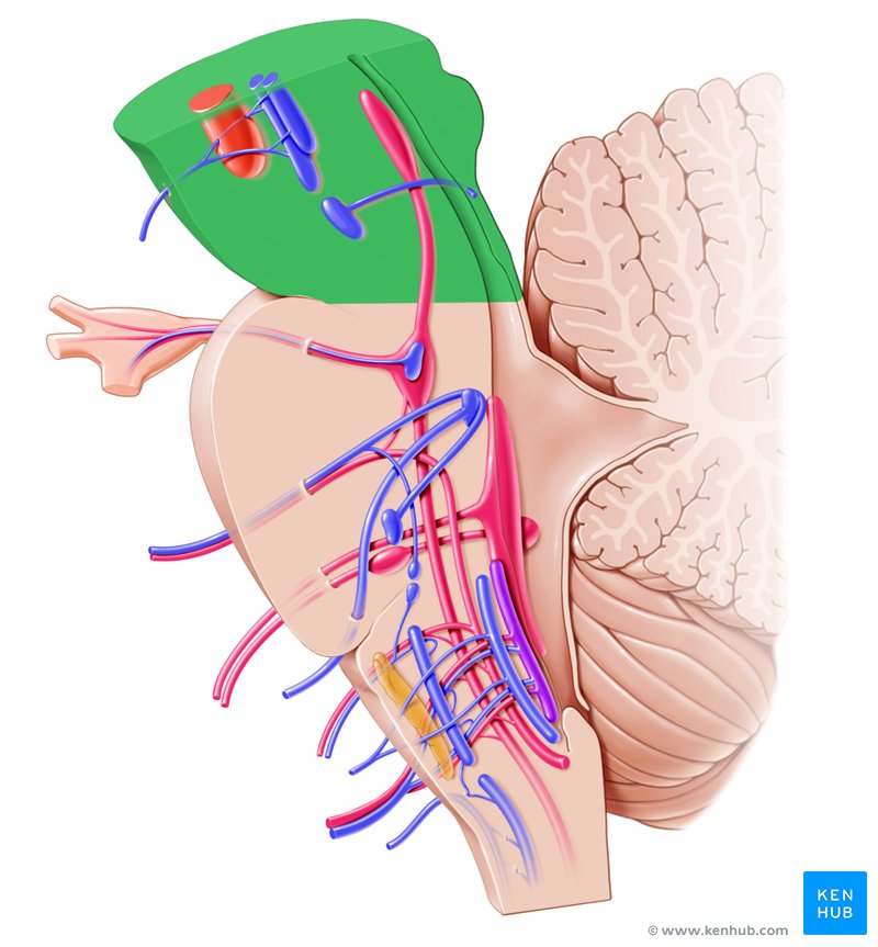
<p>topmost area of brainstem important for motor control and sensory processing</p>
10
New cards

Spinal Cord

cylindrical bundle of nerves that travel down the spine, carrying information between the muscles and the brain
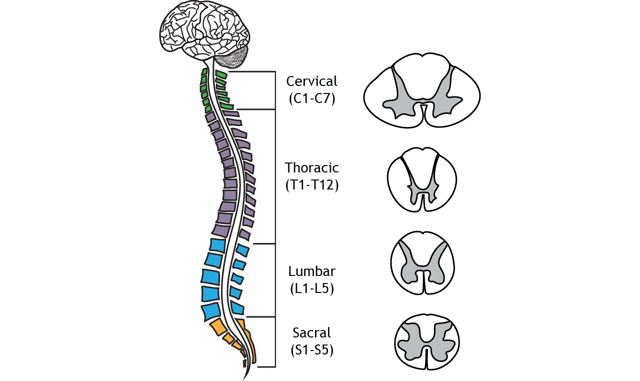
<p>cylindrical bundle of nerves that travel down the spine, carrying information between the muscles and the brain</p>
11
New cards

Dorsal Root

Area that branches of the spinal cord that sends signals from the PNS to the CNS (brain)
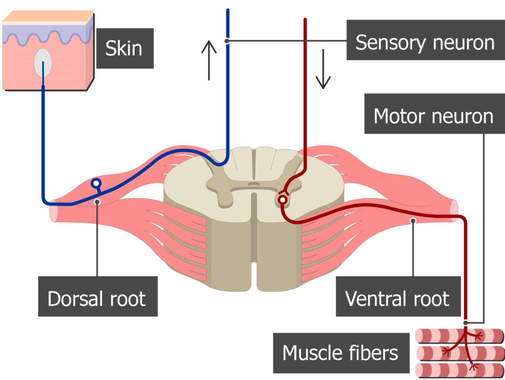
<p>Area that branches of the spinal cord that sends signals from the PNS to the CNS (brain)</p>
12
New cards

Ventral Root

Area that branches off the spinal cord that sends signals from the CNS to the PNS
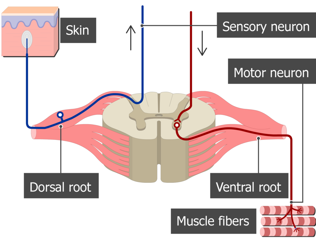
<p>Area that branches off the spinal cord that sends signals from the CNS to the PNS</p>
13
New cards

Gyri (gyrus)

hilly areas of the cortex

<p>hilly areas of the cortex</p>
14
New cards

Sulci (sulcus)

valley-like areas of the cortex

<p>valley-like areas of the cortex</p>
15
New cards

Frontal Lobe

Rostral most lobe of the brain. Important for higher order thinking, motor control, and language. Contains pre motor and primary motor cortices, and brocas area. Seperated from parietal by central sulcus and from temporal by lateral fissure
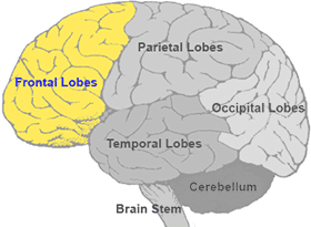
<p>Rostral most lobe of the brain. Important for higher order thinking, motor control, and language. Contains pre motor and primary motor cortices, and brocas area. Seperated from parietal by central sulcus and from temporal by lateral fissure</p>
16
New cards

Parietal Lobe

Central-most lobe of the brain. Important for sensory processing and integrating them into other cognitive functions. Contains primary somatosensory cortex. Seperated from the frontal lobe by the central sulcus
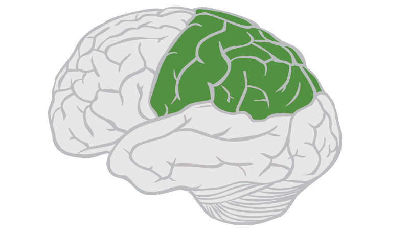
<p>Central-most lobe of the brain. Important for sensory processing and integrating them into other cognitive functions. Contains primary somatosensory cortex. Seperated from the frontal lobe by the central sulcus</p>
17
New cards

Temporal Lobe

Ventral-most lobe of the brain. Important for auditory processing and encoding of memory. Includes superior temporal gyrus and fissure.
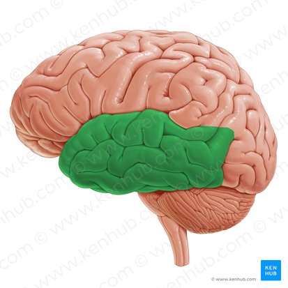
<p>Ventral-most lobe of the brain. Important for auditory processing and encoding of memory. Includes superior temporal gyrus and fissure.</p>
18
New cards

Occipital Lobe

Caudal most lobe of the cortex. Important for processing visual information. Includes primary visual cortex
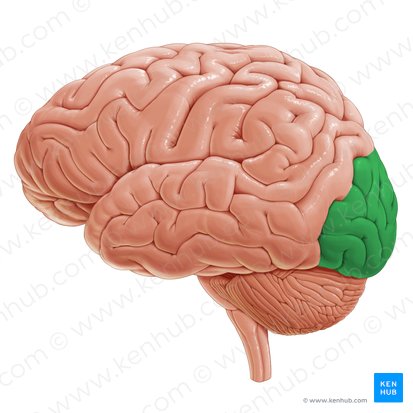
<p>Caudal most lobe of the cortex. Important for processing visual information. Includes primary visual cortex</p>
19
New cards

Lateral Fissure/Sulcus

Large fissure that separates the temporal lobe from the frontal and parietal
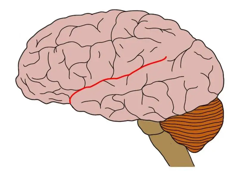
<p>Large fissure that separates the temporal lobe from the frontal and parietal</p>
20
New cards

Central Sulcus / Fissure of Ronaldo

Large fissure that separates the frontal lobe from the occipital lobe. Precentral gyrus contains primary motor cortex and postcentral gyrus contains primary somatosensory cortex
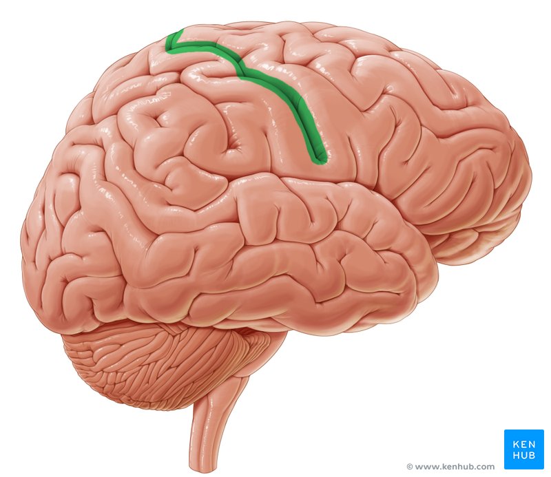
<p>Large fissure that separates the frontal lobe from the occipital lobe. Precentral gyrus contains primary motor cortex and postcentral gyrus contains primary somatosensory cortex</p>
21
New cards

Superior Temporal Sulcus

Sulcus located superiorly on the temporal lobe
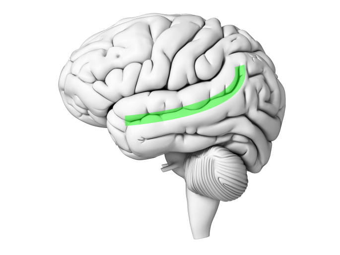
<p>Sulcus located superiorly on the temporal lobe</p>
22
New cards

Olfactory Sulcus

Sulcus located on the bottom of the frontal lobe near the olfactory bulb
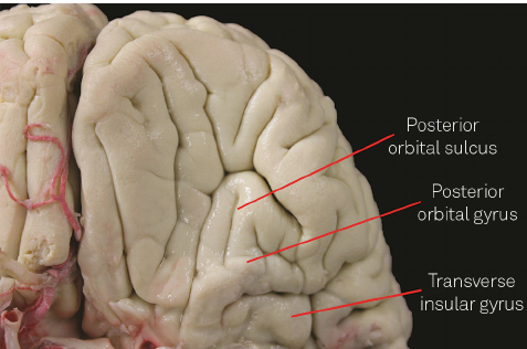
<p>Sulcus located on the bottom of the frontal lobe near the olfactory bulb</p>
23
New cards

Superior Temporal Gyrus

Gyrus located in the temporal lobe directly above the superior temoral sulcus
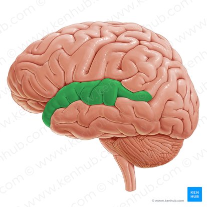
<p>Gyrus located in the temporal lobe directly above the superior temoral sulcus</p>
24
New cards

Precentral Gyrus

anterior side of the central gyrus, located in the frontal lobe. Contains the primary motor cortex
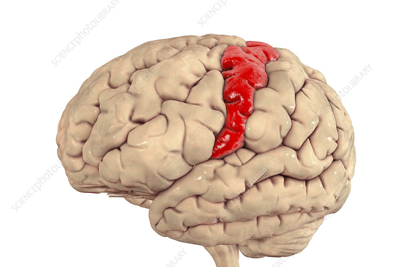
<p>anterior side of the central gyrus, located in the frontal lobe. Contains the primary motor cortex</p>
25
New cards

Postcentral Gyrus

Posterior side of the central gyrus. Located in the Parietal lobe. Contains the primary somatosensory cortex
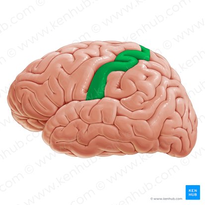
<p>Posterior side of the central gyrus. Located in the Parietal lobe. Contains the primary somatosensory cortex</p>
26
New cards

Prefrontal Cortex

Anterior portion of the frontal lobe. Important for personality
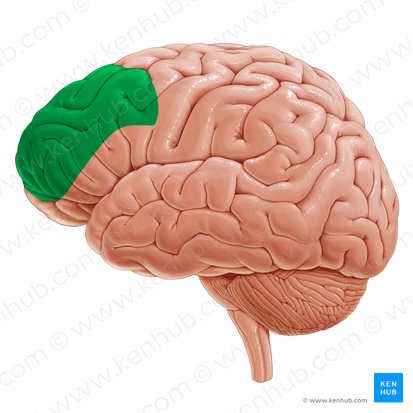
<p>Anterior portion of the frontal lobe. Important for personality</p>
27
New cards

Ventromedial Prefrontal Cortex

Ventral area of the frontal lobe important for cognitive, social, and emotional behaviors
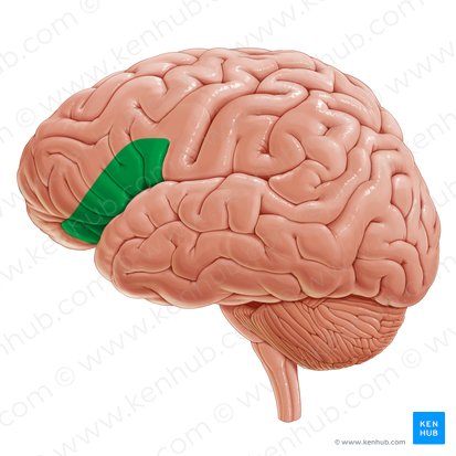
<p>Ventral area of the frontal lobe important for cognitive, social, and emotional behaviors</p>
28
New cards

Striate Cortex / Primary Visual Cortex

First area of the brain to recieve visual information. Processes visual information and spreads it throughout the cortex
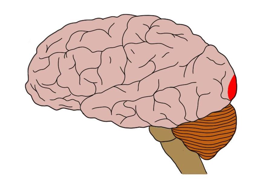
<p>First area of the brain to recieve visual information. Processes visual information and spreads it throughout the cortex</p>
29
New cards

Extrastriate Cortex

Area of occipital lobe surrounding the primary visual cortex.
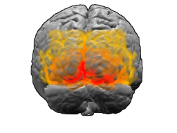
<p>Area of occipital lobe surrounding the primary visual cortex.</p>
30
New cards

Broca’s Area

Region of the frontal lobe crucial for producing speech. Found in the left hemisphere
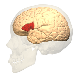
<p>Region of the frontal lobe crucial for producing speech. Found in the left hemisphere</p>
31
New cards

Wernicke’s Area

Area of the posterior superior temporal gyrus crucial for comprehending language. Found in the left hemisphere
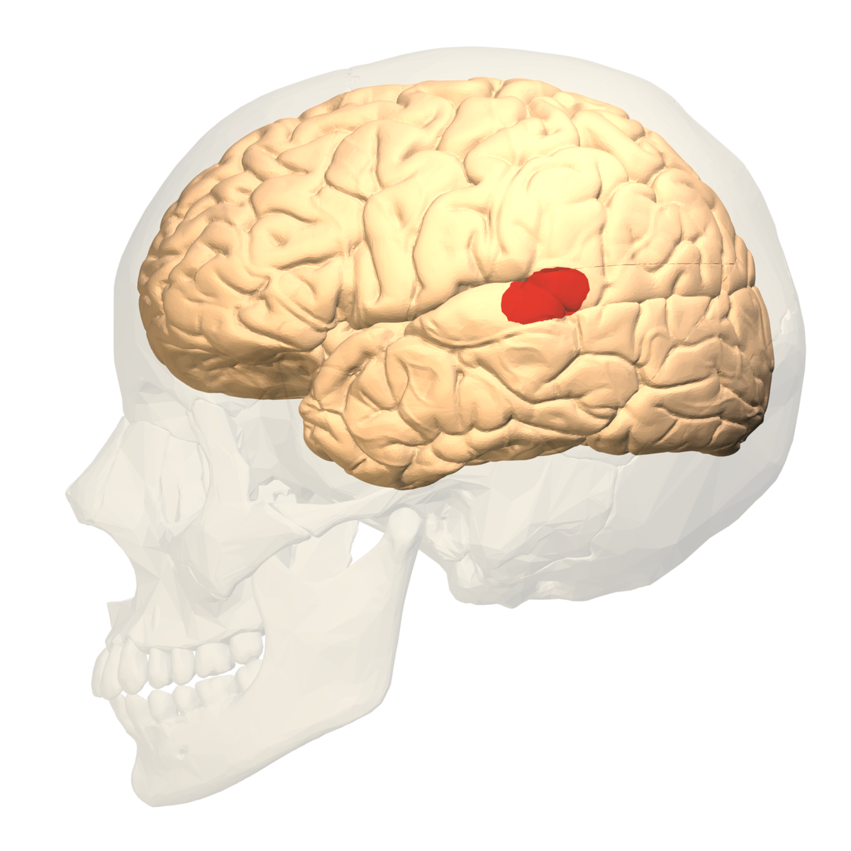
<p>Area of the posterior superior temporal gyrus crucial for comprehending language. Found in the left hemisphere</p>
32
New cards

Cerebellar Vermis

Connects the two cerebellums hemispheres together. Spans vertically down the midsagittal plane
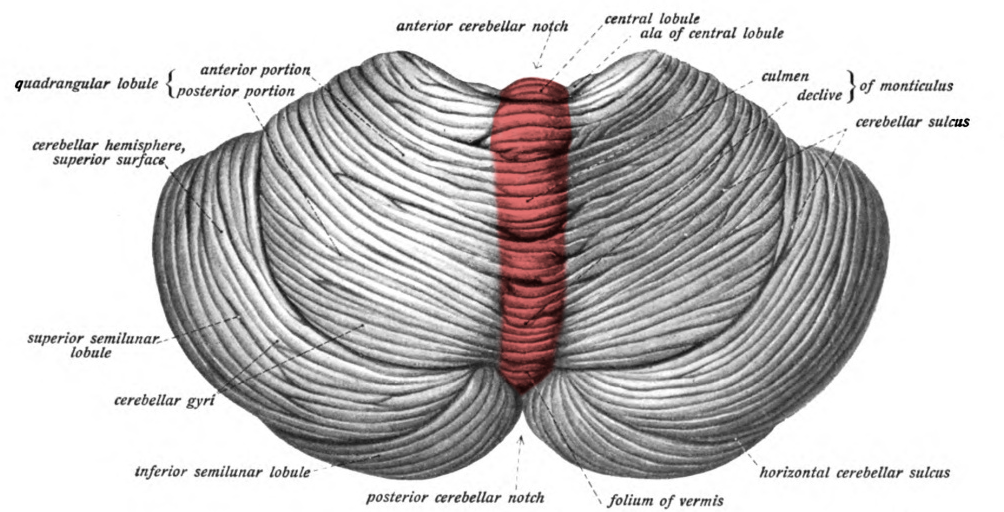
<p>Connects the two cerebellums hemispheres together. Spans vertically down the midsagittal plane</p>
33
New cards

Ventricles

cavernous spaces in the brain that contain Cerebrospinal Fluid (CSF) - help cushion the brain, reduce pressure, and regulates chemical properties.
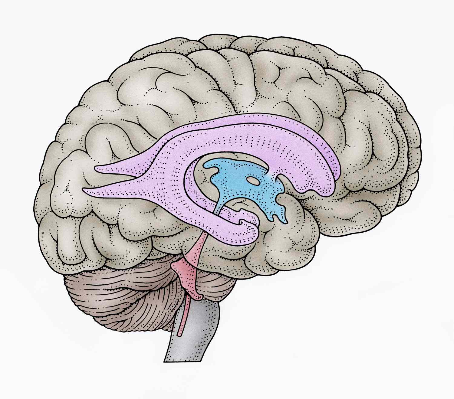
<p>cavernous spaces in the brain that contain Cerebrospinal Fluid (CSF) - help cushion the brain, reduce pressure, and regulates chemical properties. </p>
34
New cards

Choroid Plexus

Structure that secretes CSF into the ventricles. Spongy blood vessel like structure.
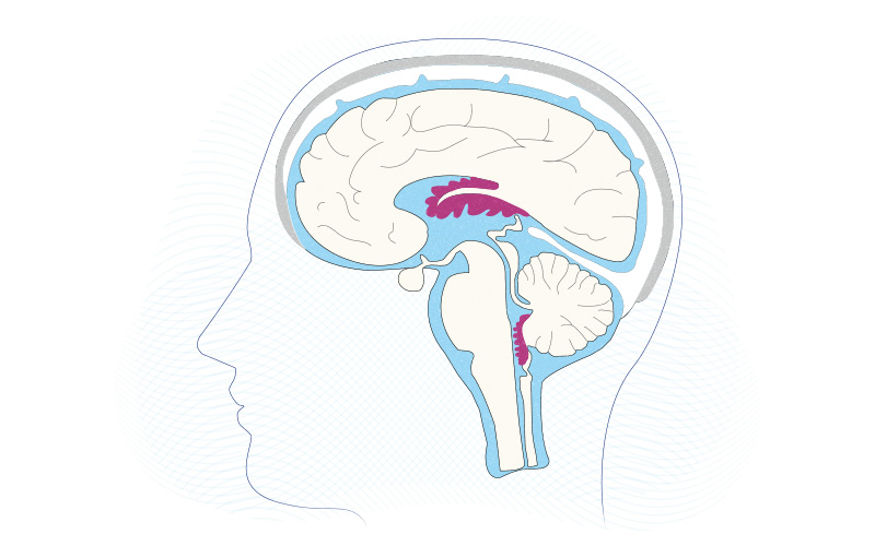
<p>Structure that secretes CSF into the ventricles. Spongy blood vessel like structure.</p>
35
New cards

Massa Intermedia

structure that attaches the left thalamus to the right thalamus by passing through the third ventricle.
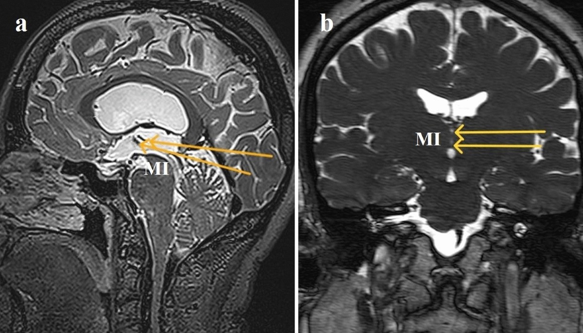
<p>structure that attaches the left thalamus to the right thalamus by passing through the third ventricle.</p>
36
New cards

Foramen

term used to represent a hold or opening in the ventricles that allow CSF to pass from one area to another
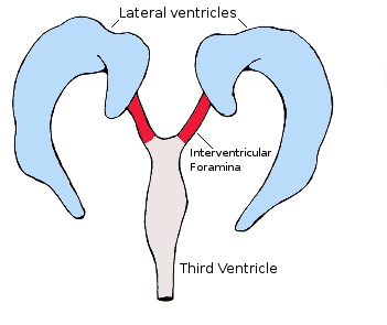
<p>term used to represent a hold or opening in the ventricles that allow CSF to pass from one area to another</p>
37
New cards

Intraventricular Foramen / Foramen of Monroe

a small opening that connects the lateral ventricles to the third ventricle, allowing CSF into it and beyond
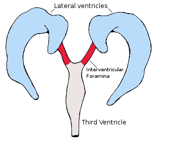
<p>a small opening that connects the lateral ventricles to the third ventricle, allowing CSF into it and beyond</p>
38
New cards

Frontal/Coronal

Vertical plane that runs side to side, splitting the body/brain into a front and back
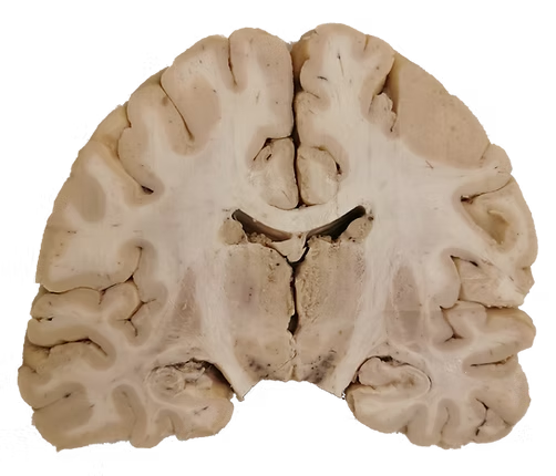
<p>Vertical plane that runs side to side, splitting the body/brain into a front and back</p>
39
New cards

Sagittal/Lateral

Vertical plane that runs front to back, splitting the body/brain into a left and right
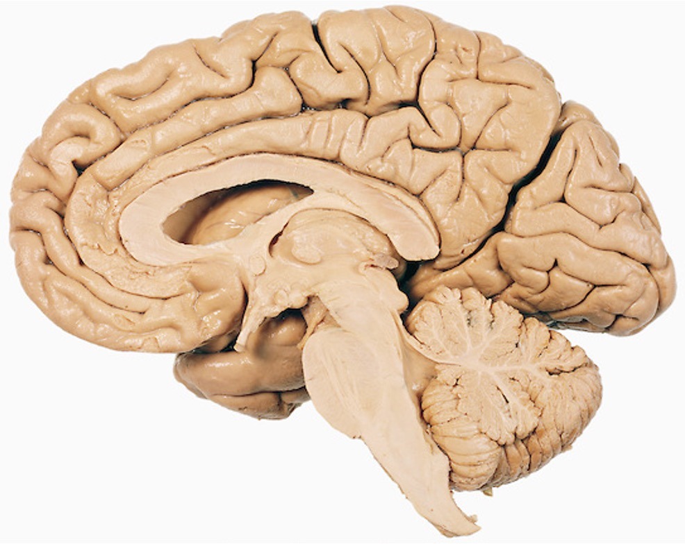
<p>Vertical plane that runs front to back, splitting the body/brain into a left and right</p>
40
New cards

Horizontal/Axial/Transverse

Horizontal plane that runs front to back, splitting the body/brain into a top and bottom
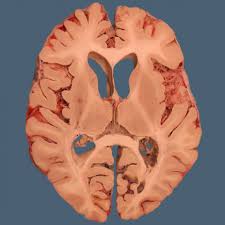
<p>Horizontal plane that runs front to back, splitting the body/brain into a top and bottom</p>
41
New cards

Superior

above

42
New cards

Inferior

Below

43
New cards

Anterior

infront of

44
New cards

Posterior

Behind

45
New cards

Medial

Toward the middle

46
New cards

Lateral

Toward the side

47
New cards

Dorsal

Top

48
New cards

Ventral

Bottom

49
New cards

Basilar Artery

Large blood vessel located on the bottom of the brain
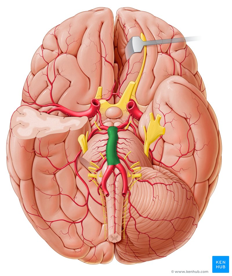
<p>Large blood vessel located on the bottom of the brain</p>
50
New cards

Vertebral Artery

Large blood vessels on the bottom of the brain that come together to form the basilar artery
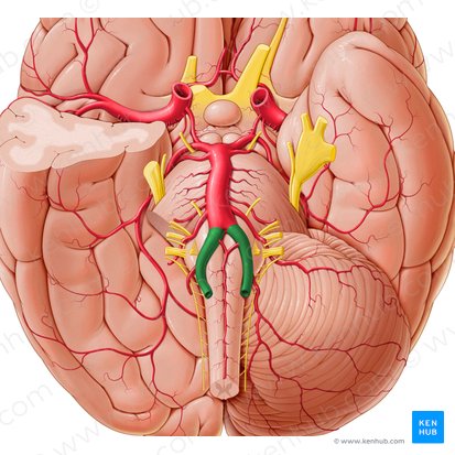
<p>Large blood vessels on the bottom of the brain that come together to form the basilar artery</p>
51
New cards

Pineal Gland

Gland located in the diencephalon, just behind the thalamus. Important for secreting melatonin and regulating sleep cycle. Directly above (superior to) superior colliculi.
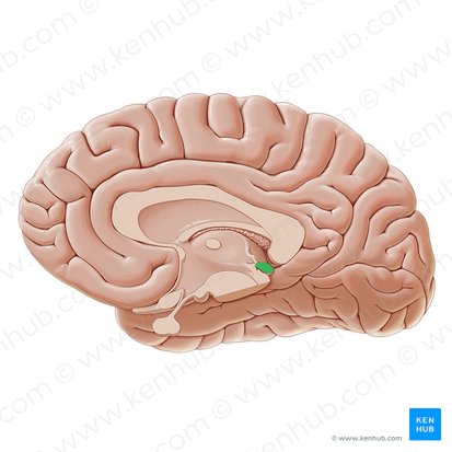
<p>Gland located in the diencephalon, just behind the thalamus. Important for secreting melatonin and regulating sleep cycle. Directly above (superior to) superior colliculi.</p>
52
New cards

Pituitary Gland/Infundibulum

Gland that regulates bodily functions by secreting hormones. Located directly below hypothalamus
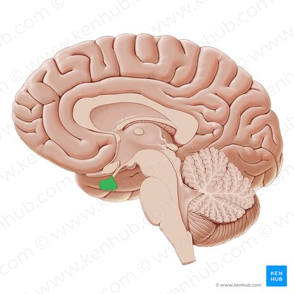
<p>Gland that regulates bodily functions by secreting hormones. Located directly below hypothalamus</p>
53
New cards

Superior Colliculus

Located in the midbrain and is part of the dorsal brain stem. Important for visual attention and eye movements
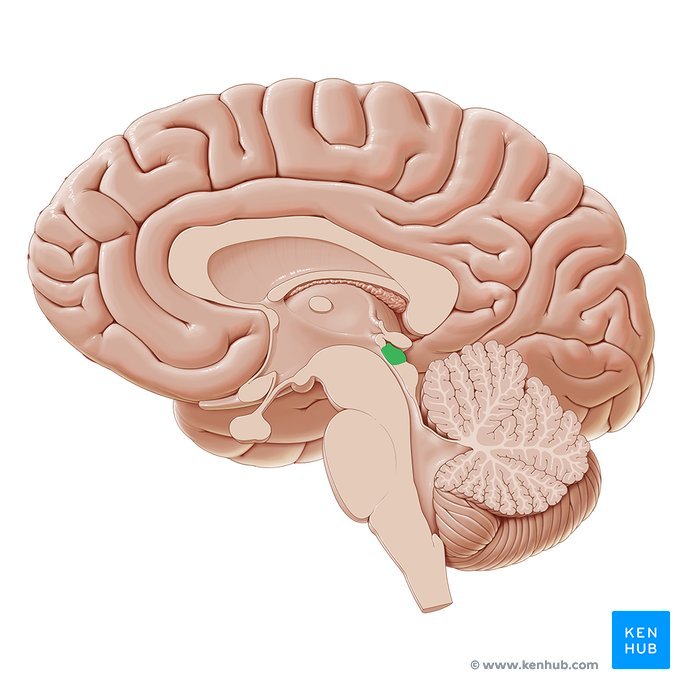
<p>Located in the midbrain and is part of the dorsal brain stem. Important for visual attention and eye movements</p>
54
New cards

Inferior Colliculus

Located in the midbrain directly below the superior colliculus. Important for auditory cues
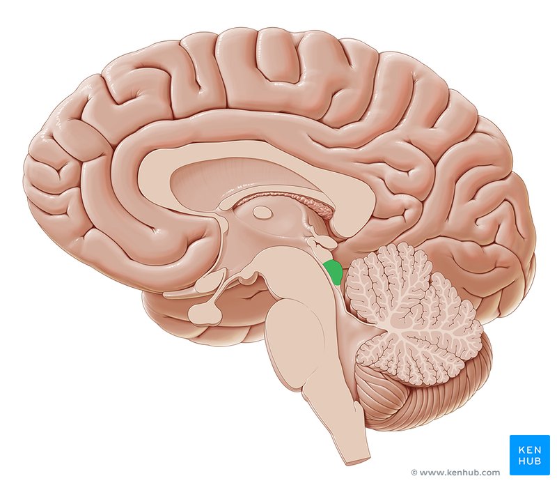
<p>Located in the midbrain directly below the superior colliculus. Important for auditory cues</p>
55
New cards

Optic Nerve

Nerve that sends visual info from the retina to the optic chiasm
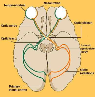
<p>Nerve that sends visual info from the retina to the optic chiasm</p>
56
New cards

Optic Chiasm

Area where optic nerve diverges and visual info heads to the two different hemispheres
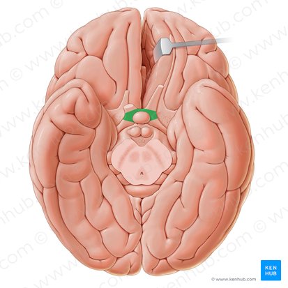
<p>Area where optic nerve diverges and visual info heads to the two different hemispheres</p>
57
New cards

Optic Tract

Post-Optic Chiasm nerve that sends visual info to the thalamus (LGN)
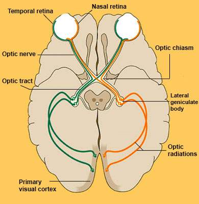
<p>Post-Optic Chiasm nerve that sends visual info to the thalamus (LGN)</p>
58
New cards

Mammilary Body

Small structures located under the brain in the posterior hypothalamus. Important for memory consolidation and spatial navigation
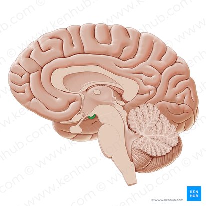
<p>Small structures located under the brain in the posterior hypothalamus. Important for memory consolidation and spatial navigation</p>
59
New cards

Olfactory Nerve (Bulb and Tract)

Nerve that carries olfactory information to the brain. Begins in the bulb where odorants turn into electrical signals, travelling down the olfactory tract
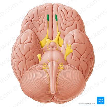
<p>Nerve that carries olfactory information to the brain. Begins in the bulb where odorants turn into electrical signals, travelling down the olfactory tract</p>
60
New cards

Crus Cerebri/Cerebral Peduncle

bundle of nerve in the midbrain that connect the cerebral hemispheres to the cerebellum and other parts of the brainstem. Mickey Ears

<p>bundle of nerve in the midbrain that connect the cerebral hemispheres to the cerebellum and other parts of the brainstem. Mickey Ears</p>
61
New cards

Vagus Nerve

Nerve that begins in the medulla oblongata, sending motor commands to the body
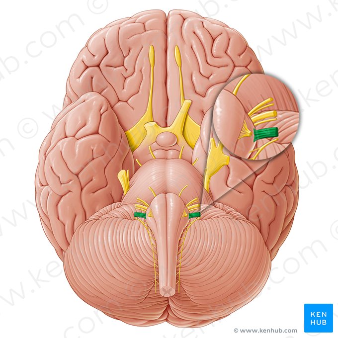
<p>Nerve that begins in the medulla oblongata, sending motor commands to the body</p>
62
New cards

Vestibulocochlear Nerve

Nerve that carries auditory info from the ears to the opposite hemisphere of the brain. Located on the bottom of the brain
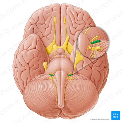
<p>Nerve that carries auditory info from the ears to the opposite hemisphere of the brain. Located on the bottom of the brain</p>
63
New cards

Amygdala

Almond shaped structure located in the temporal lobe in the limbic system. Involved in emotion, memory, and motivation
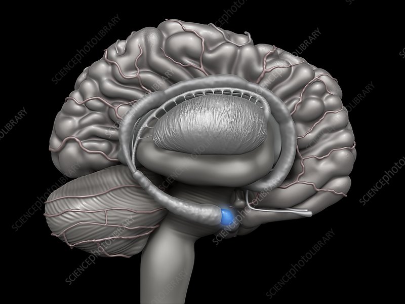
<p>Almond shaped structure located in the temporal lobe in the limbic system. Involved in emotion, memory, and motivation</p>
64
New cards

Fornix

Area in the limbic system responsible for being the primary output pathway for the hippocampus. Connects hippocampus to mammillary bodies and hypothalamus
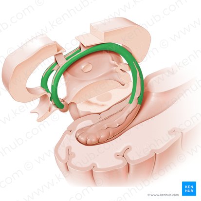
<p>Area in the limbic system responsible for being the primary output pathway for the hippocampus. Connects hippocampus to mammillary bodies and hypothalamus</p>
65
New cards

Hippocampus

Seahorse shaped structure in the limbic system in the temporal lobe important for consolidation of memory and memory.
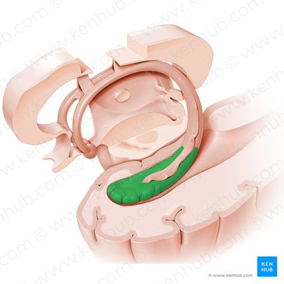
<p>Seahorse shaped structure in the limbic system in the temporal lobe important for consolidation of memory and memory.</p>
66
New cards

Caudate Nucleus

C-shaped structure in the basal ganglia important for movement, learning, and decision-making (green in photo)
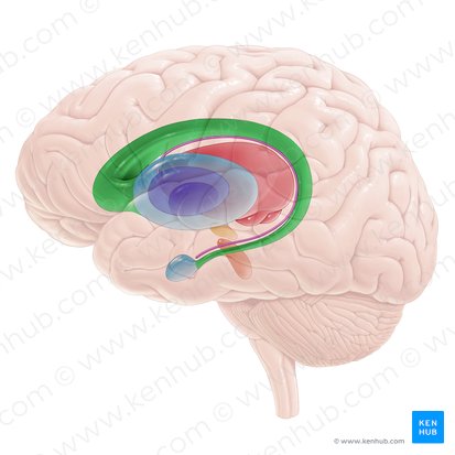
<p><strong><em><u>C</u></em></strong>-shaped structure in the basal ganglia important for movement, learning, and decision-making (green in photo)</p>
67
New cards

Corpus Callosum

Thick band of nerve fibers that connects the two hemispheres with each other. Largest white matter area in the brain
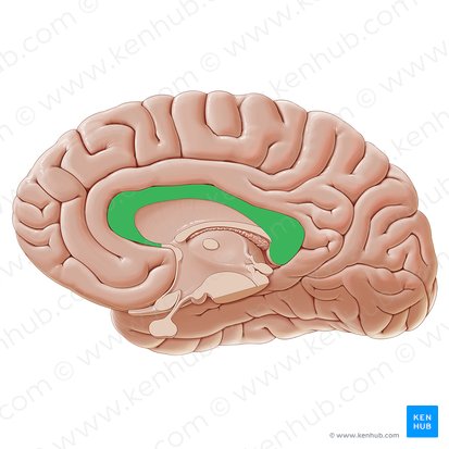
<p>Thick band of nerve fibers that connects the two hemispheres with each other. Largest white matter area in the brain</p>
68
New cards

Anterior Commissure

Bundle of nerve fibers that connects the two hemispheres of the brain at the midline. Primarily connects the olfactory bulbs and temporal lobes. Located below the corpus callosum
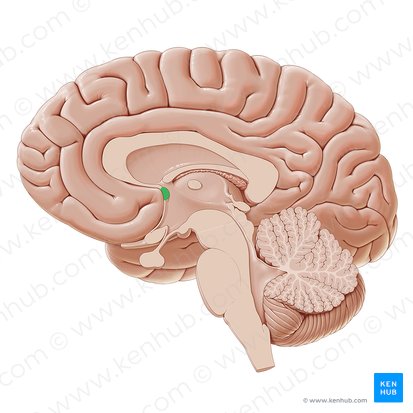
<p>Bundle of nerve fibers that connects the two hemispheres of the brain at the midline. Primarily connects the olfactory bulbs and temporal lobes. Located below the corpus callosum</p>
69
New cards

Posterior Commissure

Bundle of nerve fibers that connects the two hemispheres of the brain. Located dorsally to the midbrain.
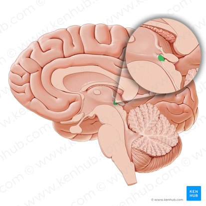
<p>Bundle of nerve fibers that connects the two hemispheres of the brain. Located dorsally to the midbrain.</p>
70
New cards

Globus Pallidus

Structures in the basal ganglia important for regulating movement, reward, and motivation. Laterally surrounded by the putamen
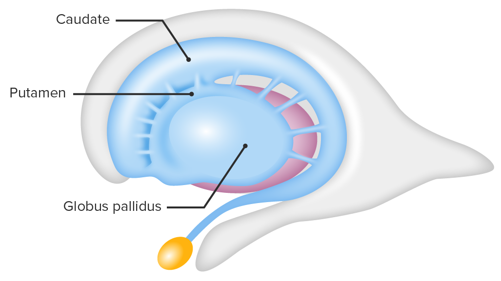
<p>Structures in the basal ganglia important for regulating movement, reward, and motivation. Laterally surrounded by the putamen</p>
71
New cards

Hypothalamus

Structure located under the thalamus that is important for vital bodily functions like sleeping and waking. Part of the diencephalon
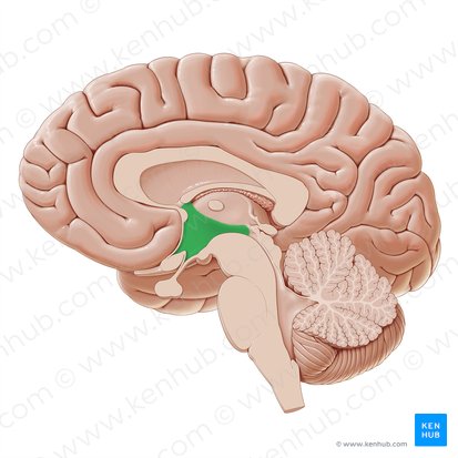
<p>Structure located under the thalamus that is important for vital bodily functions like sleeping and waking. Part of the diencephalon</p>
72
New cards

Insular Cortex (Insula)

Cortical area located in the lateral sulcus. Important for pain, attention, and language. Often called the “fifth lobe”
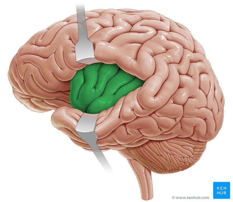
<p>Cortical area located in the lateral sulcus. Important for pain, attention, and language. Often called the “fifth lobe”</p>
73
New cards

Putamen

Basal ganglia structure involved in motor control, cognition, and reward processing. Laterally surrounds the globus pallidus
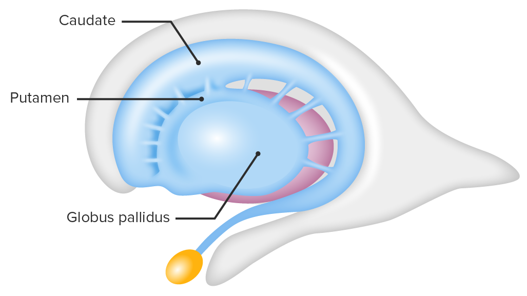
<p>Basal ganglia structure involved in motor control, cognition, and reward processing. Laterally surrounds the globus pallidus</p>
74
New cards

Red Nucleus

Large gray matter area in the upper midbrain important for controlling muscle tone and limb movement. Located in the ventral midbrain tectum
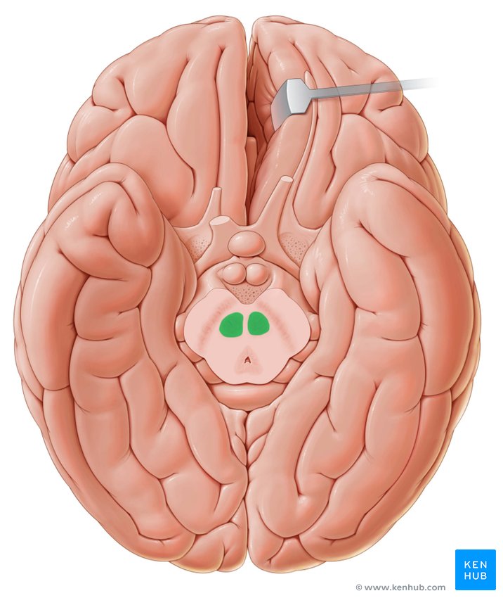
<p>Large gray matter area in the upper midbrain important for controlling muscle tone and limb movement. Located in the ventral midbrain tectum</p>
75
New cards

Substantia Nigra

Area of midbrain important for controlling movement and reward pathways. Disfunctional ones can lead to Parkinsons
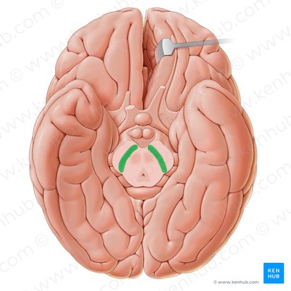
<p>Area of midbrain important for controlling movement and reward pathways. Disfunctional ones can lead to Parkinsons</p>
76
New cards

Thalamus

Structure that is the first to recieve sensory info and relays it to the rest of the brain. Located in the middle of the brain. Superior to the midbrain
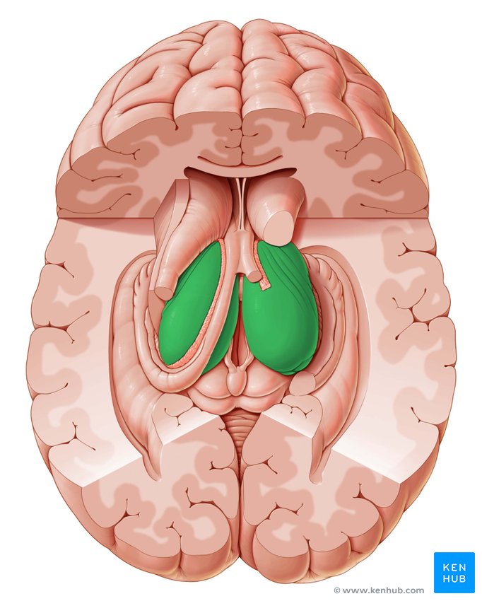
<p>Structure that is the first to recieve sensory info and relays it to the rest of the brain. Located in the middle of the brain. Superior to the midbrain</p>
77
New cards

Arbor Vitae

a network of white matter tracts within the cerebellum. Responsible for coordinating movement and balance. Looks like the roots/branches of a tree
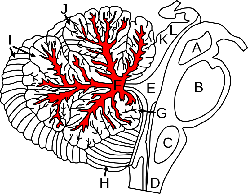
<p><span>a network of white matter tracts within the cerebellum. Responsible for coordinating movement and balance. Looks like the roots/branches of a tree</span></p>
78
New cards

Folia

The leaf-like folds that coveres the surface of the cerebellum. Increases surface area of the cerebellum, allowing for a greater number of neurons to be present
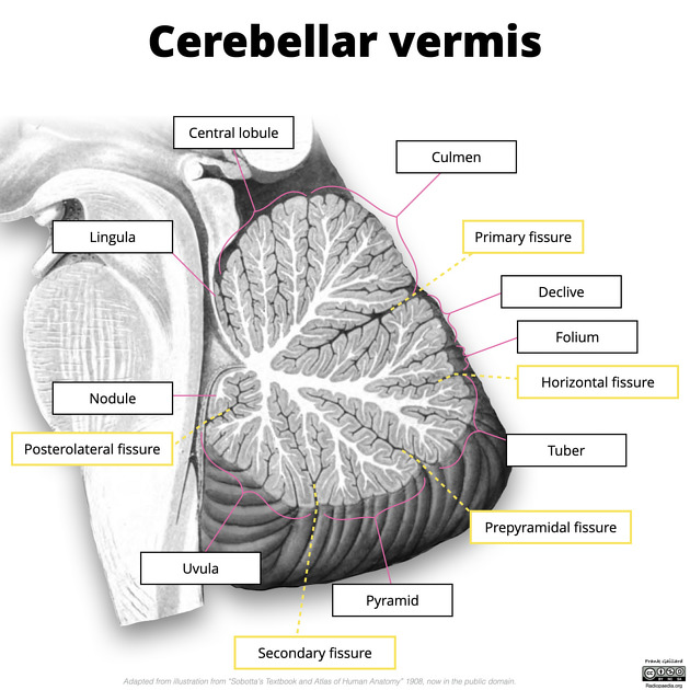
<p>The leaf-like folds that coveres the surface of the cerebellum. Increases surface area of the cerebellum, allowing for a greater number of neurons to be present</p>
79
New cards

Cerebellum Peduncles

three pairs of nerve tracts that connect the cerebellum to the brainstem. They play a crucial role in transmitting information between the cerebellum and other parts of the central nervous system
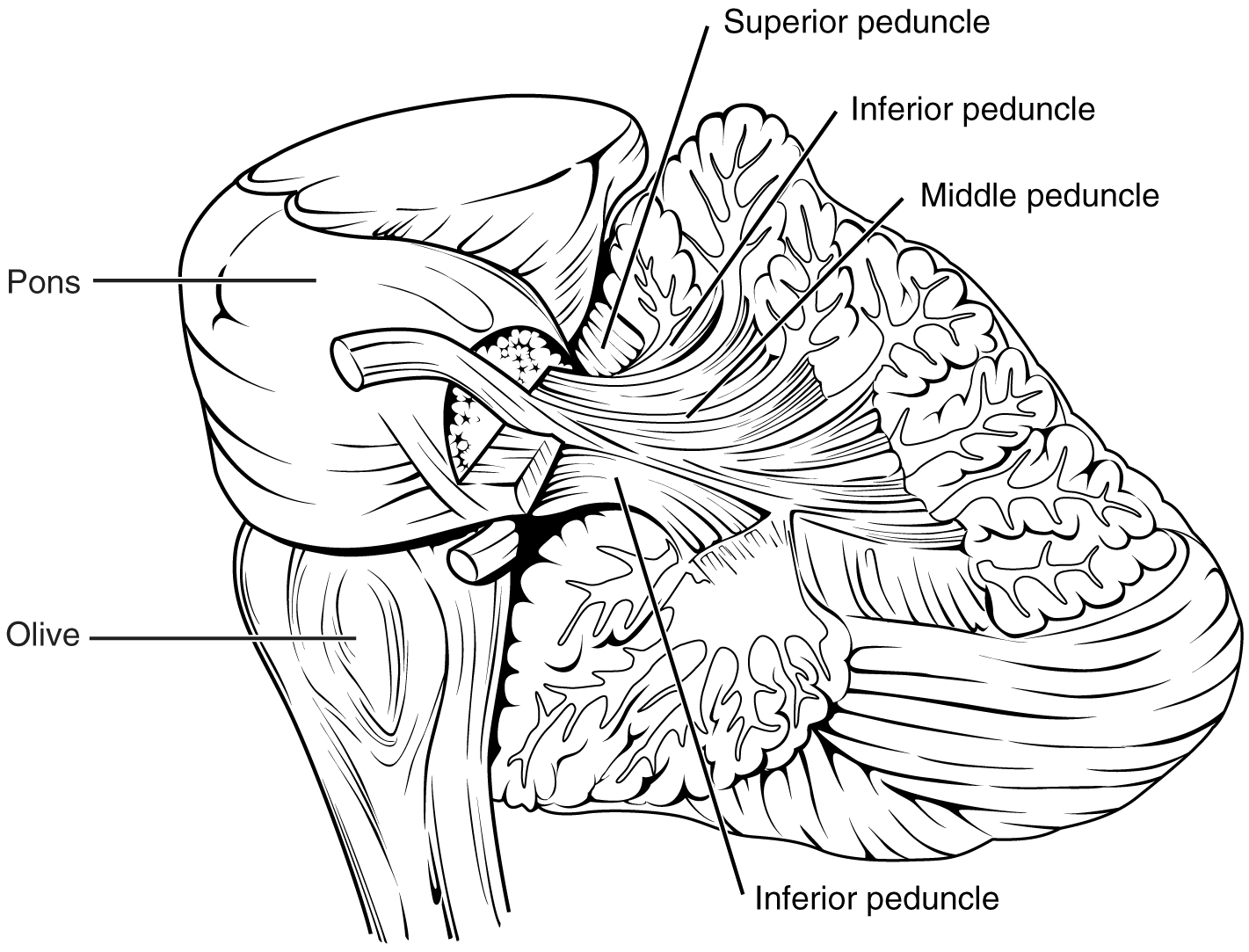
<p><span>three pairs of nerve tracts that connect the cerebellum to the brainstem. They play a crucial role in transmitting information between the cerebellum and other parts of the central nervous system</span></p>
80
New cards

Nucleus Accumbens

Small brain structure in the basal ganglia located in the ventral striatum. Lies below the anterior commisure. Key part in the brains reward system
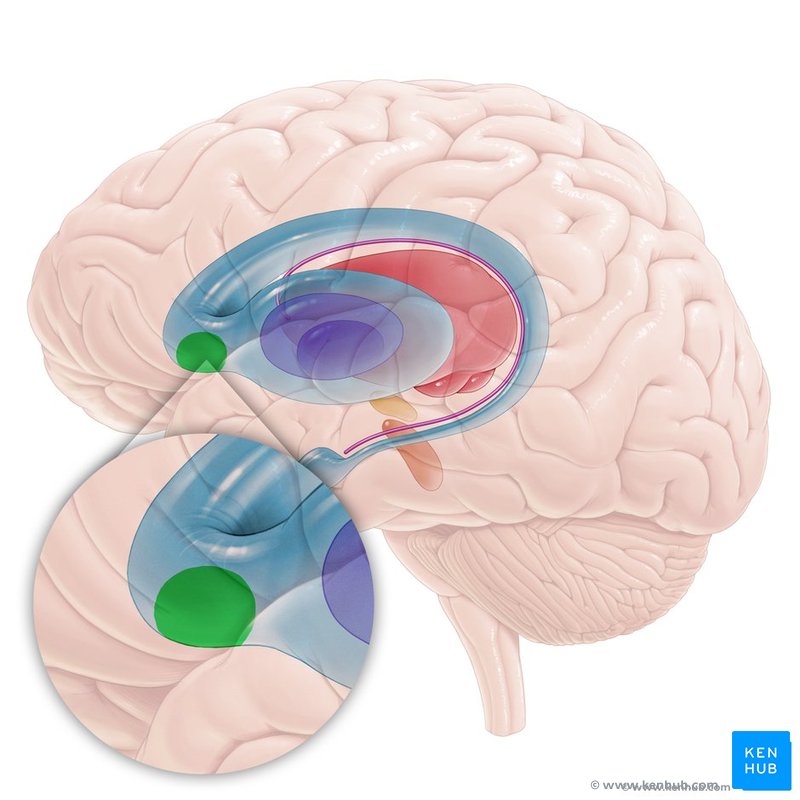
<p>Small brain structure in the basal ganglia located in the ventral striatum. Lies below the anterior commisure. Key part in the brains reward system</p>
81
New cards

Medial Geniculate Nucleus

a part of the thalamus that relays auditory information from the midbrain and hindbrain to the auditory cortex
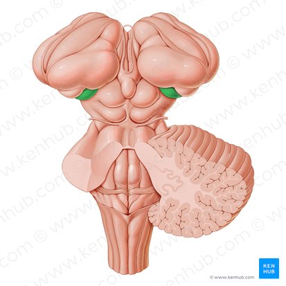
<p><span>a part of the thalamus that relays auditory information from the midbrain and hindbrain to the auditory cortex</span></p>
82
New cards

Lateral Geniculate Nucleus

Area of the thalamus that relays visual info to the cortex and superior colliculus. Located in posterior portion of thalamus
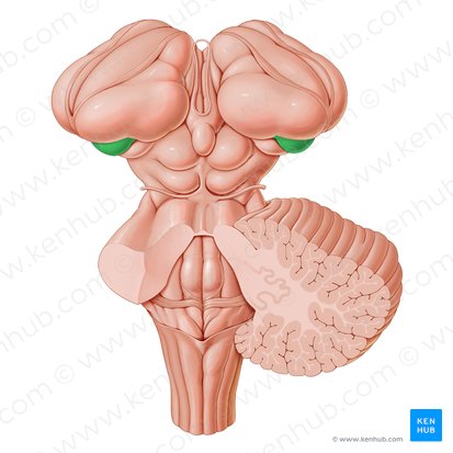
<p>Area of the thalamus that relays visual info to the cortex and superior colliculus. Located in posterior portion of thalamus</p>
83
New cards

Optic Radiations

White matter tracts that carry visual info from lateral geniculate nucleus (LGN) in the thalamus to the primary visual cortex.
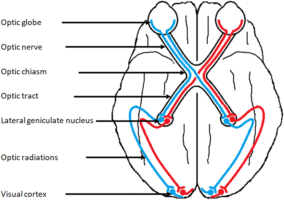
<p>White matter tracts that carry visual info from lateral geniculate nucleus (LGN) in the thalamus to the primary visual cortex.</p>
84
New cards

Periaqueductal Gray (PAG)

gray matter surrounding the cerebral aqueduct important in autonomic function and pain modulation
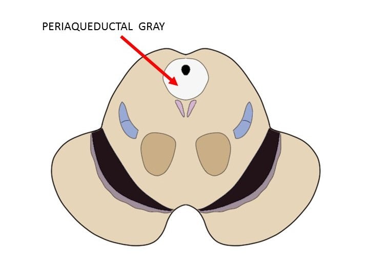
<p>gray matter surrounding the cerebral aqueduct important in autonomic function and pain modulation</p>
85
New cards

Septum Pellucidum

Thin membrane that separates the anterior horns of the lateral ventricles medially
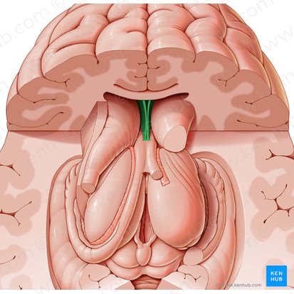
<p>Thin membrane that separates the anterior horns of the lateral ventricles medially</p>
86
New cards

Cingulate Gyrus

Gyrus located on top of the corpus callosum. Plays an important role in the limbic system
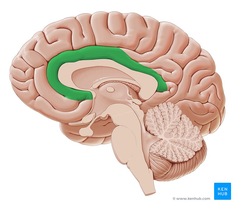
<p>Gyrus located on top of the corpus callosum. Plays an important role in the limbic system</p>
87
New cards

Calcerine Fissure

Large sulcus in the occipital lobe where the primary visual cortex is located. Divides the occipital lobe in half, separating upper and lower half of vision
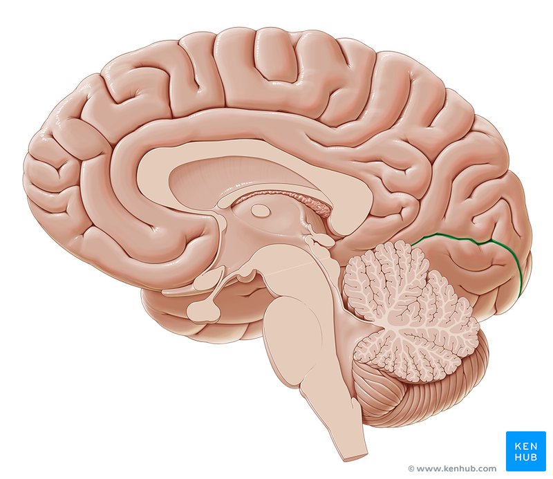
<p>Large sulcus in the occipital lobe where the primary visual cortex is located. Divides the occipital lobe in half, separating upper and lower half of vision</p>
88
New cards

Locus Coeruleus

small cluster of neurons in the brainstem, located in the dorsal pons, near the floor of the fourth ventricle. Important for physiological responses to stress and panic
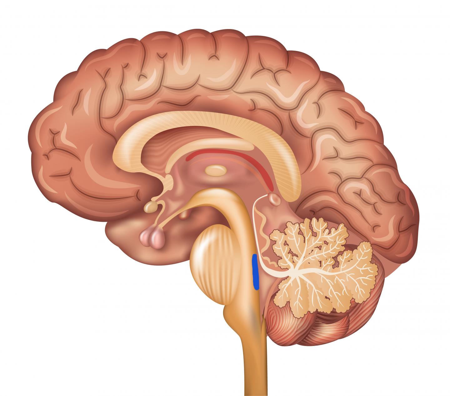
<p>small cluster of neurons in the brainstem, located in the dorsal pons, near the floor of the fourth ventricle. Important for physiological responses to stress and panic</p>
89
New cards

Uncus

area in temporal lobe that plays a role in emotion, memory, and smells. Anterior part that covers amygdala and posterior is apart of hippocampal formation
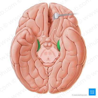
<p>area in temporal lobe that plays a role in emotion, memory, and smells. Anterior part that covers amygdala and posterior is apart of hippocampal formation</p>
90
New cards

Parahippocampal Place Area (PPA)

gray matter cortical area that surrounds hippocampus and apart of the limbic system. Important for memory encoding and retrieval
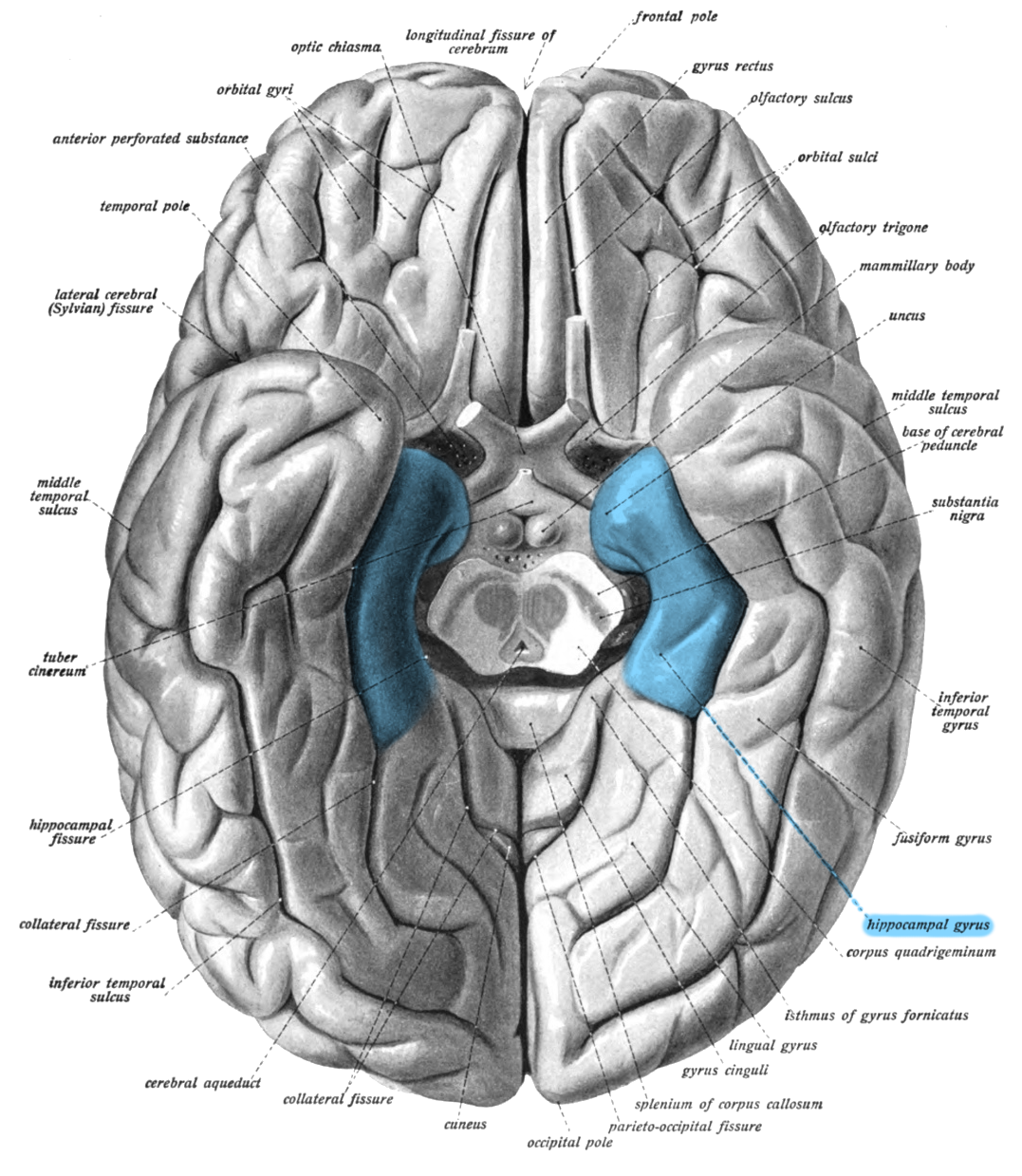
<p>gray matter cortical area that surrounds hippocampus and apart of the limbic system. Important for memory encoding and retrieval</p>
91
New cards

Fusiform Face Area (FFA)

area of the brain located in the inferior temporal lobe of the brain, in the fusiform gyrus. Important for detecting faces
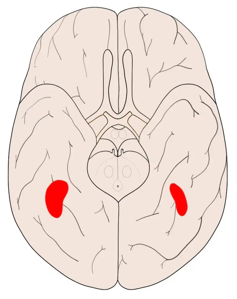
<p>area of the brain located in the <span>inferior temporal lobe of the brain, in the fusiform gyrus. Important for detecting faces </span></p>
92
New cards

Claustrum

thin sheet of gray matter in the brain that's involved in many functions, including attention, memory, and consciousness. It's highly connected to other parts of the brain, including the limbic, cortical, and subcortical structures. 
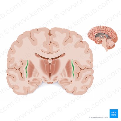
<p><span>thin sheet of gray matter in the brain that's involved in many functions, including attention, memory, and consciousness. It's highly connected to other parts of the brain, including the limbic, cortical, and subcortical structures.&nbsp;</span></p>
93
New cards

Dentate Nucleus

Deep cerebellar nucleus located in the posterior cerebellum. Plays a crucial role in motor coordination and control
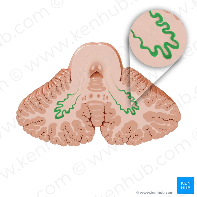
<p>Deep cerebellar nucleus located in the posterior cerebellum. Plays a crucial role in motor coordination and control</p>
94
New cards

Striatum

Cluster of nuclei in basal ganglia. Made up of caudate nucleus, putamen, and globus pallidus
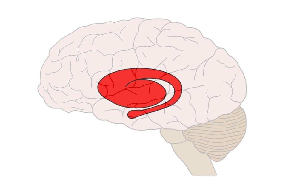
<p>Cluster of nuclei in basal ganglia. Made up of caudate nucleus, putamen, and globus pallidus</p>
95
New cards

Internal Capsule

white matter structure of the brain lateral to the thalamus and caudate nucleus. Important for connecting cortical regions to sub-cortical regions and the brainstem.
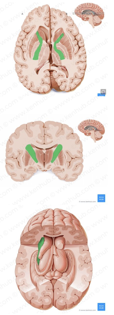
<p>white matter structure of the brain lateral to the thalamus and caudate nucleus. Important for connecting cortical regions to sub-cortical regions and the brainstem.</p>
96
New cards

External Capsule

Network of nerve fibers medial to claustrum and lateral to putamen. Thought to be important for processing sensory information and coordinating movement
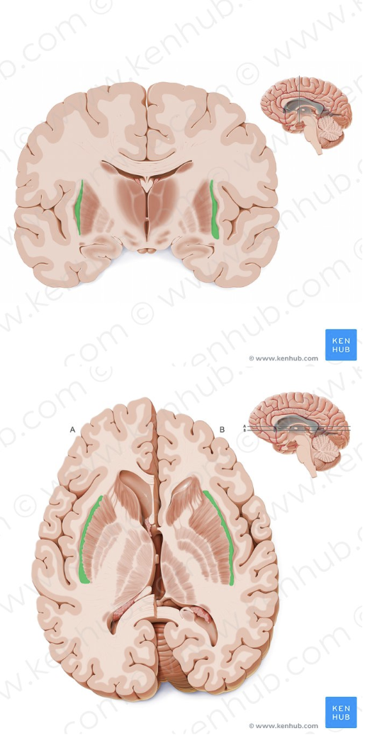
<p>Network of nerve fibers medial to claustrum and lateral to putamen. Thought to be important for processing sensory information and coordinating movement</p>
97
New cards

Extreme Capsule

Nerve fibers medial to insula and lateral to claustrum.
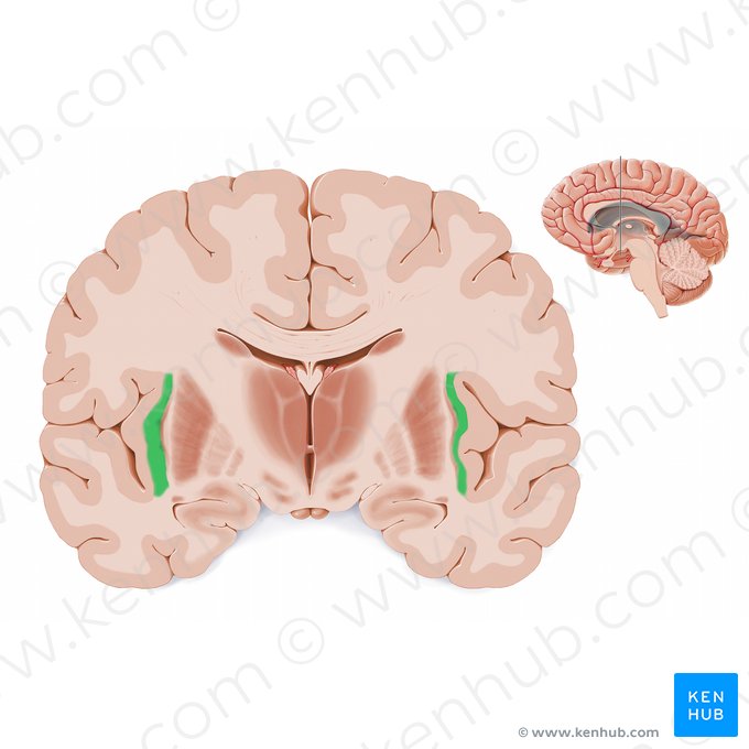
<p>Nerve fibers medial to insula and lateral to claustrum. </p>