4.3 Gas Exchange
1/30
There's no tags or description
Looks like no tags are added yet.
Name | Mastery | Learn | Test | Matching | Spaced | Call with Kai |
|---|
No analytics yet
Send a link to your students to track their progress
31 Terms
What is the structure of an alveolus and its blood supply? (3)
- An alveolus is a tiny air sac with a wall made from a single layer of flattened epithelial cells, known as Type I pneumocytes.
- It is surrounded by a dense network of capillaries, the walls of which are also only one cell thick.
- The inner surface of the alveolus is coated with a thin layer of fluid that contains lung surfactant.
How does gaseous exchange occur in the alveoli? (3)
- Gas exchange occurs by simple diffusion between the air in the alveoli and the blood in the capillaries, down a concentration gradient.
- The concentration of oxygen is higher in the alveoli than in the blood, so oxygen diffuses into the blood.
- The concentration of carbon dioxide is higher in the blood than in the alveoli, so carbon dioxide diffuses into the alveoli.
Why do single-celled organisms not require a specialised gas exchange system? (3)
- They have a large surface area to volume ratio.
- The diffusion distance from the external environment to any part of the cell is very short.
- This allows simple diffusion to be sufficient to meet their metabolic needs for nutrients and waste removal.
Why do large, multicellular organisms require specialised exchange surfaces? (3)
- They are made of billions of cells and have a small surface area to volume ratio.
- Diffusion alone is too slow to efficiently transport substances over the long distances from the outside to cells deep within the body.
- They generally have a higher metabolic rate, creating a greater demand for oxygen and for the removal of waste products.
What are the common features of an efficient respiratory surface? (3)
- They have a large surface area to maximise the rate of exchange and are very thin to ensure a short diffusion pathway.
- The surface is moist, as gases must dissolve in water before they can diffuse across a membrane.
- There is a mechanism, such as a rich blood supply, to maintain a steep concentration gradient.
What are the key tissues found in the wall of the trachea? (3)
- The trachea is lined by a pseudostratified columnar epithelial layer that contains cilia and mucus-secreting goblet cells.
- The wall is supported by C-shaped rings of cartilage that provide flexibility and support.
- Between the epithelium and the cartilage, there are layers of smooth muscle and elastic fibres.
Why are the cartilage rings in the trachea incomplete? (2)
- The cartilage provides structural support, which prevents the trachea from collapsing when air pressure is low.
- The incomplete C-shape allows the oesophagus, which is located behind the trachea, to expand when food is swallowed.
What are the roles of smooth muscle and elastic fibres in the airways? (2)
- The involuntary contraction of smooth muscle can constrict the airways, which is a protective reflex against harmful substances.
- The recoil of the stretched elastic fibres helps to redilate the airways after constriction.
How do cilia and mucus function in the trachea and bronchi? (2)
- Goblet cells in the epithelial lining secrete a sticky mucus which traps dust, pollen, and other inhaled particles.
- Cilia are small, hair-like structures that beat in a coordinated wave, moving the mucus upwards and away from the lungs.
Why are elastic tissue and lung surfactant important for lung function? (2)
- Elastic tissue stretches during inhalation and recoils during exhalation to help force air out of the lungs.
- Lung surfactant is a phospholipid that coats the inner surface of the alveoli, reducing surface tension and preventing them from collapsing.
How does air travel from the nasal cavity to the alveoli? (3)
- Air enters through the nasal cavity, where it is filtered, warmed, and moistened, before passing down the trachea.
- The trachea divides into two bronchi, which are lined with mucus-secreting cells and cilia to trap and remove dust.
- Each bronchus leads to smaller tubes called bronchioles, which end in microscopic air sacs called the alveoli, where gas exchange occurs.
What are the main components of the insect gas exchange system? (3)
- Spiracles are openings on the surface of the thorax and abdomen through which gases enter and leave.
- Spiracles lead to a system of internal tubes called tracheae, which are supported by rings of chitin to prevent them from collapsing.
- The trachea branch into a vast network of smaller, thin-walled tubes called tracheoles that extend to all tissues.
What are the functions of spiracles and tracheoles in insects? (3)
- Spiracles are regulated by sphincters, which can open or close to control gas exchange and reduce water loss.
- Tracheoles are the main sites of gas exchange, as their walls are thin, moist, and permeable to gases.
- They branch extensively to provide a large surface area and deliver oxygen directly to respiring cells.
How does mechanical ventilation occur in an insect? (3)
- Mechanical ventilation involves the rhythmical flattening and expanding of the thorax and abdomen.
- The expansion of the abdomen increases its volume, which decreases the internal pressure.
- This creates a pressure gradient that draws air into the tracheal system through the spiracles.
How does a fish take in water for ventilation? (3)
- The floor of the buccal cavity is lowered, and its sides are widened.
- This action increases the volume and decreases the pressure inside the buccal cavity.
- With the opercular valves closed, the reduced internal pressure causes water to enter the mouth.
How is water forced over the gills in a fish? (3)
- The floor of the buccal cavity is raised, and its sides are narrowed.
- This decreases the volume and increases the pressure inside the buccal cavity.
- The increased pressure forces the opercular valves to open, and water is pushed out over the gills.
How are fish gills adapted to provide a large surface area? (3)
- Each gill consists of many filaments, which are thin, finger-like projections.
- The surface of each filament is folded into numerous microscopic plates called lamellae.
- The lamellae contain a dense network of blood capillaries, which maximises the surface available for diffusion.
Why is the counter-current exchange system in fish important? (3)
- In this system, the flow of water over the lamellae is in the opposite direction to the flow of blood inside them.
- This arrangement maintains a steep concentration gradient for oxygen across the entire length of the lamella.
- It ensures that an equilibrium is not reached, which maximises the efficiency of gas exchange from water to the blood.
How does inspiration occur? (3)
- The external intercostal muscles contract, moving the ribcage upwards and outwards, while the diaphragm contracts and flattens.
- These movements increase the volume of the thorax.
- The increase in volume decreases the pressure in the lungs to below atmospheric pressure, causing air to flow in.
How does expiration occur? (3)
- The external intercostal muscles relax, allowing the ribcage to fall downwards and inwards, while the diaphragm relaxes and domes upwards.
- These movements decrease the volume of the thorax.
- The decrease in volume increases the pressure in the lungs to above atmospheric pressure, forcing air to flow out.
How are mammalian lungs adapted for efficient gas exchange? (3)
- The lungs contain millions of microscopic air sacs called alveoli, which collectively create a very large surface area.
- The walls of the alveoli are composed of a single layer of flattened squamous epithelial cells, creating a short diffusion distance.
- The surrounding capillaries also have walls that are only one cell thick and are very narrow, reducing the diffusion pathway further.
How is a steep concentration gradient for diffusion maintained in the lungs? (3)
- Ventilation constantly replenishes the air in the alveoli, ensuring a high partial pressure of oxygen.
- The continuous flow of blood through the capillaries rapidly transports oxygen away from the lungs.
- Haemoglobin in red blood cells readily binds with oxygen, which further lowers the partial pressure of oxygen within the blood.
How does gas exchange become more efficient in a very active insect? (3)
- During vigorous activity, the build-up of lactic acid in the muscle cells lowers their water potential.
- Water then moves by osmosis from the fluid-filled ends of the tracheoles into the muscle cells.
- This increases the surface area within the tracheoles that is available for direct gas exchange with the tissues.
What adaptations of fish gills ensure a short diffusion distance? (3)
- The walls of the lamellae are made of thin squamous epithelium and are only one cell thick.
- The walls of the blood capillaries inside the lamellae are also only one cell thick.
- The capillaries are positioned very close to the surface of the lamellae, which minimises the distance gases have to travel between the water and the blood.
How does having many alveoli increase the efficiency of diffusion in the lungs? (2)
- The large number of alveoli provides a massive total surface area for gas exchange.
- This allows for a higher rate of diffusion, meaning more oxygen can enter the blood and more carbon dioxide can be removed at any one time.
What other features of alveoli enable efficient diffusion in the lungs? (3)
- The walls of the alveoli and the surrounding capillaries are each only one cell thick, creating a very short diffusion pathway for gases.
- A constant flow of blood through the extensive capillary network maintains a steep concentration gradient between the alveolar air and the blood.
- The inner surface of the alveoli is coated with a thin layer of moisture, which allows oxygen to dissolve before diffusing into the blood.
Why do people with cystic fibrosis have a low oxygen concentration in their blood? (3)
- The thick, sticky mucus characteristic of the condition builds up in the airways, causing them to become narrowed or blocked.
- This obstruction reduces the amount of oxygen that can reach the alveoli.
- This in turn lowers the concentration gradient and decreases the rate of diffusion of oxygen into the bloodstream.
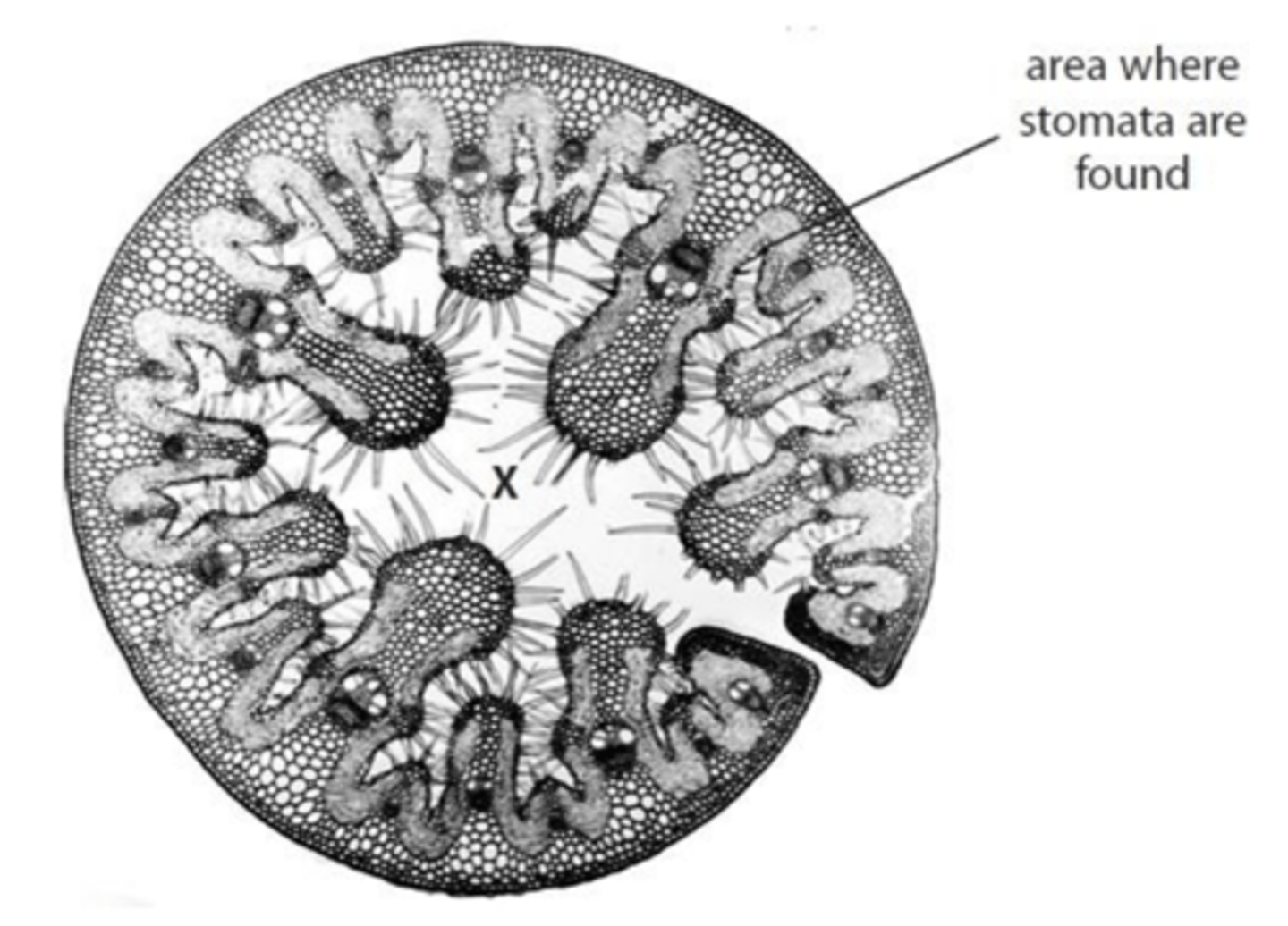
How does the structure of this leaf ensure the water potential at X remains high? (3)
- The leaf is curled with the stomata located on the inner surface, and these stomata are sunken in pits which are lined with fine hairs.
- This structure traps a layer of humid air, which is saturated with water vapour, close to the stomata.
- This reduces the water potential gradient between the inside of the leaf and the air outside, thereby lowering the rate of water loss by transpiration.
What features of gas exchange surfaces are common to both insects and mammals? (3)
- Both have gas exchange surfaces with a large surface area, provided by many tracheoles in insects and alveoli in mammals.
- They both feature a short diffusion pathway for gases, as the walls of the tracheoles and alveoli are very thin.
- The gas exchange surfaces in both organisms are moist, which allows oxygen to dissolve before it can diffuse into the tissues or blood.
Why do plants in water-deficient environments need extra xylem in their leaves? (2)
- Xylem tissue transports water to the leaf cells, which is essential for photosynthesis and for maintaining cell turgidity.
- The rigid structure of the xylem also provides mechanical support, holding the leaf in place to ensure it can absorb a maximum amount of light.
Why can a beetle prolong its time underwater using an air bubble? (3)
- The beetle uses the oxygen within the air bubble for aerobic respiration, which gradually depletes the initial oxygen supply.
- Initially, oxygen diffuses from the bubble into the water down a concentration gradient, causing the bubble to shrink.
- As the oxygen concentration in the bubble falls below that of the surrounding water, oxygen will then diffuse from the water into the bubble.