Descriptive Terminology
1/35
There's no tags or description
Looks like no tags are added yet.
Name | Mastery | Learn | Test | Matching | Spaced | Call with Kai |
|---|
No analytics yet
Send a link to your students to track their progress
36 Terms
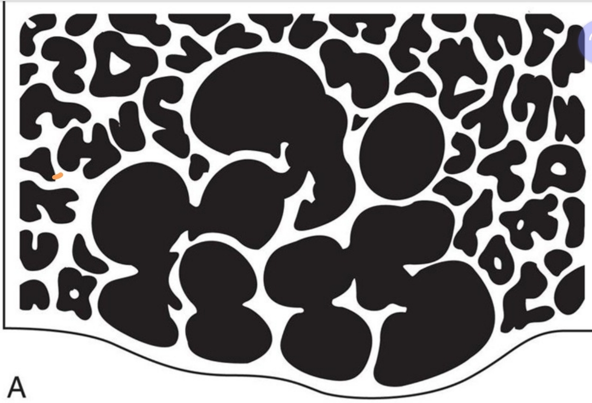
1 compartment
Tend to be small and nonexpansile
Borders may appear corticated or noncorticated on image
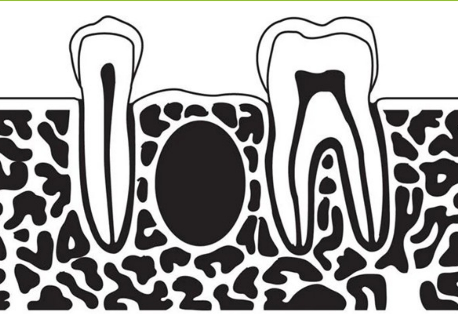
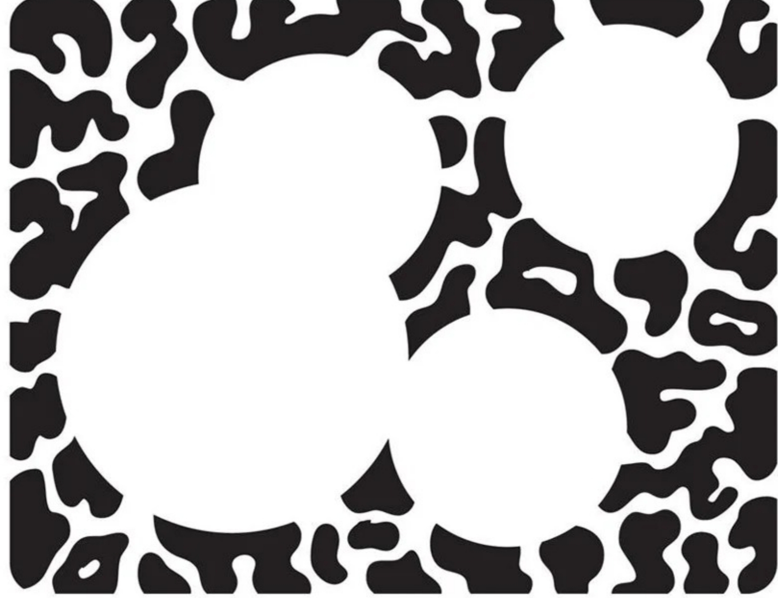
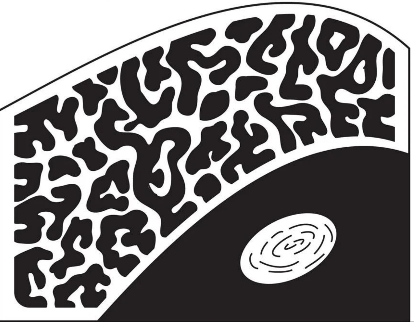
Unilocular Radiolucent lesions

Unilocular lesion, __________ borders
Lesion exibits a thin, well demarcated radiopaque rim of bone at periphery
Usually indicative of a benign, slow growing process
Corticated
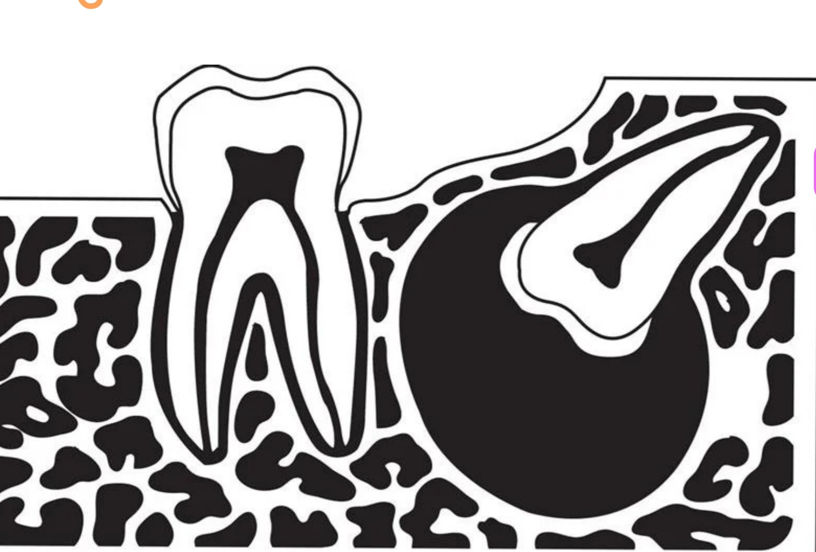
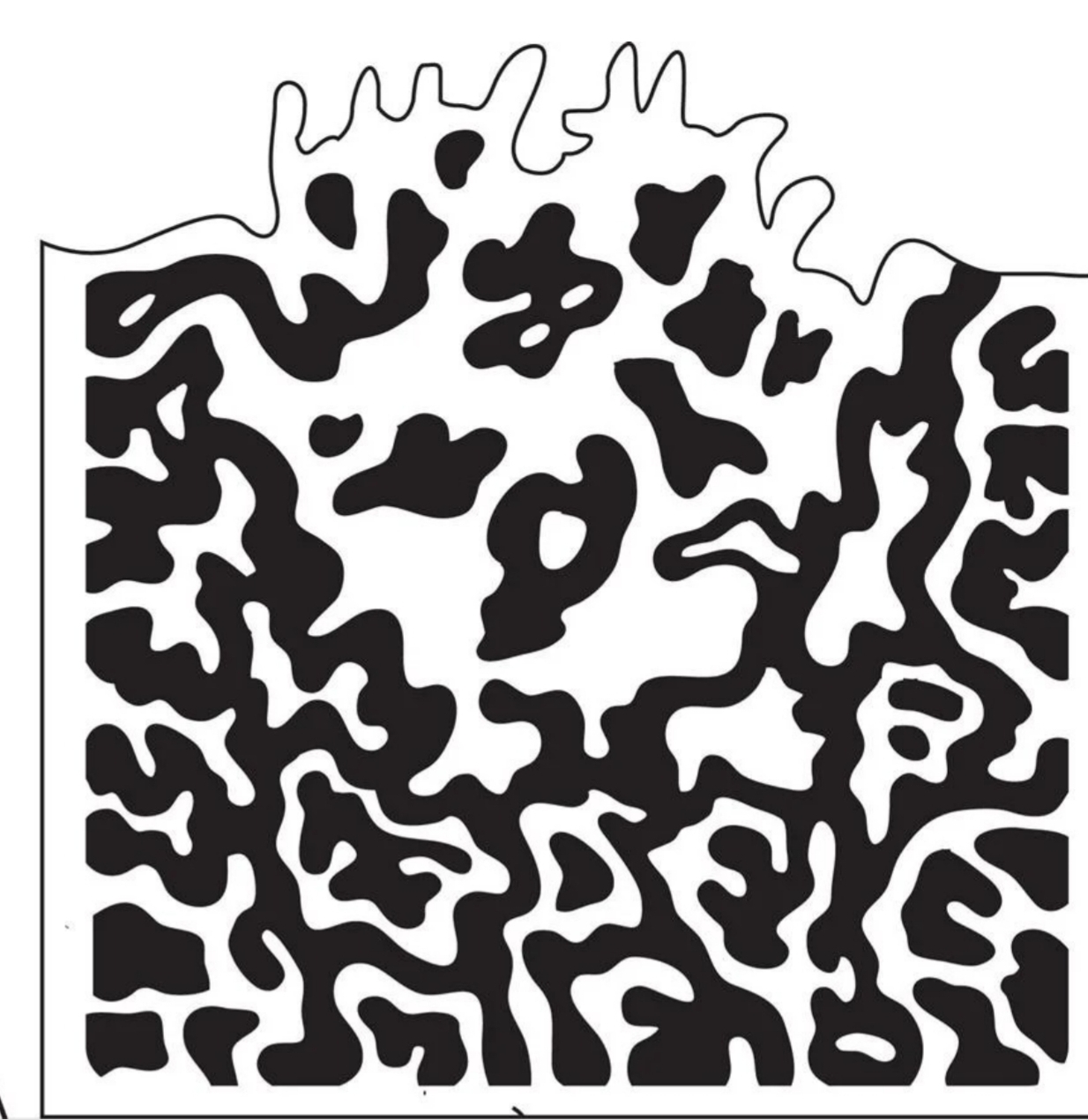
Unilocular lesion, _____________ Borders
Lesion does not exhibit thin radiopaque rim of bone at periphery
Periphery appears fuzzy or poorly defined
May represent either a benign or a malignant process
Noncorticated
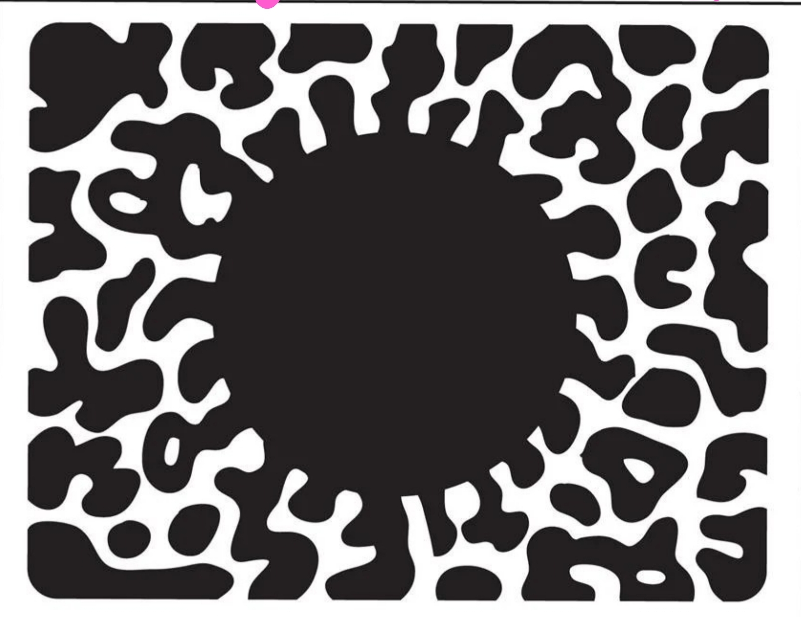
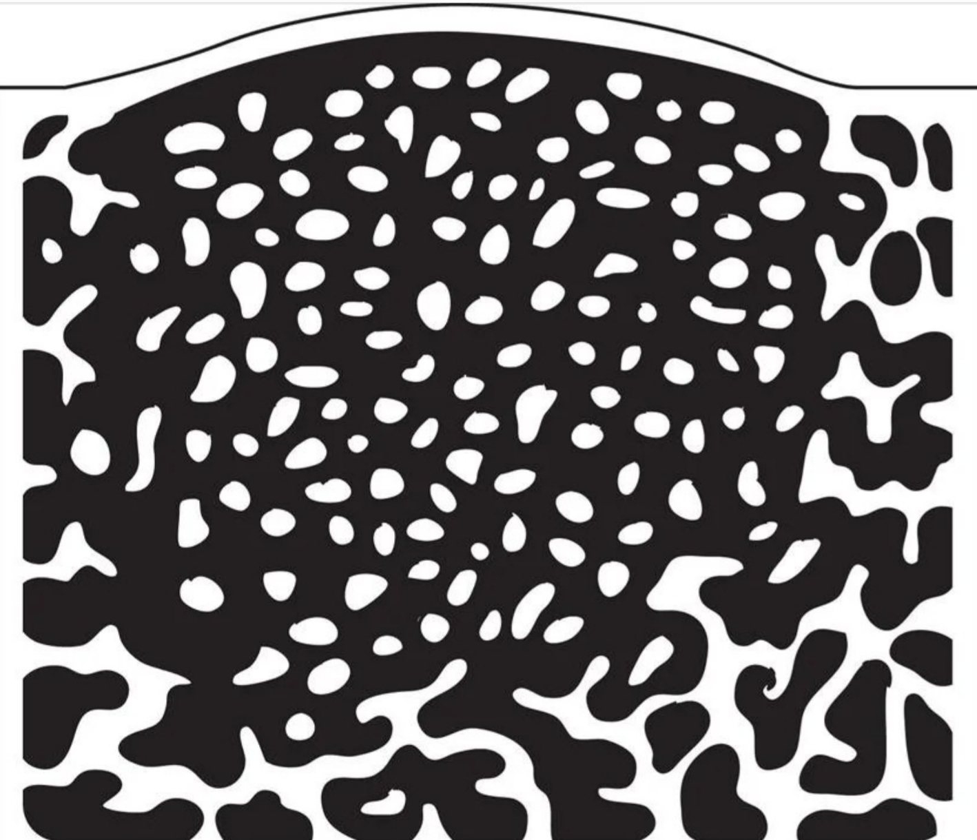
Exhibits multiple radiolucent compartments
frequently expansile (grows)
Typically benign lesions with aggressive growth potential
Multilocular radiolucent lesions
Types of Locations of Radiolucent Lesions:
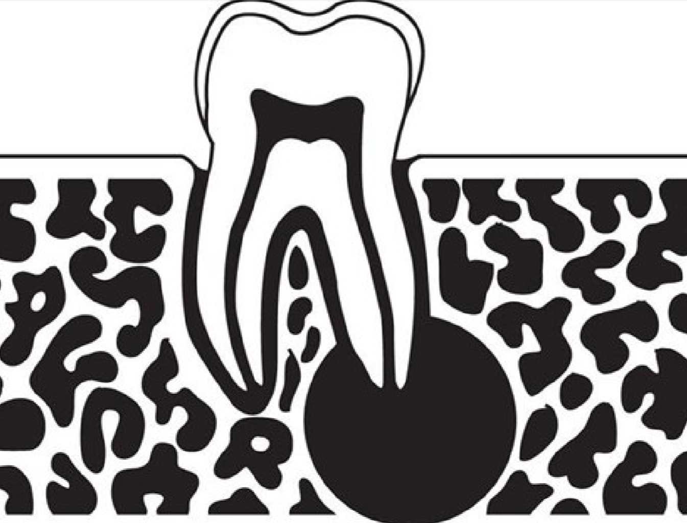
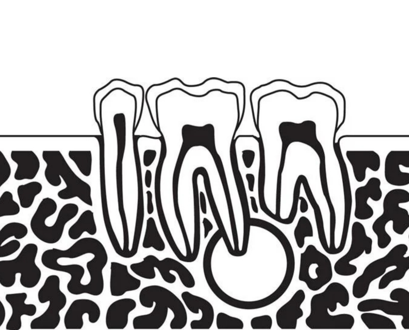
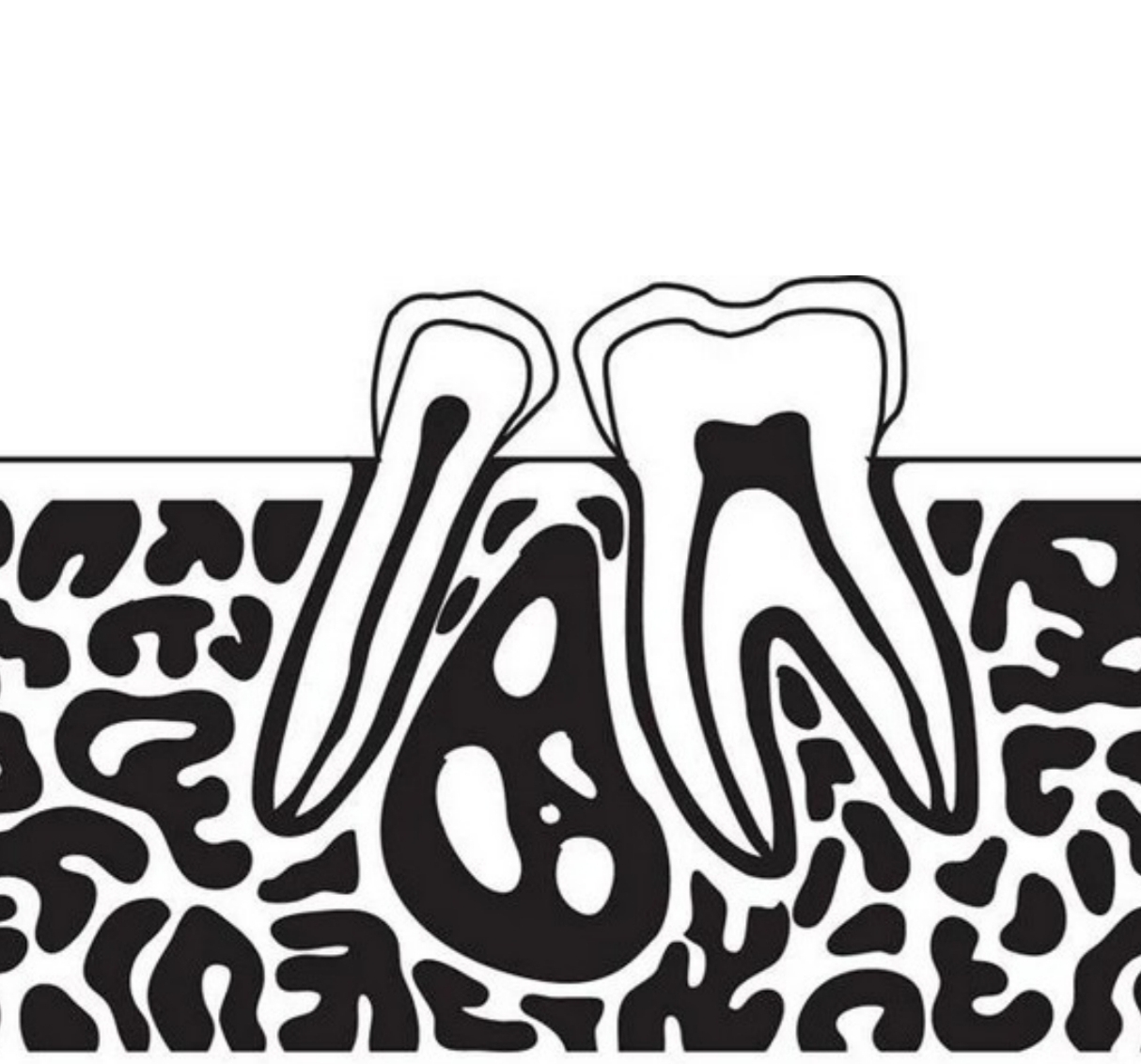
-Lesion located around apex of tooth
Periapical location
What is an example of a radiolucent lesion that has a periapical location?
Pulpal cyst secondary (caused by) to pulpal necrosis
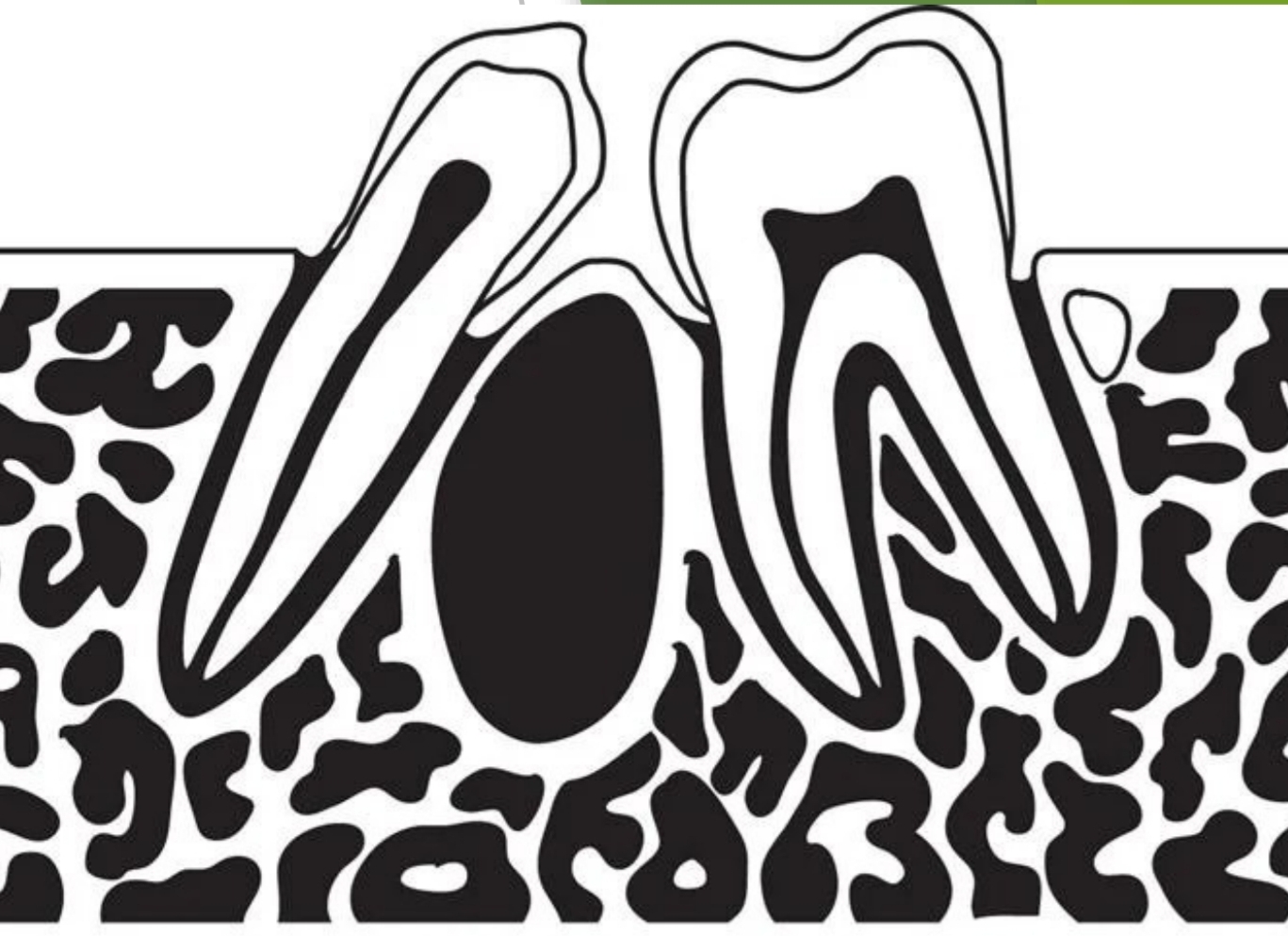
Types of Locations of Radiolucent Lesions:
Lesion located between roots of adjacent teeth
Inter-radicular Location
What is an example of a radiolucent lesion that has an Inter-radicular location?
Lateral periodontal cyst
Types of Locations of Radiolucent Lesions:
Lesion located in an area without teeth
Variety of radiolucent lesions may occur in this type of zone
Edentulous Zone
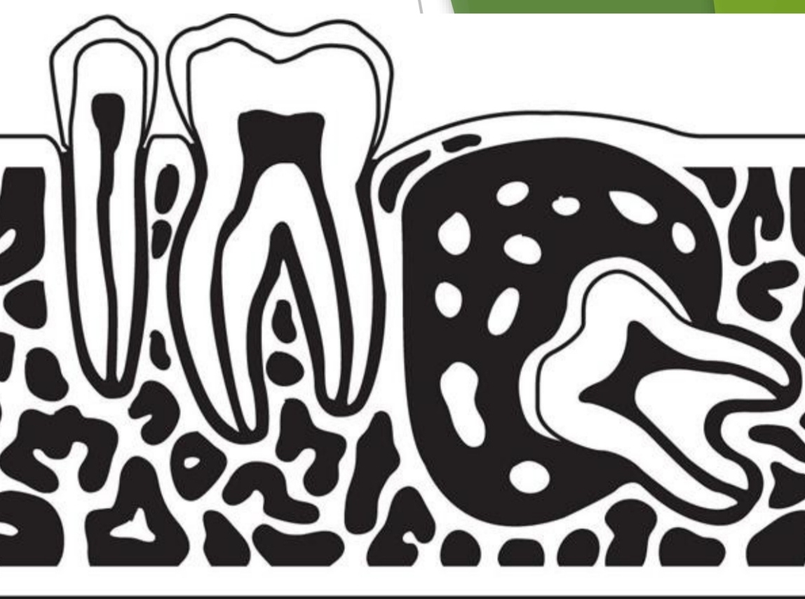
Types of Locations of Radiolucent Lesions:
Radiolucent lesion located around crown of an impacted tooth
Pericoronal Location
What is an example of a radiolucent lesion with a Pericoronal location?
Dentigerous cyst
Types of Locations of Radiolucent Lesions:
Loss of bone in maxilla or mandible that surrounds and supports teeth
Appears radiolucent
Alveolar Bone Loss
What are three terms used to describe radiopaque lesions?
Appearance
Location
Size
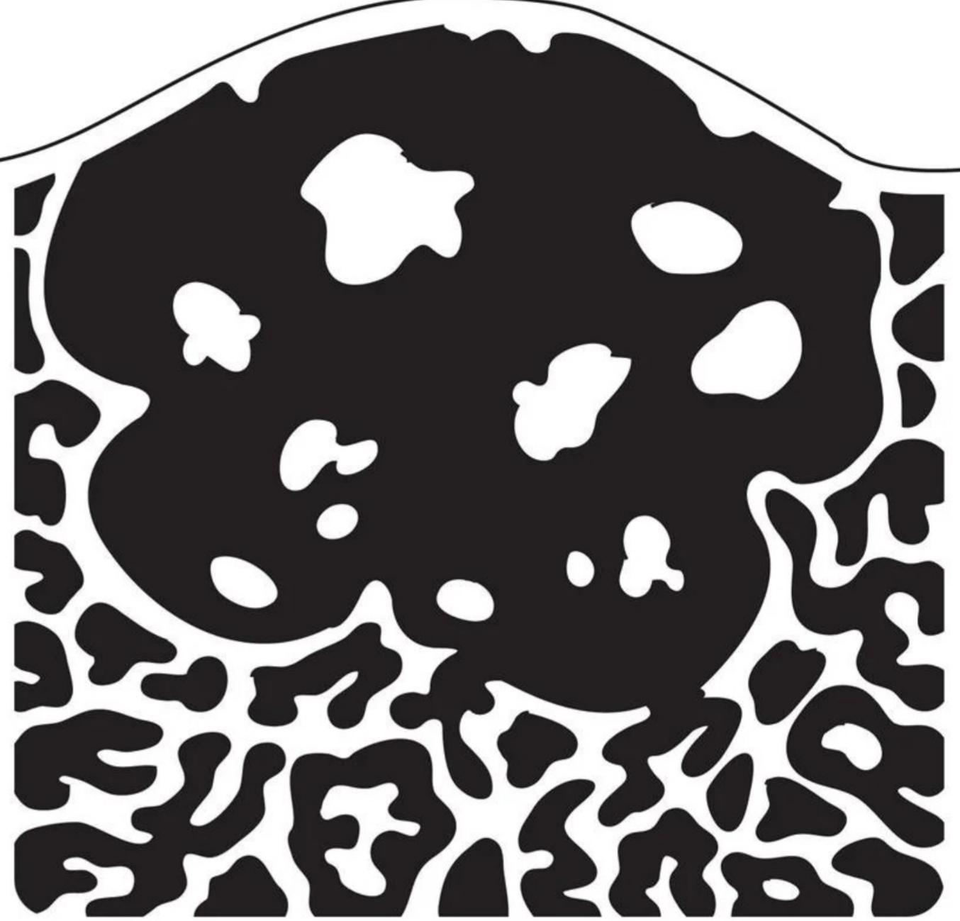
Appearance of Radiopaque Lesions:
Well-defined, localized radiopaque lesions on an image
Focal Opacity
What is an example of a radipaque lesion described by Focal Opacity?
Condensing Osteitis

Appearance of Radiopaque Lesions:
Well-defined, localized radiopaque area surrounded by a uniform radiolucent halo
Target Lesion
What is an example of a radiopaque lesion that is described as a Target Lesion?
Benign cementoblastoma
Appearance of Radiopaque Lesions:
Multiple radiopacities appear to overlap or flow together
This description of a lesion that involve multiple quadrants of jaws usually represent benign fibro-osseous disorders
Multifocal Confluent Pattern
What is an example of a radiopaque lesion that has a Multifocal Confluent Pattern?
Oseitis deformans, florid osseous dysplasia
Appearance of Radiopaque Lesions:
Radiopacity may exhibit an irregular, poorly defined pattern
It may represent a malignant condition
Irregular/Ill-defined Opacity
What is an example of a radiopaque lesion with an Irregular/Ill-defined Opacity?
Osteosarcoma and chondrosarcoma
Appearance of Radiopaque Lesions:
Granular pebbled radiopacity resembling pulverized glass
Often said to resemble appearance or texture of an orange peel
Ground Glass Opacity
What is an example of a radiopaque lesion with Ground Glass Opacity?
Fibrous dysplasia, Osteitis deformans, Osteopetrosis
Appearance of Radiopaque Lesions:
Exhibits both a radopaque and a radiolucent component
Often represents calcifying tumors
Mixed Lucent-Opaque Lesion
What is an example of a radiopaque lesion that can be described as Mixed Lucent-Opaque?
Compound odontoma
Appearance of Radiopaque Lesions:
Appears as well-defined, radiopaque area located in soft tissue
Soft Tissue Opacity
What is an example of a radiopaque lesion that has Soft Tissue Opacity?
Sialolith, Calcified lymph node
Locations of Radiopaque Lesions:
Radiopaque lesion located around apex of a tooth
Periapical Location
What is an example of a radiopaque lesion that has a Periapical Location?
Benign cememtoblastoma
Appearance of Radiopaque Lesions:
Radiopaque lesion between roots of adjacent teeth
Inter-radicular Location
What is an example of a radiopaque lesion that has an Inter-radicular Location?
Sclerotic Bone
Appearance of Radiopaque Lesions:
Radiopaque lesion located in an area without teeth
Edentulous Zone
What is an example of a radiopaque lesion in an edentulous zone?
Complex Odontoma
Appearance of Radiopaque Lesions:
Radiopaque lesion located around crown of an impacted tooth
Pericoronal Location
What is an example of a radiopaque lesion with a pericoronal location?
Adenomatoid odontogenic tumor
What can the size of both radiolucent and radiopaque lesions be?
several millimeters to several centimeters in diameter