Chapter 13: The Spinal Cord & Spinal Nerves
1/60
There's no tags or description
Looks like no tags are added yet.
Name | Mastery | Learn | Test | Matching | Spaced | Call with Kai |
|---|
No analytics yet
Send a link to your students to track their progress
61 Terms
What are the 3 functions of the spinal cord?
Processes reflexes
Integrates EPSPs & IPSPs
Conducts sensory impulses to the brain & motor impulses to effectors
what is the cauda equina?
roots of the lower spinal nerves
What is the spinal cord protected by?
bone (vertebrae)
connective tissue (meninges)
fluid (cerebrospinal fluid)
What are the meninges?
3 connective tissue membranes covering the spinal cord and brain:
Dura mater (outermost; tough)
Arachnoid mater (middle; cobweb-like)
Pia mater (innermost; thin)
What is the name for a group of interconnected nerves that facilitate communication?
plexus
what are the plexuses of the spinal cord?
cervical plexus (C1-C5)
brachial plexus (C5-T1)
lumbar plexus (L1-L4)
sacral plexus (L4-S4)
Where does the spinal cord begin and terminate (end)?
begins: medulla oblongata
terminates: L2
How many spinal bones and spinal nerves of each section are there?
Bones:
7 cervical
12 thoracic
5 lumbar
5 sacrum
1 coccygeal
Nerves:
8 cervical
12 thoracic
5 lumbar
5 sacrum
1 coccygeal
what is the filum terminale?
(“Strand terminal”) an extension of the pia mater that extends inferiorly and blends with the arachnoid and dura to anchor the spinal cord to the coccyx
what is the conus medullaris?
tapered terminal end of the spinal cord
what is the first cervical vertebrae?
atlas
what is the second cervical vertebrae?
axis
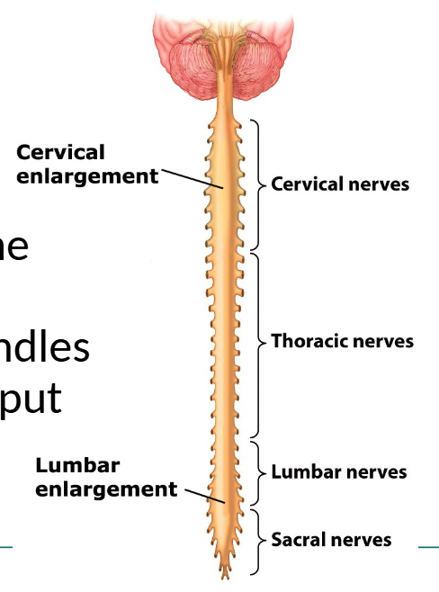
What are the 2 enlargements of the spinal cord and what do each of them handle?
cervical enlargement (C4-T1) → sensory input & motor outpute to the UPPER extremities
lumbar enlargement (T9-T12) → motor output & sensory input TO & FROM the LEGS
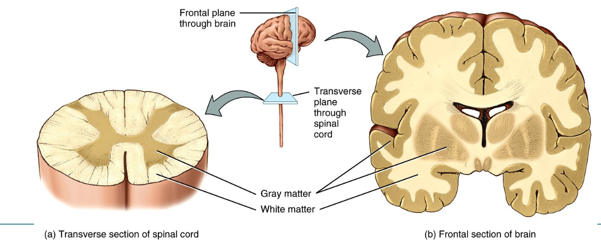
Where is white and gray matter in the spinal cord VS. the brain?
spinal cord:
white matter → OUTSIDE
gray matter → INSIDE
brain:
white matter → INSIDE
gray matter → OUTSIDE
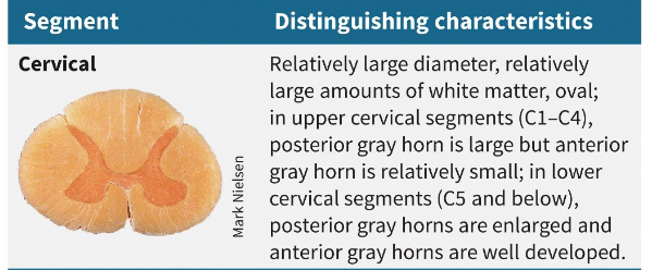
Distinguish the cervical segment of a spinal cord
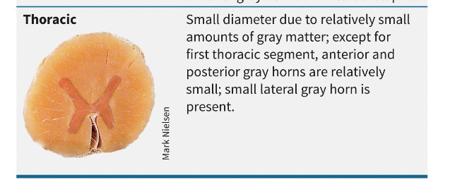
Distinguish the thoracic segment of a spinal cord
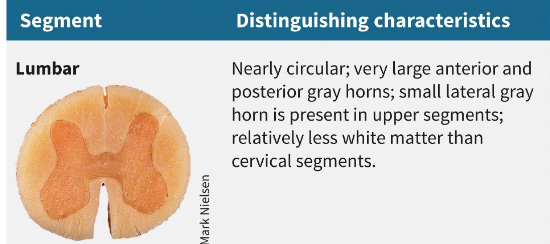
Distinguish the Lumbar segment of the spinal cord
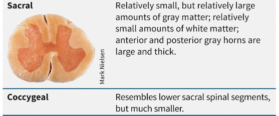
Distinguish the Sacral and Coccygeal segments of the spinal cord
What is the function of spinal nerves and how many pairs are there?
function: connect the CNS to sensory receptors, muscles, & glands and are part of the PNS
31 pairs (62 total)
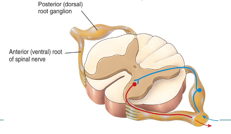
Describe how spinal nerves are connected to the spinal cord
roots (2 bundles of axons) connect each spinal nerve to a seegment of the cord by even smaller bundles of axons called ROOTLETS
Contrast posterior (dorsal) and anterior (ventral) roots/rootlets
posterior (dorsal):
contains only SENSORY axons
conducts nerve impulses from sensory receptors in skin, muscles, & organs to CNS
has GANGLION; contains cell bodies of sensory neurons
anterior (ventral):
contain only MOTOR axons
conducts nerve impulses from the CNS to effectors
what is a lumbar puncture?
needle inserted into subarachnoid space for the purpose of withdrawing CSF (for diagnosis or to reduce pressure or to introduce a drug or contrast agent)
CSF often collected for meningitis or other CNS diseases
what does the gray matter of the spinal cord consist of ?
dendrites
cell bodies of neurons
unmyelianted axons
neuroglia
Internal spinal cord anatomy: Locate/describe the anterior median fissure
wide groove on anterior (ventral) side

Locate/describe the posterior median sulcus
narrow furrow on the posterior (dorsal) side

locate/describe the gray commissure
froms crossbar of the H

locate/describe the central canal
small space in the center of the gray commissure
extends the entire length of the spinal cord
filled with CSF

locate/describe the anterior white commisure
anterior to the gray commissure
connects the white matter of the right and lefct sides of the spinal cord

locate/describe the posterior gray horns
contain axons of incoming sensory neurons & cell bodies & axons of interneurons

locate/describe the anterior gray horns
contain soma\tic motor nuclei — provide nerve impulses for contraction of skeletal muscles
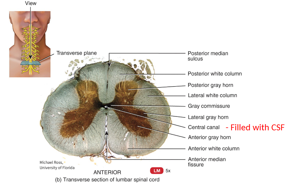
locate/describe the lateral gray horns
between posterior/anterior gray horns
only present in thoracic & upper lumbar & mid-sacral segments
contain autonomic motor nuclei — regulate cardiac muscle, smooth muscle, & gland activity

locate and describe funiculi
3 broad areas of white matter; contains bundles of acons (tracts
anterior white funiculi
posterior white funiculi
lateral white funiculi

Contrast tracts & nerves
tracts → bundles of axons in CNS
sensory tracts: ascend to brain
motor tracts: descend from brain
nerves → bundles of axons in PNS
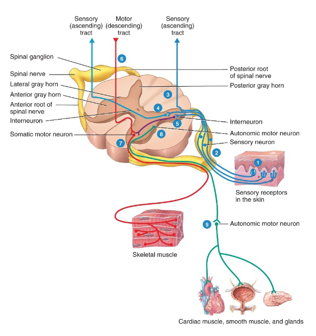
Trace the processing of sensory/motor output by the spinal cord.
sensory receptors detect a sensory stimulus
sensory neurons convey sensory input along axons from sensory receptors → spinal nerves → posterior root. Then proceeds along 3 possible paths (3, 4, or 5)
SENSORY (ASCENDING) TRACT: axons of sensory neurons may enter the posterior gray horn then extend into white matter then ascend to brain
SENSORY (ASCENDING) TRACT: axons of sensory neurons may enter the posterior gray horn and synapse with interneurons whose axons extend into the white matter then ascend to the brain
MOTOR (DESCENDING TRACT): axons of sensory neurons may enter the posterior gray horn and synapse with interneurons that synapse with somatic motor neurons which are involved in spinal reflex pathways
Motor output from the spinal cord to skeletal muscles involves somatic motor neurons of the anterior gray horn.
when activated, somatic motor neurons convey motor output in the form of nerve impulses along their axons, which sequentially pass through the anterior gray horn and anterior root to enter the spinal nerve. from the spinal nerve, axons of somatic motor neurons extend to skeletal muscles of the body
motor output from the spinal cord to cardiac & smooth muscle, & glands involves autonomic motor neurons of the lateral gray horn. when activated, autonomic motor neurons convey motor output along their axons, which then pass through the lateral gray horn, anterior gray horn,, ande anterior root to enter the spinal nerve
from the spinal nerve, axons of autonomic neurons from the spinal cord synapse with another group of autonomic motor neurons located in the PNS. the axons of the 2nd group in turn synapse with with cardiac muscle, smooth muscle, & glands.
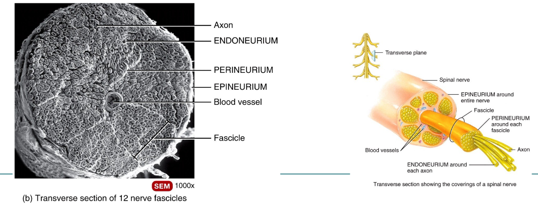
____ wrap each axon which are arranged in bundles called ___ surrounded by a ___, with the entire nerve sheathed by a CT ____
endoneurium
fascicles
perineurium
epineurium
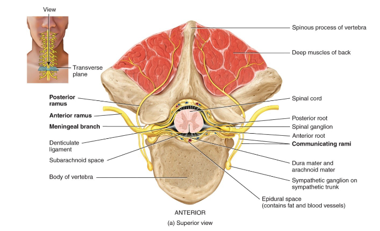
When the spinal nerves exit the CNS into the PNS, they almost immediately split into 3 major branches:
anterior ramus → serves the muscles & structures of the upper and lower limbs and the skin of the lateral and anterior surfaces of the trunk (form the cervical, brachial, etc. plexuses)
posterior ramus → serves deep muscles & skin of posterior surface of trunk
rami communicantes
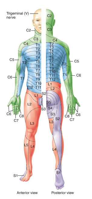
what is a dermatome
cutaneous area of skin that provides sensory input to the CNS via the posterior roots of one pair of spinal nerves or via the trigeminal (V) nerve
Describe the cervical plexus and its major nerve
C1-C5
supplies the skin and muscles of the head, neck, superior portion of the shoulders & chest, & diaphragm
contains phrenic nerve (origin: C3-C5) — causes contraction diaphragm
Describe the brachial plexus and its major nerves
C5-T1
provides almost entire nerve supply to shouldeers & upper limbs
major nerves
long thoracic; origin C5-C7
serratus anterior muscle
median; origin C5-T1
lesion = carpal tunnel syndrome
digits
radial; origin C5-T1
digits
ulnar; origin C8-T1
digits
“funny bone”
What is Erb’s palsy?
paralysis of the arm that most often occurs as an infant’s head & neck are pulled toward the side at the same time as the shoulders pass through the birth canal
Describe the lumbar plexus and its major nerves
L1-L4
supplies antrolateral abdominal wall, external gentials, part of lower limbs
major nerves:
femoral (L2-L4): largest lumbar plexus nerve, distributed to flexor muscles of hip joint & extensor muscles of knee, skin over anteior & medial aspect of thight, & medial side of leg & foot
obturator (L2-L4): adductor muscles of hip joint, skin over medial aspect of thigh
Describe the sacral plexus & it's major nerve
L4-S4
supplies buttocks, perineum, lower legs
major nerve:
sciatic nerve (L4-S3): 2 NERVES — tibial & common fibular bound tgt by sheath of CT (Splits into 2 at knee). sends branches to hamstrings and adductor magnus
How does info travel in the spinal cord?
WHITE matter tracts conduct nerve impulses to and from the brain
GRAY matter receives & integrates incoming/outgoing info to perform spinal reflexes
what is the origin and destination of the spinothalamic tract? which kind of tract is it?
origin: spine
destination: thalamus
therefore: sensory ascending tract
What is a reflex?
fast, involuntary, unplanned response to a stimulus
maintains homeostasis
gray matter of spinal cord = integrating center for spinal reflexes
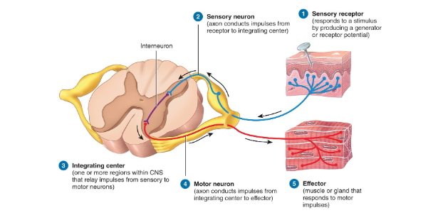
List and breifly explain the 5 components of a reflex arc
Sensory Receptor → resp. to stimulus
Sensory Neuron → axon conduscts impulses
Integrating Center → 1+ regions within CNs that relay impulses from sens. to motor neurons
Motor Neuron → axon conducts impulses from integrating center to effector
Effector → muscle or gland that responds to motor impulses
List and differentiate the 6 types of reflex arcs
ipsilateral → sensory neuron ENTERING & motor neuron EXITING on the SAME SIDE of the integrating center
contralateral → sensory neuron entering ONE side & motor neuron exiting on the OTHER
monosynaptic → single synapse between sensory and motor neuron
polysynaptic → 1+ association neurons relaying messages to other association/motor neurons
intersegmental → signal of a single sensory neuron activates several motor neurons via the association neurons in several segments of the spinal cord
reciprocal innervation → neural circuit simultaneously contracts one muscle & relaxes its antagonists
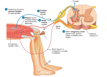
Explain what a stretch reflex is
causes contraction & relief of a muscle that has been stretched
stimulates receptors in muscle spindle of the agonist (stretched muscle)
monosynaptic & ipsilateral
stretching stims sensory receptor (muscle spindle)
sensory neuron excited
integrating center: sensory neuron activates motor neuron
motor neuron excited
effector: same muscle contracts and relieves the stretching
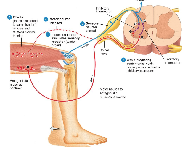
Explain what a tendon reflex is
causes relaxation of the muscle attached to the stimulated tendon
polysynaptic & ipsilateral
prevents injury due to excess muscle tension or over-contraction
increased tension stimulates sensory receptor (tendon organ)
sensory neuron excited
integrating center: neuron activates inhibitory neuron
motor neuron inhibited
agonist muscle reflazes and relieves excess tension, antagonist muscles contract
Explain what a Flexor (withdrawal) reflex is
causes withdrawal of a limb to avoid injury or pain (i.e pulling hand away from stove)
polysynaptic, ipsilateral, & intersegmental
stepping on tack stims sensory receptor (dendrites of pain-sensitive neuron)
sensory neuron excited
integrating center: sensory neuron activates interneurons in several spinal cord segments
motor neurons excited
effectors: flexor muscles contract & withdraw leg
Explain what a crossed-extensor reflex is
maintains balance during a withdrawal reflex
polysynaptic, contralateral, intersegmental
flexor muscles contract/withdraw right leg
stepping on tack stims sensory receptor
sensory neuron excited
integrating center: spinal cord; sensory neuron activates several interneurons
motor neuron excited
effectors: extensor muscles contract and extend left leg
Describe the patellar reflex
leg extends in response to stretch of the patellar tendon
can be blocked by damage in corticospinal tracts from diabetes, neurosyphilis, or damage to the lumbar region
describe the achilles reflex
causes contraction of the calf when a force is applied to the achilles tendon
absent after damage to the lower cord or lumbosacral plexus
describe the babinski/plantar flexion reflex
normal in adults if they flex (curl) big toe when sole of foot stimulated (negative babinski)
if extend/point up → damage to corticospinal tract (positive babinski) (normal in infants
Indicate the extent of paralysis from traumatic injury for each sement of the spinal cord
Cervical → no function from the neck down
Thoracic → some arm and chest muscle control
Lumbar → most thigh muscles
Sacral → most leg muscles
What does “transection” of the spinal cord mean?
ascending and descending tracts are partially or completely severed
at base of skull → death by asphyxiation
upper cervical area → quadriplegia (paralysis of four limbs)
between cord enlargements results in some form of paraplegia (paralysis of both lower limbs)
List the 4 major spinal disorders
spinal cord compression
degenerative diseases
shingles
poliomyelitis
What is spinal cord compression
the spinal cord may be compressed by bone, blood (hematomas), pus (abscesses), tumors (Cancerous or not), or a ruptured or herniated disk
What are degenerative diseases?
mulitple sclerosis → deymyelination of olgidendroglia
amyotropic lateral sclerosis (ALS) → progressive nervous system disease that affects nerve cells in the brain and spinal cord, causing loss of muscle control
What is shingles?
acute infection of the PNS caused by herpes zoster virus (also causes chicken pox)
causes pain, discoloration of the skin and line of skin blisters
never crossses mid-line; only on one side of the body
What is poliomyelitis (Polio)
caused by the poliovirus
virus spreads from person to person & can infect a person’s spinal cord, causing paralysis