ANAT322 midterm notes
1/373
There's no tags or description
Looks like no tags are added yet.
Name | Mastery | Learn | Test | Matching | Spaced | Call with Kai |
|---|
No analytics yet
Send a link to your students to track their progress
374 Terms
Where do humoral (hormones, antibodies, etc.) outputs of the hypothalamus go?
To the pituitary
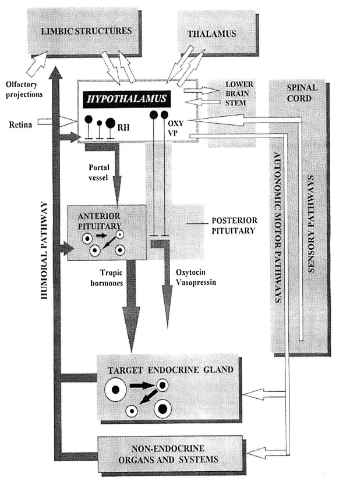
Humoral inputs of the hypothalamus
steroids, peptide hormones at the MBH, cytokines, neuropeptides (ultrashort loop feedback)

Neural outputs of the hypothalamus go to?
mainly to limbic structures, thalamus and brain stem

Neural inputs of the hypothalamus come from?
Amygdala, hippocampus, septum, thalamus, basal ganglia, cortex, lower brain stem, spinal cord, retina.
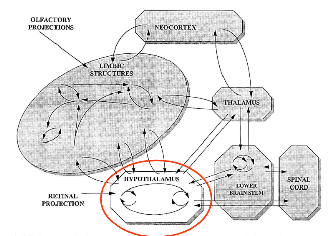
What major areas of the brain is the hypothalamus connected to?
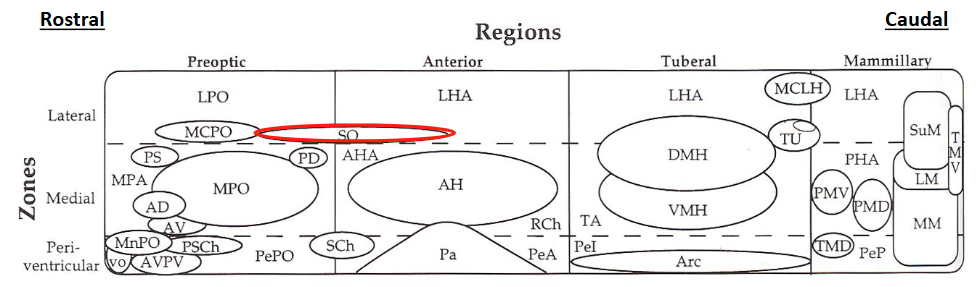
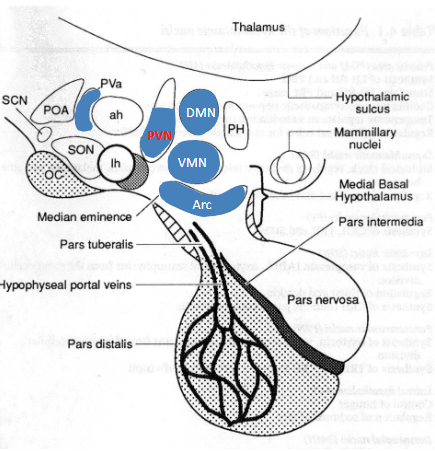
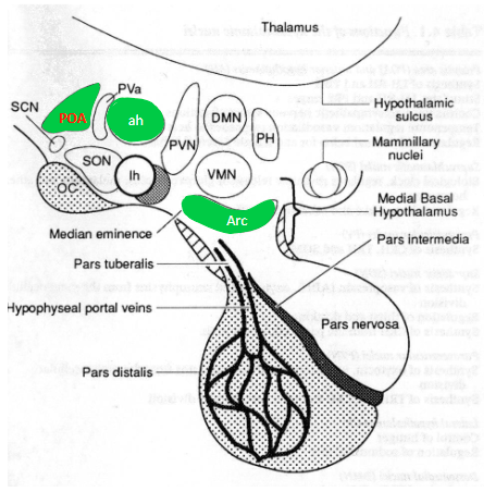
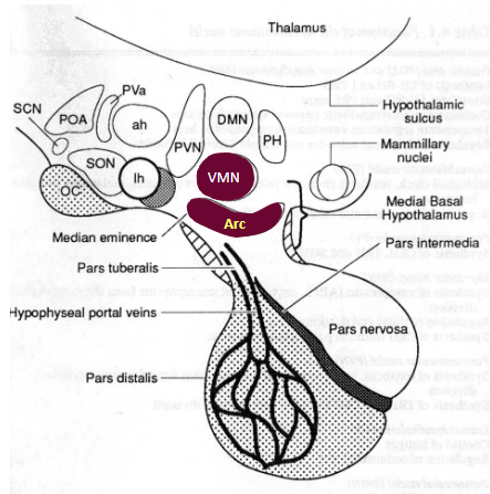
Nuclear groups and zones of the hypothalamus (not a question memorize the diagram)
Identify the 2 types of cells in the hypothalamus
Periventricular zone contains?
Majority of the neuroendocrine nuclei, other cell groups closely connected to the neuroendocrine cells, predominantly intrahypothalamic connections
Close to 3rd ventricle (probably exchange of molecules)
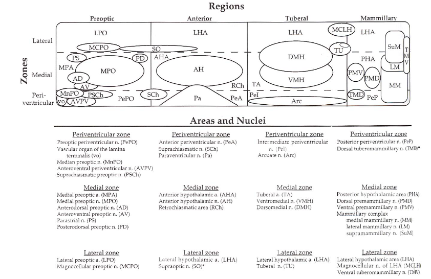
Medial zone contains (function?)
Large nuclei involved in the initiation of motivated behaviours such as aggressive or appetitive behaviour, extensive connections throughout the brain, many sensory inputs, frequently via the limbic system (amygdala, hippocampus, etc.) and through brainstem nuclei relaying visceral information (Nucleus of the solitary tract, NTS)

Lateral zone function?
Functional importance has been difficult to establish because of its structural complexity (is traversed by the Medial Forebrain Bundle, MBF), has been associated with the processing of hunger/thirst, aggression, reproduction, general arousal and sensory sensitization associated with motivated behaviours

What is the pituitary gland? What are its lobes?
Endocrine gland weighing about 0.5 g, composed of an anterior, intermediate and posterior lobe.
How is the pituitary gland connected to the hypothalamus?
Connected with the brain (hypothalamus) via the median eminence and the infundibulum.
Where is the pituitary gland located (hint: ANAT214)
Located in a concave pouch of the sphenoid bone named “sella turcica”.
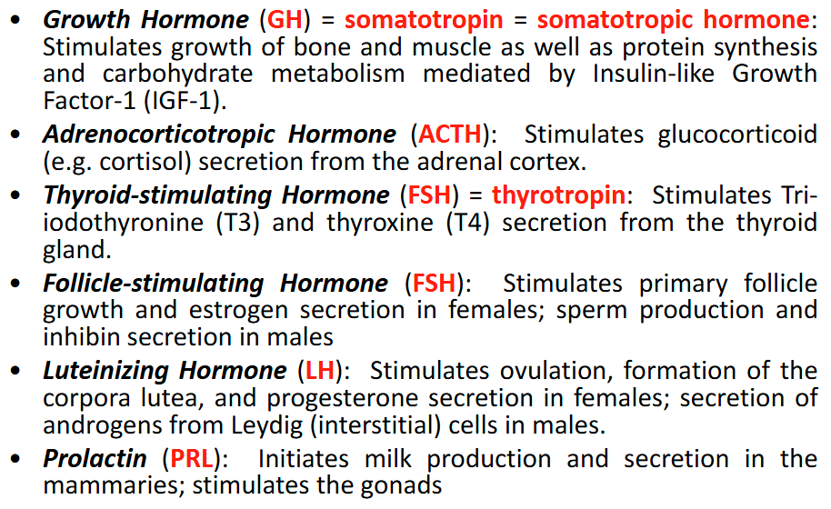
Hormones of the anterior lobe of the pituitary gland
GH
ACTH
TSH
FSH
LH
PRL
Does the posterior pituitary produce any hormones?
No, only anterior and intermediate lobe
Are the two lobes of the pituitary derived from the same or distinct source?
Derived from two distinct embryological sources

Where does the anterior lobe of the pituitary derive from?
The adenohypophysis (anterior lobe, intermediate lobe, pars tuberalis) arises from Rathke’s pouch in the roof of the developing pharynx (stomatic ectoderm)
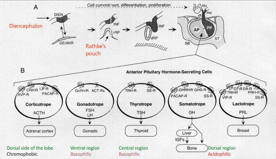
Where does the posterior lobe of the pituitary derive from?
The neurohypophysis (posterior pituitary, infundibular stem and median eminence) originates from the base of the diencephalon (neuroectoderm).


Understand that the pituitary has many different cells that create the different hormones. Not all are created by one cell type.
What is the intermediate lobe of the pituitary?
The pars intermedia is situated between the anterior and posterior lobes. It is poorly developed in humans. It consists of several colloid-filled follicles (“cysts”) and strands of basophilic cells which may in- vade the lobus nervosus due to ameboid motility (“basophilic infiltration“).
Intermediate lobe of the pituitary function? (i.e. what hormones does it secrete and their function?)
• Melanocyte Stimulating Hormone (MSH): Stimulates dispersion of melanin granules in melanocytes, i.e. skin darkening.
• β-Endorphin: Neuromodulator, circulating analgesic

Posterior lobe of the pituitary function? (i.e. what hormones does it store -NOT secrete- and their function?)
• Oxytocin (OT): Stimulates smooth muscle contraction (e.g. uterine contractions)
• Vasopressin (VP) = Antidiuretic Hormone (ADH): Promotes water reabsorption in the kidneys and elevates blood pressure

How is the posterior pituitary formed? How are hormones released?
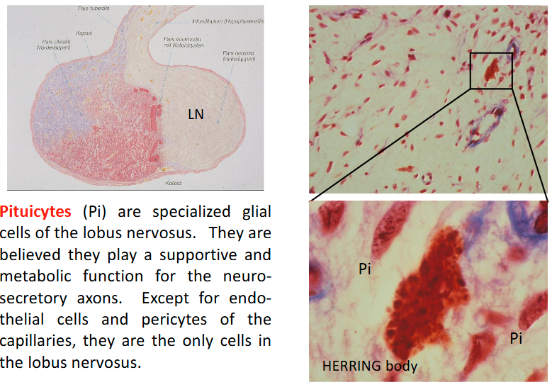
The bulk of the lobus nervosus is formed by the terminations of neurosecretory axons from the hypothalamo-hypophyseal tract and fenestrated capillaries into which the neurohormones are released.
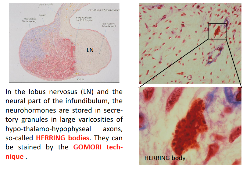
Herring bodies function?
In the lobus nervosus (LN) and the neural part of the infundibulum, the neurohormones are stored in secretory granules in large varicosities of hypothalamic-hypophyseal axons, so-called HERRING bodies.
They can be stained by the GOMORI technique.

Are herring bodies and pituicytes found in the anterior pituitary?
No, only in the posterior pituitary
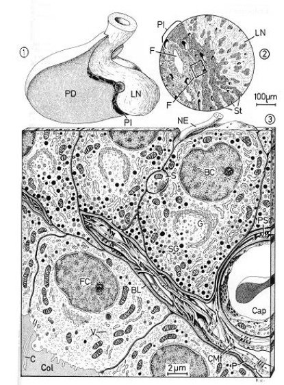
Pituicytes (Pi)
Neurosecretory material in the posterior lobe LN HERRING body Pituicytes (Pi) are specialized glial cells of the lobus nervosus. They are believed they play a supportive and metabolic function for the neuro- secretory axons. Except for endo- thelial cells and pericytes of the capillaries, they are the only cells in the lobus nervosus.
Helps the secretion of neuro-hormones
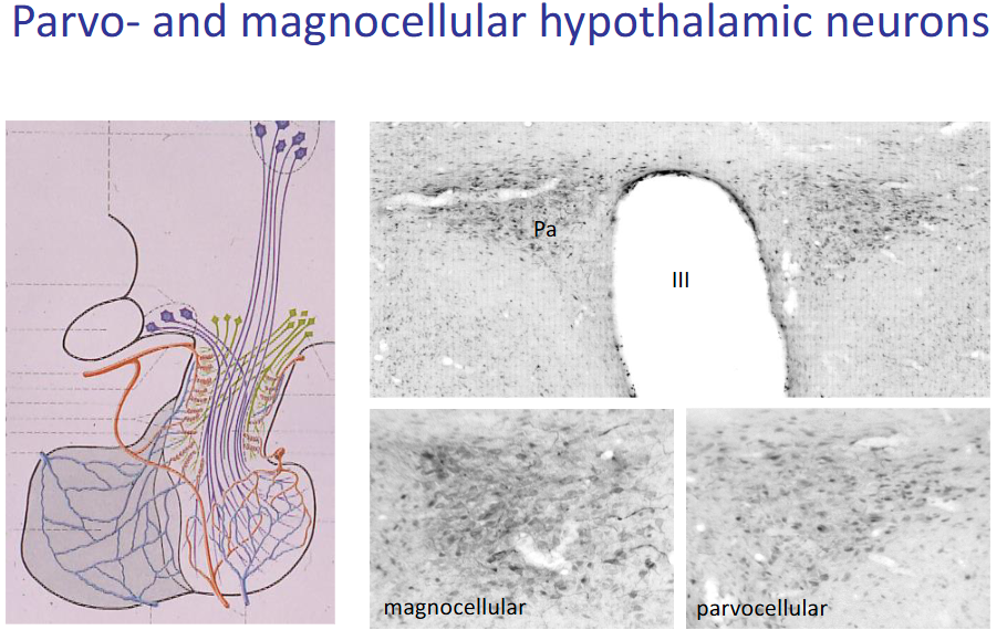
The parvocellular system
• Neurons are typically small (diameter < 20 μm)
• Synthesis of hypophysiotropic hormones (releasing and release-inhibiting hormones)
• Distributed throughout various hypothalamic nuclei
• They extend their axons (thin, unmyelinated) to the median eminence (ME), where they release their neurohormones into the hypophyseal portal veins → anterior pituitary
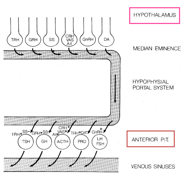
Definition of a hypophysiotropic hormone
1. The hormone is present in the median eminence.
2. Its concentration is higher in the hypophyseal portal system than in the general circulation.
3. Its level in the portal blood is correlated with the rate of release of (a) particular hypophyseal hormone(s).

Corticotropin Releasing Hormone
A large peptide containing 41 amino acid residues.
Potent stimulator of ACTH (and endorphin) release from corticotropes. Synergistic action of AVP and opioids (glucocorticoids.)
Distinct diurnal (day-night) pattern of release.
Thyrotropin Releasing Hormone
Tripeptide stimulating synthesis and release of TSH as well as PRL.
Plays also a neuromodulator role, i.e. in the control of the sympathetic branch of the autonomic nervous system
Gonadotropin Releasing Hormone
Decapeptide increasing release and synthesis of both LH and FSH in the gonadotrophs.
GnRH release is pulsatile. This is a pre-requisite for the maintenance of LH secretion and proper gonadal function.
Growth Hormone Releasing Hormone
Increases production and release of GH in somatotrophs.
Somatostatin
Inhibits synthesis and release of GH , TSH, and PRL. Antagonist to GHRH and TRH.
Very widespread throughout the brain outside the hypothalamus and in the periphery, e.g. in the digestive tract (δ-cells of pancreatic islets!). Acts mostly as an inhibitor of secretion.
Dopamine
May be the inhibitor of Prolactin release.
Also acts on the release of MSH.
Median eminence – pituitary stalk
The median eminence (ME) is a relatively small region in the mediobasal hypothalamus (MBH); composed of internal and external layer, blood brain barrier is open (fenestrated capillaries): circumventricular organ.
The ME is a neurohemal organ where neurosecretory cells release their neurohormone into the circulation.
Median eminence - internal layer
The internal layer contains 20-25,000 fibers originating from magnocellular neurons in transit directed to the posterior pituitary and subependymal layer (OT, VP, but also CRH, SS, PRL, Enk, etc.).
Median eminence - external layer
The external layer is comprised of about 60% of nerve fibers and terminals (40-50,000 axons) from parvocellular neurons (POMC, GHRH, NT, DA, GABA, TRH, Enk, AII, GnRH, etc). The remaining constituents are vascular elements (primary portal plexus, portal veins, etc.), glial cells and tanycytes.
Hypophysial blood supply
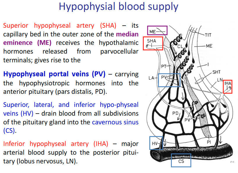
Superior hypophyseal artery (SHA)
Its capillary bed in the outer zone of the median eminence (ME) receives the hypothalamic hormones released from parvocellular terminals; gives rise to the Hypophyseal portal veins (PV)

Hypophyseal portal veins (PV)
Carrying the hypophysiotropic hormones into the anterior pituitary (pars distalis, PD).
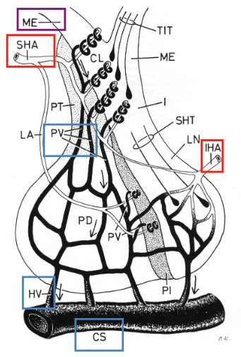
Superior, lateral, and inferior hypophyseal veins (HV)
Drain blood from all subdivisions of the pituitary gland into the cavernous sinus (CS).

Inferior hypophyseal artery (IHA)
Major arterial blood supply to the posterior pituitary (lobus nervosus, LN).

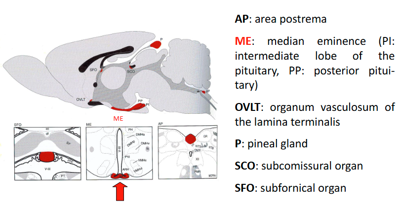
Circumventricular organs
Characterized by a lack of blood-brain barrier and, hence, free diffusion of macromolecules between plasma and interstitial fluid of brain tissue.
Modes of chemical communication
Endocrine - Hormones released into the blood to travel to distant target cells.
Paracrine – Hormone or messenger released into the
interstitial space affects neighboring target cells.
Autocrine – Hormone or messenger has a direct
feedback effect on the releasing cell.
Neurocrine – Neurotransmitter released by a
presynaptic neuron at specialized synapses.
Neuroendocrine – Neurohormones are released by
specialized (neurosecretory) nerve cells either into the
general circulation (e.g. in the posterior pituitary) or into
the hypothalamo-hypophyseal portal circulation.
What is a hormone?
Chemical messenger, produced by ductless (= endocrine) glands, released into the general circulation or the interstitial space, influences target cells through specific receptors, phylogenetically old type of intercellular communication.
Neuronal communication
Point-to-point connections; restricted number of target cells; rapid, direct communication
Neuroendocrine communication
Neurons release neurotransmitters into the bloodstream; reach a large number of target cells; indirect, relatively slow communication.
How are Neuroendocrine cells innervated? How are they stimulated/inhibited?
Neuroendocrine cells are innervated by other (integratory) neurons and stimulated or inhibited by neurotransmitters – they “transduce” a neuronal into an endocrine signal.
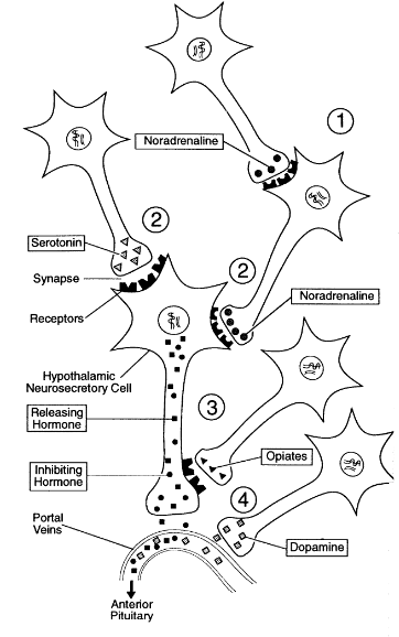
Neuronal control of hypothalamic hormone release
1. Indirect stimulation/inhibition of the neurosecretory cell via innervation of an interneuron regulating its activity
2. Direct innervation of the neurosecretory cell via axo- dendritic or axo-somatic synapses, thus influencing the cell’s activity
3. Presynaptic inhibition via axo- axonic synapses at the terminals
4. Direct release of hypothalamic dopamine into the portal blood stream
Peptides function?
- Peptides are the most common neuroendocrine messengers (except catecholamines: noradrenalin (norepinephrine) peripherally, dopamine centrally).
- They may have various effects, e.g. somatostatin (centrally: neurotransmitter, neurohormone; peripherally (e.g. in the pancreas): paracrine factor)
- Pituitary and peripheral protein and peptide hormones tend to be larger than central (hypothalamic) neuropeptides.
- Very variable: Amino acids (glutamate, GABA), biogenic amines (catecholamines, serotonin, histamin), (poly-)peptides, etc.
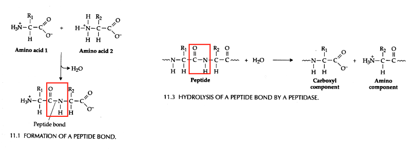
Formation and hydrolysis of peptide bonds (not a question)
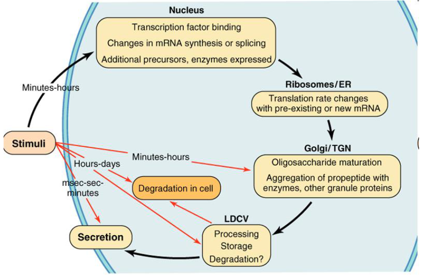
How are neuropeptides regulated?
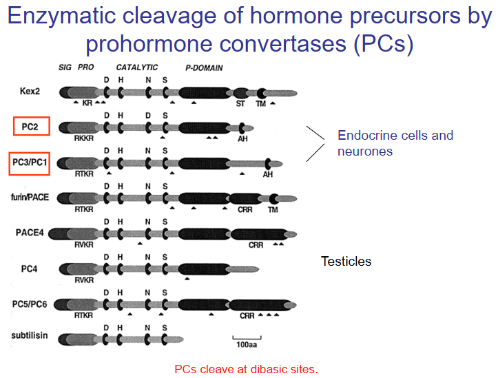
What are the prohormone convertases?
Structures of bioactive peptide precursors (not a question)

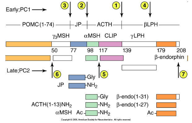
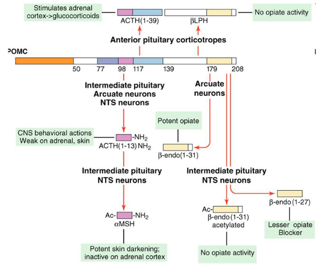
Sequential processing of pro-opiomelanocortin
Cleavage of the POMC precursor occurs at seven sites, with some of the reactions being tissue-specific. The circled numbers indicate the temporal order of cleavage in tissues where these proteolytic events occur. ACTH, adrenocorticotropic hormone; CLIP, corticotropin-like intermediate lobe peptide; JP, joining peptide; LPH, lipotropin; MSH, melanocyte-stimulating hormone; PC, prohormone convertase.
Has a pH optimum
Numbers indicate the steps of cleavage in decreasing pH conditions
Tissue-specific processing of POMC
Take-home:
Different tissues at different pHs will create a different product
If low PCs - less cleavage/processing
If high PCs, low pH and is processed in LDCV - more cleavage/processing
Large dense-core vesicles, LDCVs
Storage of peptides in secretory granules (conditions determine the processing of the peptide hormones)
White matter of a neuron
Myelin, surrounding many axons, has a white glistening colour. Therefore, those parts of the central nervous system (CNS) containing many myelinated axons are referred to as White Matter.
Grey matter of a neuron
Those parts containing aggregations of nerve cell bodies amidst delicate neuronal processes and glial cells appear gray, hence the term Gray Matter.
Bundles of axons: Tracts
Tracts – A bundle of axonal nerve fibers with a common origin and a common target area constitutes a tract, e.g. the corticospinal (or pyramidal) tract or the hypothalamo-hypophyseal tract.
The connection between a group of neurons and the target of their axons is also referred to as a projection.
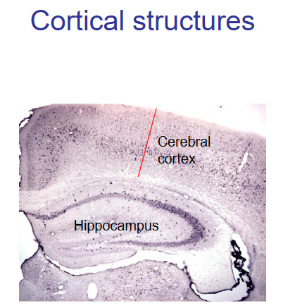
Cytoarchitectonic organization of neuronal cell bodies: Cortex
Cortex – Cortical structures such as the superficial coat of gray matter of the cerebral hemispheres and of the cerebellum are characterized by a more or less orderly arrangement of the neuronal cell bodies in layers or laminae.
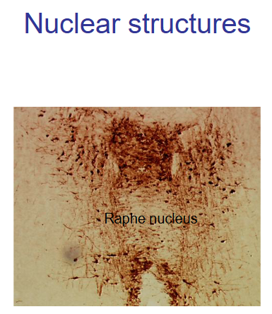
Cytoarchitectonic organization of neuronal cell bodies: Nuclei
Nuclei – Groups of nerve cells lacking this layered structure are referred to as nuclei. They are defined by topography and/or cellular morphology, connectivity, neurochemical characteristics (expression of the same transmitters, peptides, etc.).
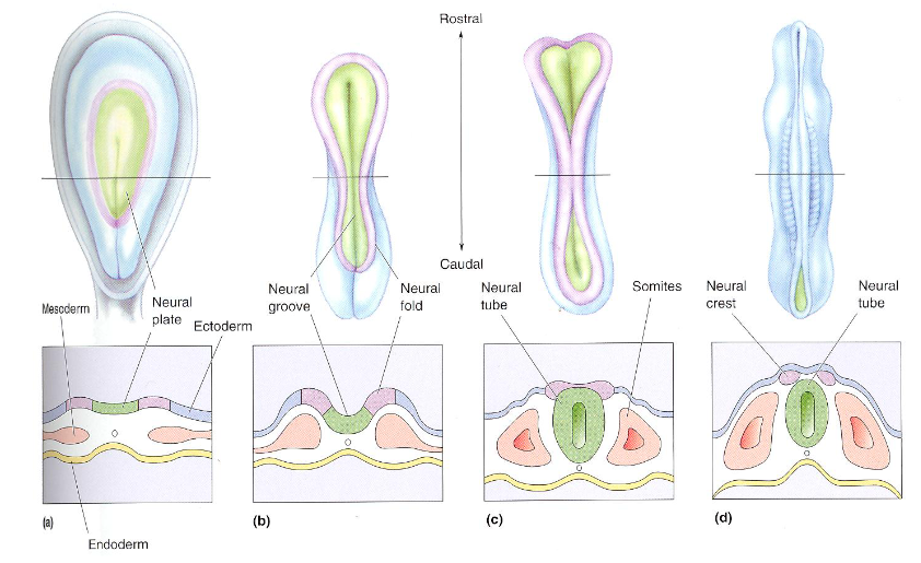
Formation of the neural tube
What is the chromophobe cell type of the anterior pituitary?
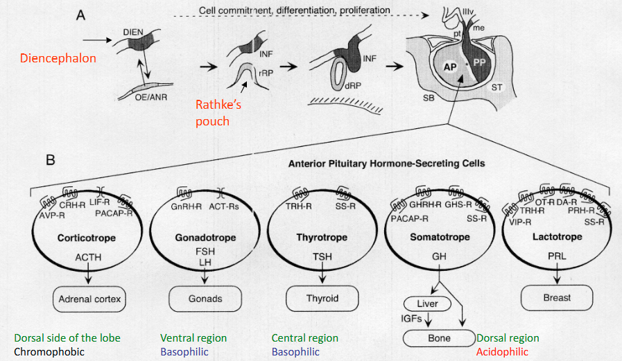
What is the basophilic cell type of the anterior pituitary?

What is the acidic cell type of the anterior pituitary?

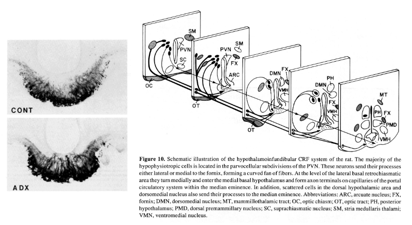
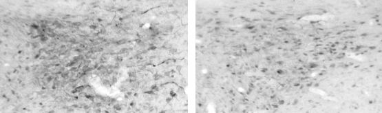
What this figure/staining show?
The inner part of the medial eminence (axons) is barely stained, while the outer part is (hormones).-
Adrenalectomy increases CRH (more staining) because there is no cortisol negative feedback. Will also promote magnocellular neurons to produce CRH (explains why the inner part of ME is stained)
Are there neuronal cell bodies in the posterior pituitary?
No, only glial cell bodies
Herring bodies in the posterior pituitary contains?
Oxytocin and/or vasopressin

Ultrastructure of the anterior pituitary: why is there a large pericapillary space?
To facilitate the many secreted hormones at once

What are pituicytes?
Glial cells
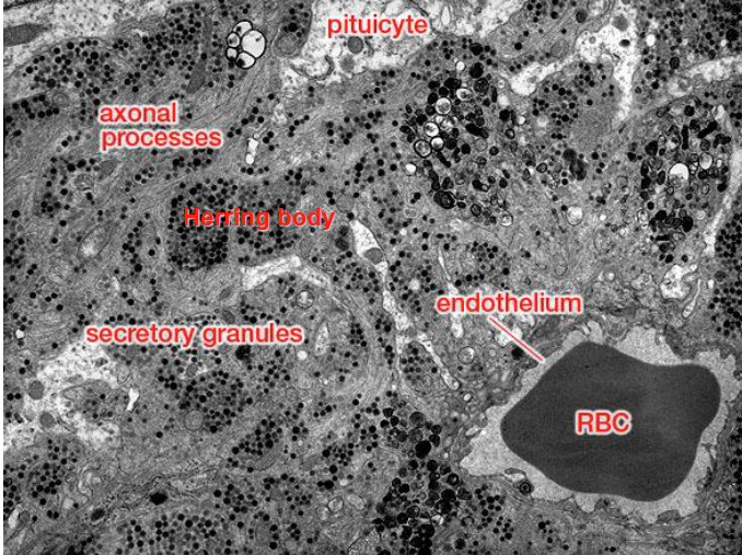
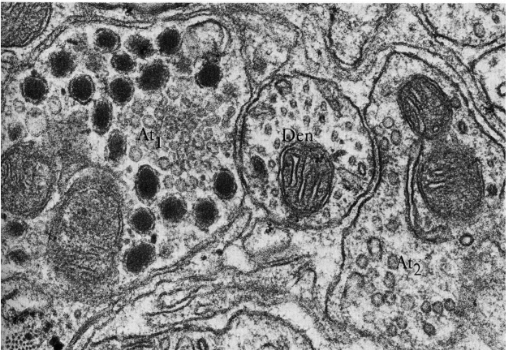
Ultrastructure of the posterior pituitary (not a question)

Long feedback control of hypothalamic hormone release
From the target organ through hormones or via sensory neuronal input

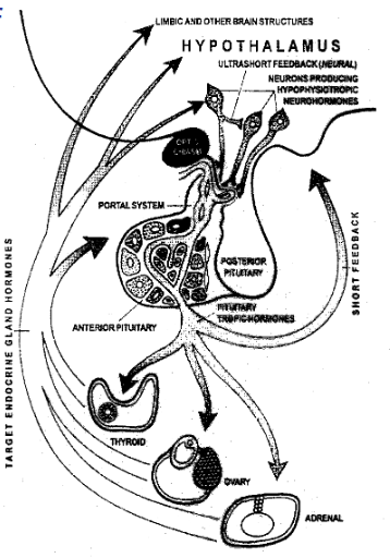
Short feedback control of hypothalamic hormone release
Through pituitary hormones acting on hypothalamic neuroendocrine neurons

Ultrashort feedback control of hypothalamic hormone release
Through auto- or paracrine action of hypothalamic hormones within one nucleus or though intra- hypothalamic projections from one neuroendocrine nucleus to another

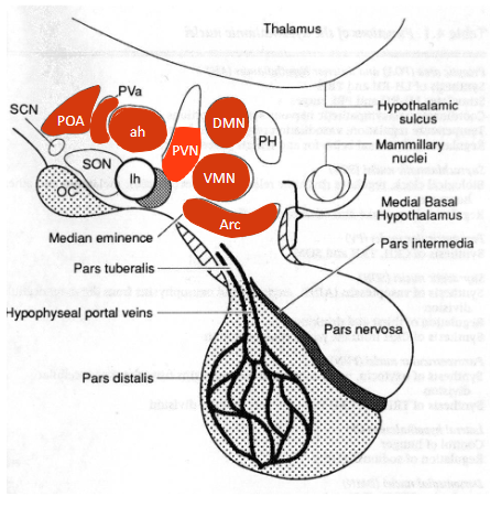
How are paracellular and magnocellular neurons differentiated?
Based on the size of the neurons, localization, and neuropeptide content, the parvocellular and magnocellular divisions are differentiated.
Parvocellular contains CRH, etc, while magnocellular contains vasopressin and oxytocin
Magnocellular neurons → Posterior pituitary. Parvocellular neurons → Medial eminence → Anterior pituitary

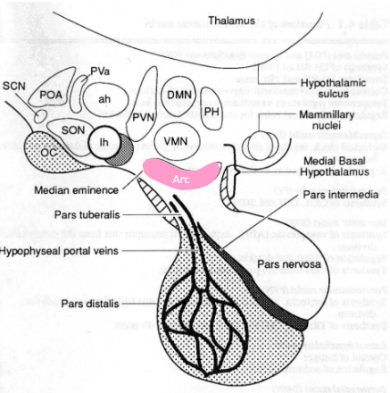
Where is CRH located?
The majority of CRH neurons are in the dorsal medial parvocellular portion of the PVN.
Approximately 2000 parvocellular neurons contain CRH (=CRF).

What creates the lateral parts of the paraventricular nucleus? What does it contain?
Magnocellular neurons containing VP and OT form the lateral limits of the nucleus.

What are the peptides CRH can synthesize?
CRH neurons can synthesize a wide variety of other peptides: Enkephalin, Cholecystokinin (CCK) and Neurotensin (NT) (which are not affected by adrenalectomy, ADX); Vasopressin and Angiotensin II (which are only observed after ADX).
Can CRH and its derivative peptides be visualized?
Yes, but you must add Colchicine to visualize them (except VP) because they have high turn-over rates.
ADX ONLY makes CRH visible (no cortisol = less negative feedback)
Colchicine depolarizes microtubules, which blocks axonal transport
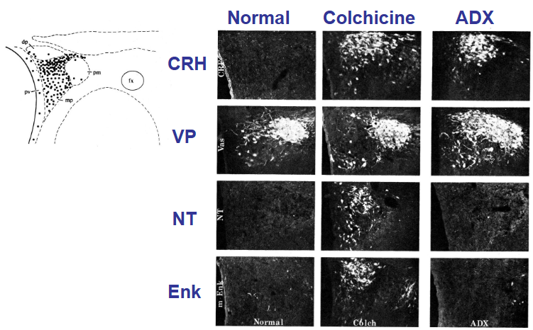
Colchicine function?
Inhibits axonal transport through depolymerization of microtubules
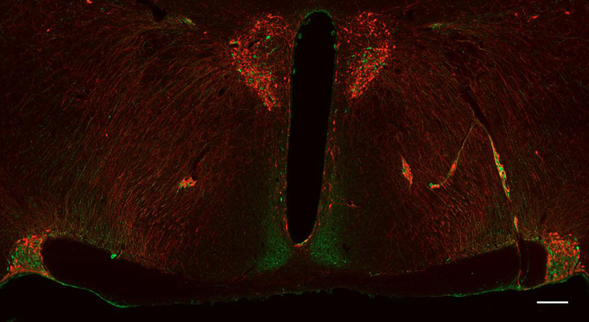
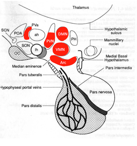
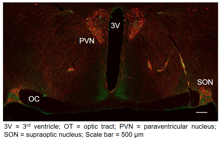
Where are the magnocellular and parvocellular neurons in this image?
In the PVN:
Magnocellular: Lateral
Parvocellular: Medial
In SON:
Magnocellular
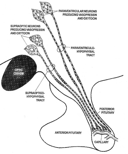
What does the magnocellular system secrete?
Mainly secretion of oxytocin (OT) and arginin-vasopressin (AVP) from neurons of the paraventricular nucleus (PVN) and supraoptic nucleus (SON).
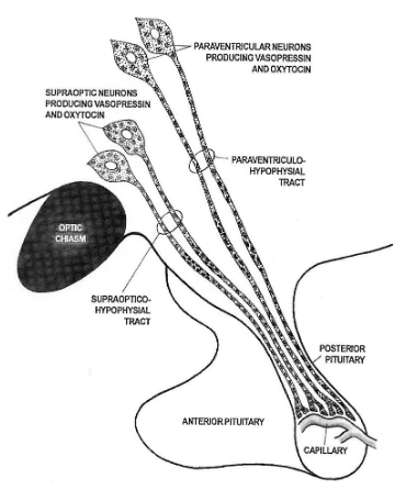
When does the magnocellular system secrete VP and OT?
Secreted in response to electrical activity. Transported down the axons by axoplasmic flow to terminals and varicosities in the posterior pituitary.
AVP and OT neurons of the PVN also project to the brain stem and spinal cord (cardiovascular control).

Does the magnocellular system terminate in the medial eminence like the parvocellular system?
No, they directly project to the posterior pituitary through the inner part of the medial eminence
Their axons make upmost of the posterior pituitary
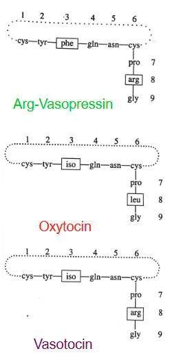
Relation between Vasopressin (VP), Oxytocin (OT), and Vasotocin?
They are nonapeptides differing in only two amino acid residues each in postitions 3 and 8.
These differences contribute to their difference in function

Structure of Vasopressin (VP), Oxytocin (OT), and Vasotocin?
The hormones are ring structures due to the formation of a disulfide (S-S) bond between Cys1 and Cys6.

Types of Vasopressin (not a question)
A few species such as pig and relatives have Lys-VP (LVP) in place of Arg-VP (AVP)

Why do higher vertebrates have both VP and OT?
The presence of both VP and OT in higher vertebrates instead of only one nonapeptide hormone is likely due to a gene duplication event early in vertebrate evolution.
What are AVP and OT derived from, and how are they made?
Each peptide is derived from a large precursor protein through proteolytic cleavage at dibasic sites by prohormone convertases (PCs).
What does AVP and OT release with?
AVP and OT are always found and released together with the so-called neurophysins, cystein-rich proteins of approx 95 amino acids, representing byproducts of the maturation cleavage. There is no known function attributed to the neurophysins.
Neurophysin I goes with OT, neurophysin II with AVP.
How were we able to find AVP and OT? (hint: neurophysins)
The neurophysins are the GOMORI-stainable material in the secretory granules of the HERRING bodies. They are, therefore, the moleculaes that led to the trails of the actual nonapeptide hormones AVP and OT.
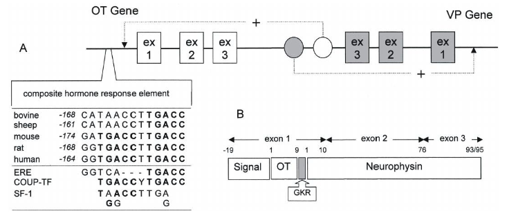
Structure of the AVP and OT genes
Both genes contain three exons. The hormone sequence is contained in exon1.
The precursor proteins contain a signal peptide, the hormone sequence proper, a consensus sequence (gly-lys-arg) for proteolytic maturation, and neurophysin. The upstream HRE is responsive to thyroid hormone, estrogen, and retinoic acid.
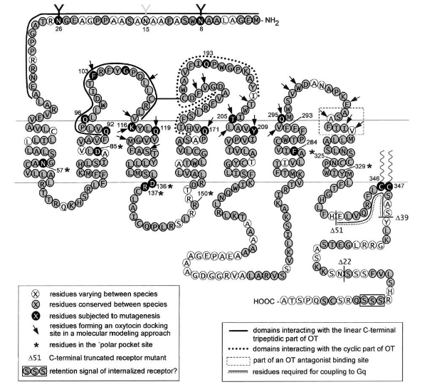
What types of receptors do OT and AVP act on?
Both OT and AVP exert their actions via binding to G-protein-coupled receptors.
How many OT and AVP-Rs have been cloned?
So far, one OT receptor (OTR) and three AVP-Rs (V1A, V1B, V2) have been cloned.
What are OTR, V1A, V1B, and V2 coupled to?
OTR, V1A, and V1B are coupled to phospholipase C – production of inositol trisphosphate and 1,2- diacylglycerol. The former triggers Ca2+ release from intracellular stores, the latter activates protein kinase C.
V2 couples to adenylate cyclase leading to increase in cAMP.
The oxytocin receptor (OTR) (not a question)
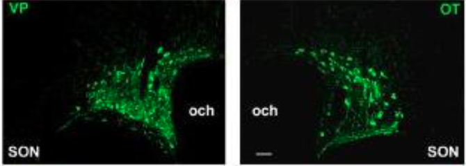
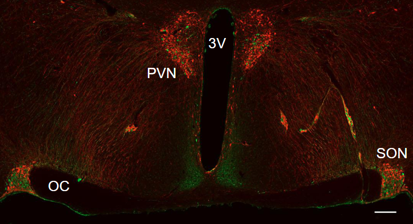
Anatomical distribution of AVP and OT - The paraventricular and supraoptic nuclei (not a question)
PVN: Contains magnocellular and parvocellular neurons
SON: Only contains magnocellular neurons
What does the supraoptic nucleus contain? Where does it project to?
The SON is very homogeneous in that all projection neurons are magnocellular and all are neurophysin-positive, i.e. they all express either OT or AVP. Virtually all of them project to the posterior pituitary.