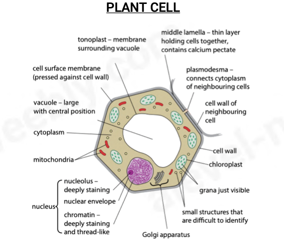
Outline the parts of a Plant cell (Light Microscope)
tonoplast - membrane surrounding vacuole
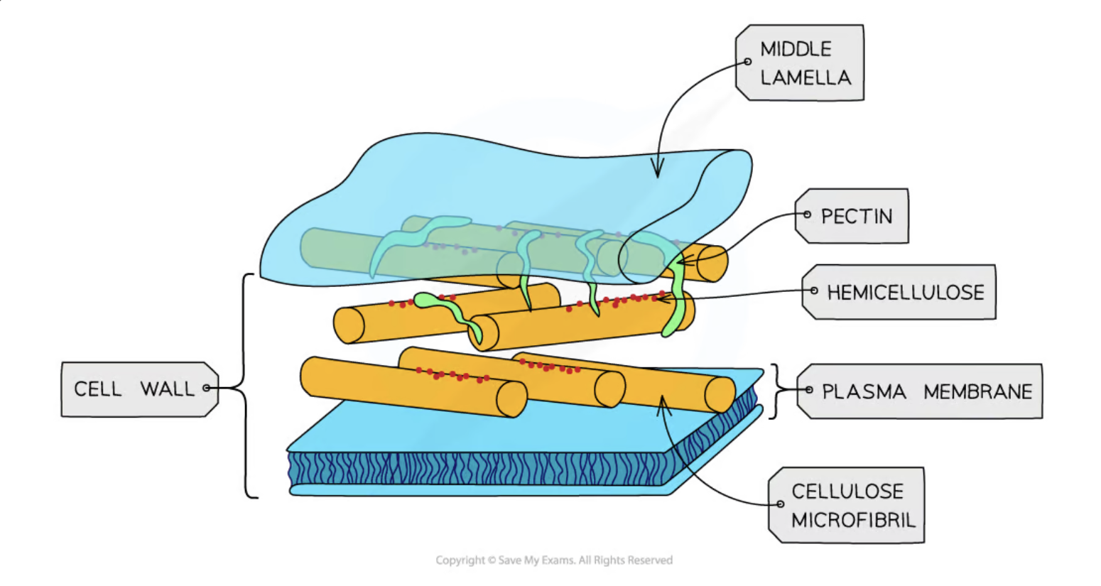
middle lamella - thin layer holding cells together, contains calcium pectate
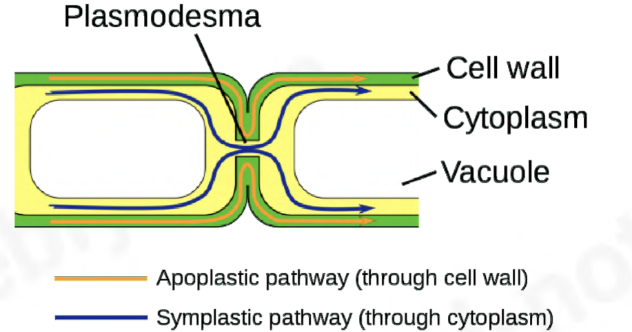
plasmodesma - connects cytoplast of neighbouring cells
cell wall
cell surface membrane(pressed against cell wall)
chloroplast
grana (within cholorplast)
small structres, difficult to identify
Golgi apparatus
nucleus - nucleolus; deeply staining, nuclear envelope & chromatin; deeply staining and thread-like
mitochondria
cytoplasm
vacuole - large with central position
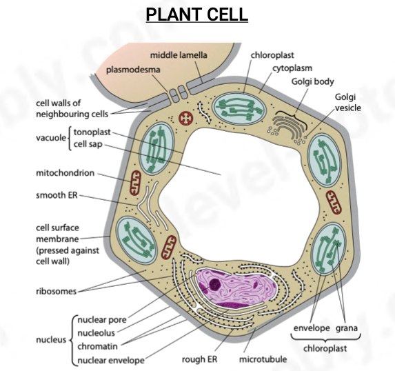
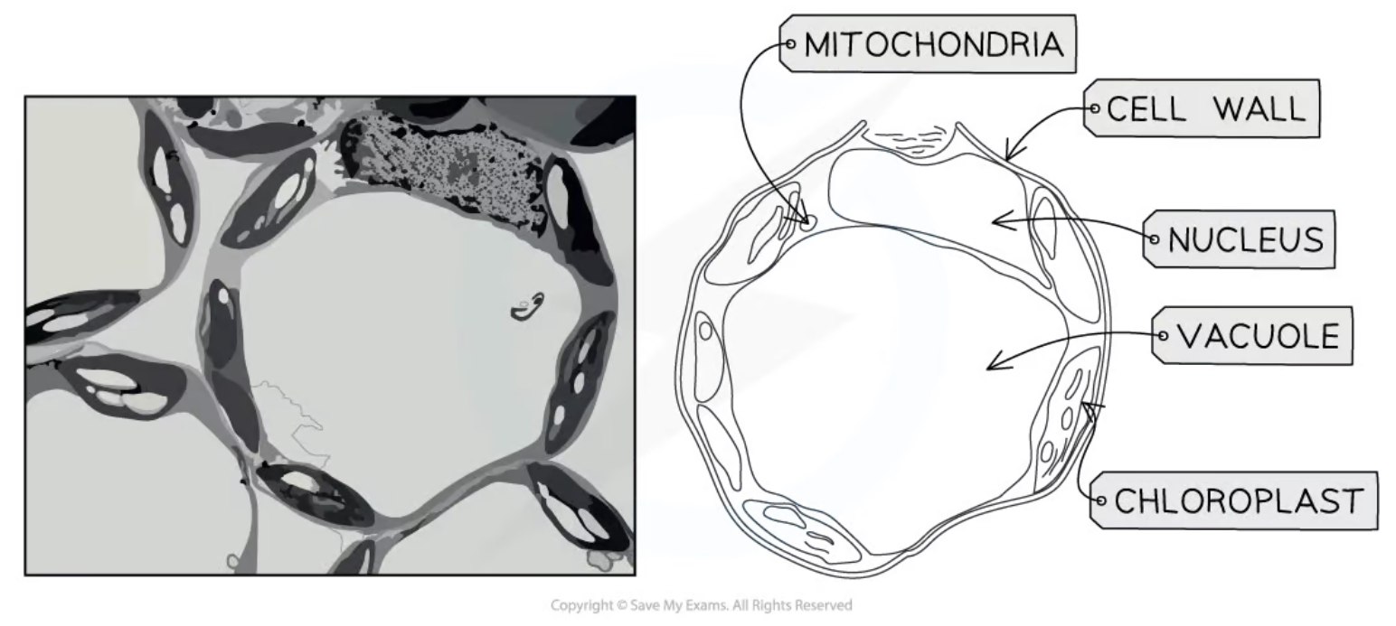
Outline the parts of a Plant cell (Electron micrograph)
plasmodesma
middle lamella
chloroplast - envelope & grana
cytoplasm
golgi body
golgi vesicle
microtubule
rough ER
nucleus - nuclear pore, nucleolus, chromatin, nuclear envelope
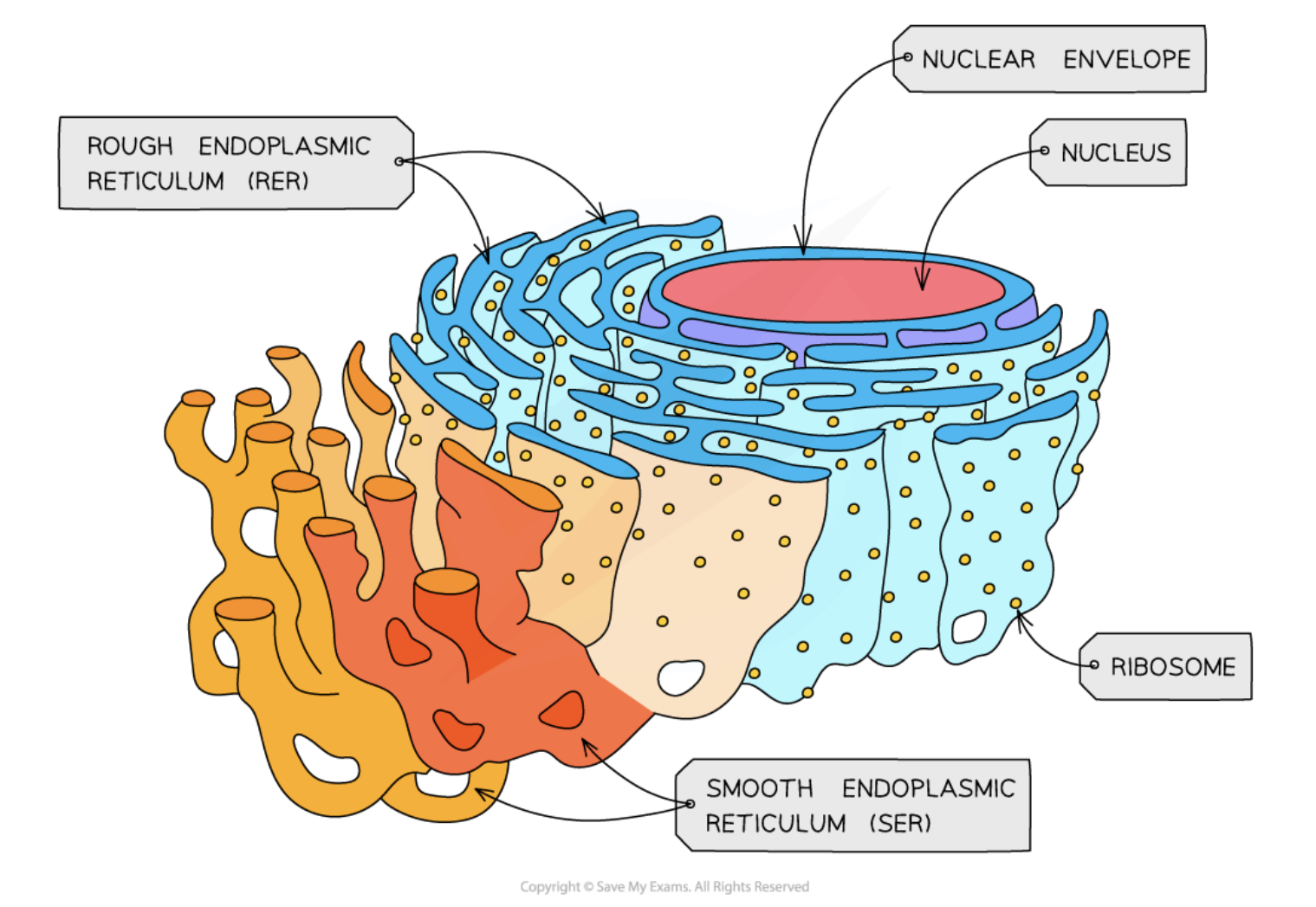
ribosomes
cell surface membrane(pressed against cell wall)
smooth ER
mitochondrion
vacuole - tonoplast , cell sap
cell wall
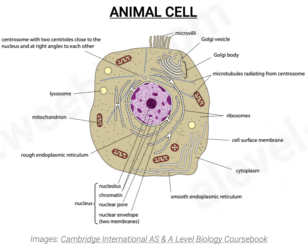
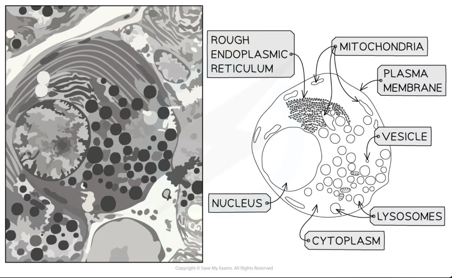
Outline the parts of the Animal cell (electron micrograph)
Microvilli
golgi vesicle
golgi body
microtubules radiating from centrosome
ribosomes
cell surface membrane
cytoplasm
smooth endoplasmic reticulum
nucleus - nucleolus, chromatin, nuclear pore,nuclear envelope(two membrane)
rough ER
mitichondrion
lysosome
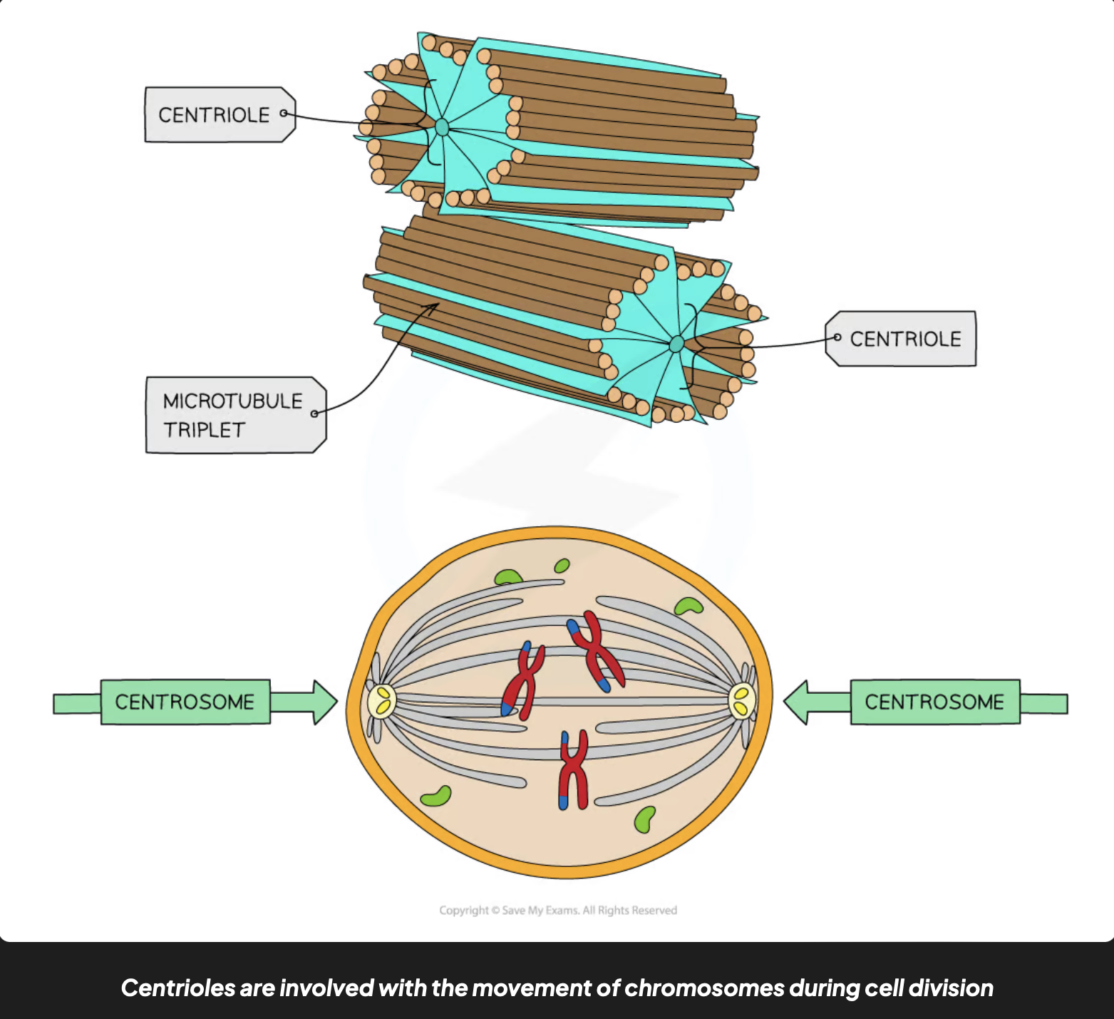
centrosome with two centrioles close to the nucleus and at right angles to each other
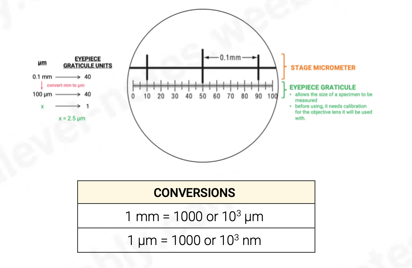
Magnification calculation
magnification = image size/actual size
What is ‘Resolution’ ?
the ability to distinguish between 2 seperate points -
as resolution increases, image clarity and detail also increase
What is ‘Magnification’ ?
how much bigger a sample appears to be under a microscope than in it is in real life
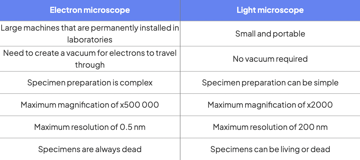
What is the Resolution and the Magnification of a Light Microscope
resolution - 200 nm
magnification - x1500
What is is the Resolution and the Magnification of an Electron Microscope
SEM - 3nm
TEM - 0.5 nm
x250,000 — x500,000
Outline the Light microscope
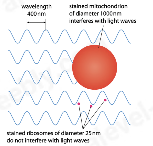
limit of resolution: half the wavelength
ribosomes (25nm) cant be seen with a light microscope as they dont interfere with the light waves
different stains are absorbed by different cell organelles so they can be observed more clearly
Outline the Electron microscope
vacuum (electrons cannot be focused without a vacuum as they will collide with air molecules and a scatter)
water boils at room temperature in a vacuum so the sample must be dehydrated(specimen has to be dead)
Advantages of light microscope over and electron microscope and their differences
can observe living tissue
more portable
easier to use - no technical training required
possible to see natural colours
observer can stain particular types of tissue for better visibility
Electron Micrograph of Plant cell
Electron Micrograph of Animal cell
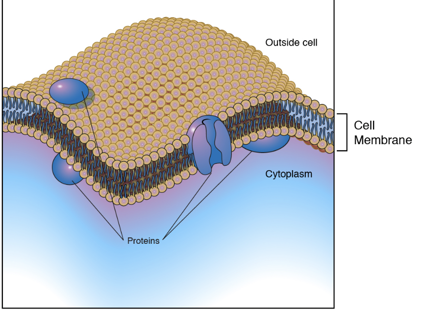
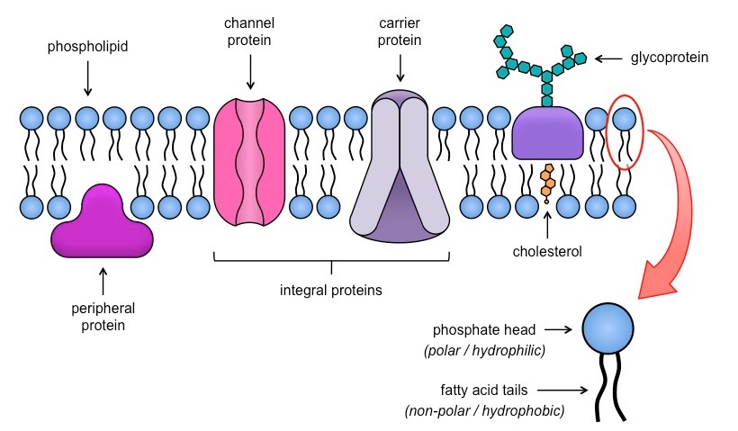
Describe the Cell surface membrane (phospholipid bilayer) (7 nm)
has a selectively permeable membrane that allows for the exchange of certain substances
is the barrier between cytoplasm and external environment
has cell recognition (surface antigens)
selects substances that enter/leave cells
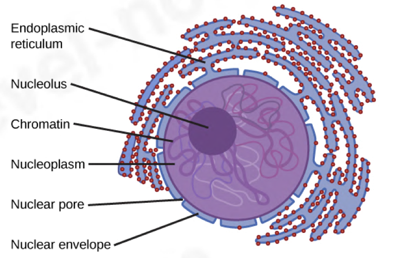
Outline the Nucleus (7 μm in diameter)
controls cell’s activities
very dense, takes up colour the most when stained
divides first during cell division
surrounded by 2 membranes, known as the nuclear envelope which is continious with the RER
contains:
a) nuclear pores: allow and control substances
entering the nucleus (protein to make ribosomes, ATP, some hormones, nucleotides)
and leaving the nucleus (mRNA, ribosomes for protein synthesis)
b) nucleolus (2.5 μm in diameter): contains loops of DNA from several chromosomes and synthesises ribosomes
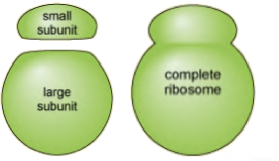
Outline Ribosomes (25 nm in diameter)
composed of 2 subunits
carry out protein synthesis
80S - found in cytoplasm
70S - found in chloroplasts & mitochondria
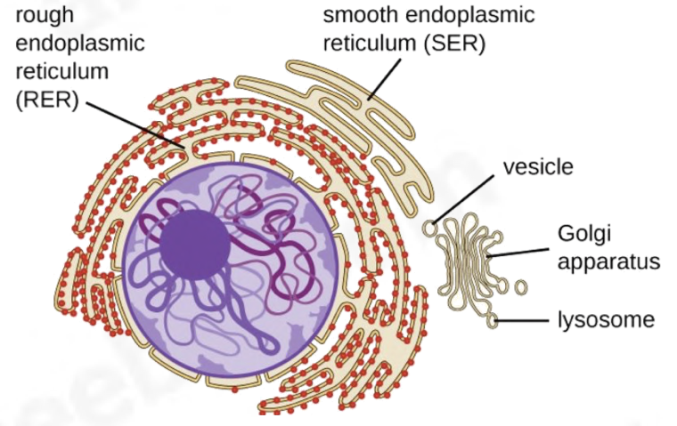
Outline the Rough endoplasmic reticulum (RER)
composed of membranes that form an extended system of fluid-filled sacs (cistern)
single membraned organelle
Attached ribosomes, therefore site of protein synthesis
proteins made by the ribosomes enter the sacs and are often modified as they go through them
(vesicles) break off from the ER and join to form the Golgi
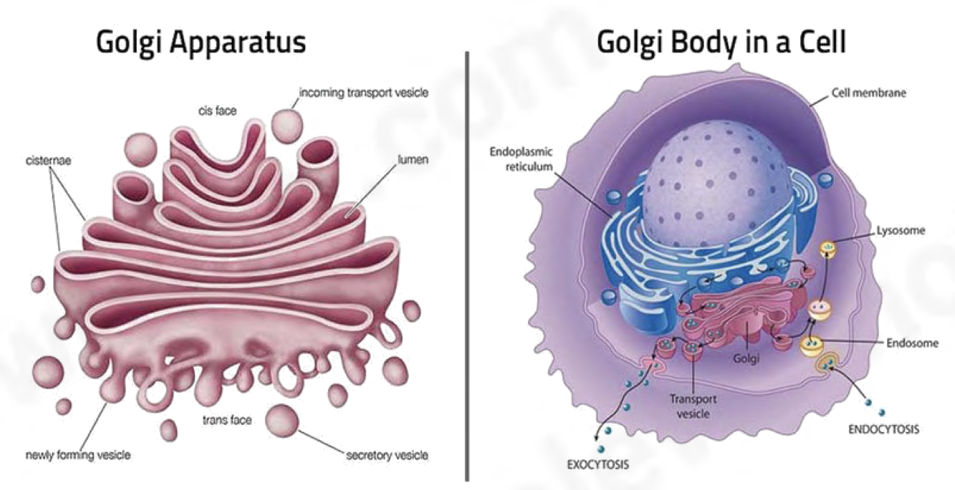
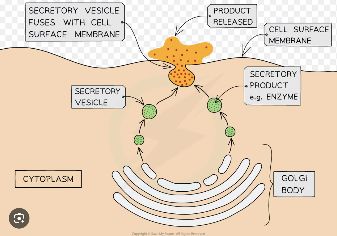
Outline the Golgi apparatus
composed of stacks of cisternae formed by the vesicles which bud off from the RER
single membraned organelle
packages substances into vesicles for transport
responsible for:
glycosylation
phosphorylating proteins
assembly of polypeptides into proteins (40 structure)
folding proteins
removing the 1st amino acid methionine to activate proteins
Outline the Smooth endoplasmic reticulum (SER)
synthesises lipids and steroids such as cholesterol and the reproductive hormones estrogen and testosterone
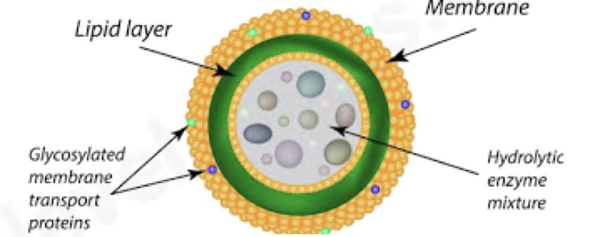
Outline Lysosoms (0.1—1μm in diameter)
spherical single membraned sacks
non permanent structures
no internal structure
contain hydrolytic enzymes
responsible for digestion/breakdown of unwanted structures e.g., old organelles
can even digest whole cells e.g., in mammary glands after the period of lactation
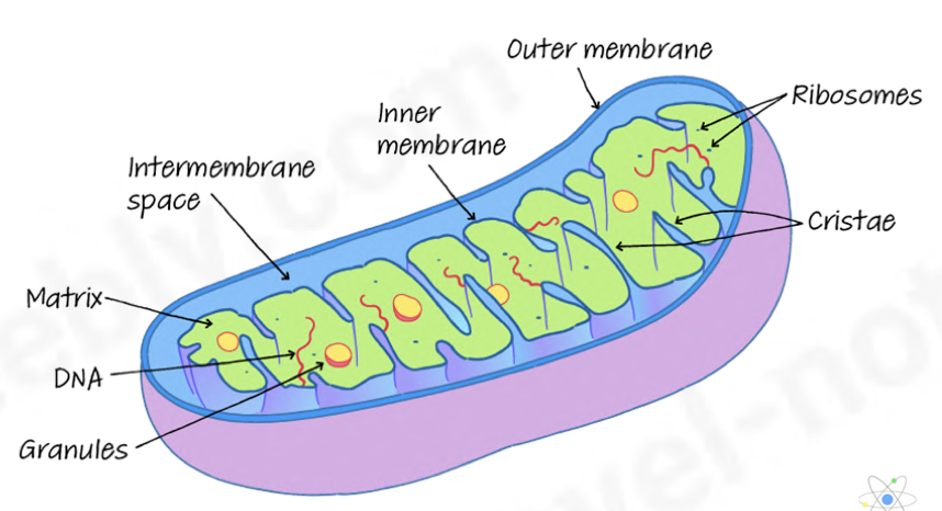
Outline the mitochondria (0.5—10μm in diameter)
carries out aerobic respiration
synthesises ATP (adenosine triphosphate)
transfers energy released from energy-rich molecules e.g, sugars and fats during respiration into ATP
more present in cells that have a higher demand for energy e.g., muscle, liver, and root hair cells
outer membrane contains the transport protein porin
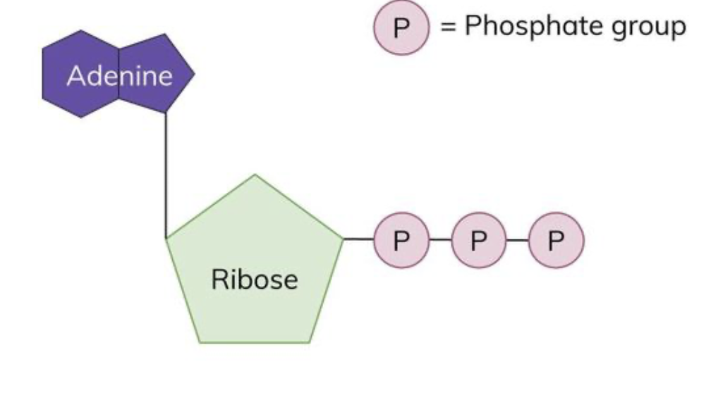
What is ATP and what is its function?
ATP is the energy-carrying molecule in all living cells
once made, ATP leaves the mitochondrion and can spread rapidly to all parts of the cell where energy is needed
its energy is released by its breakdown into ADP(adenosine diphosphate) in a hydrolysis reaction
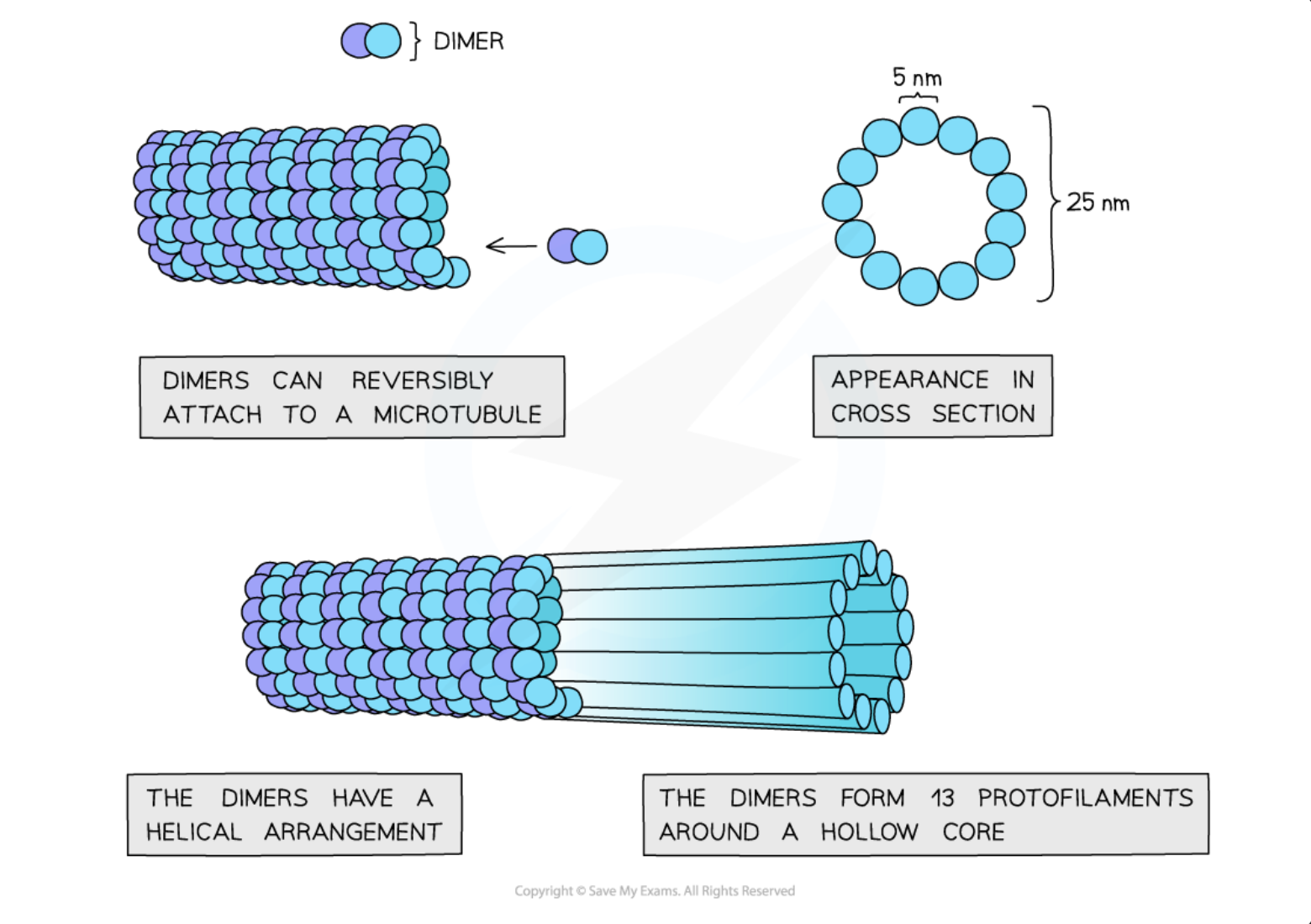
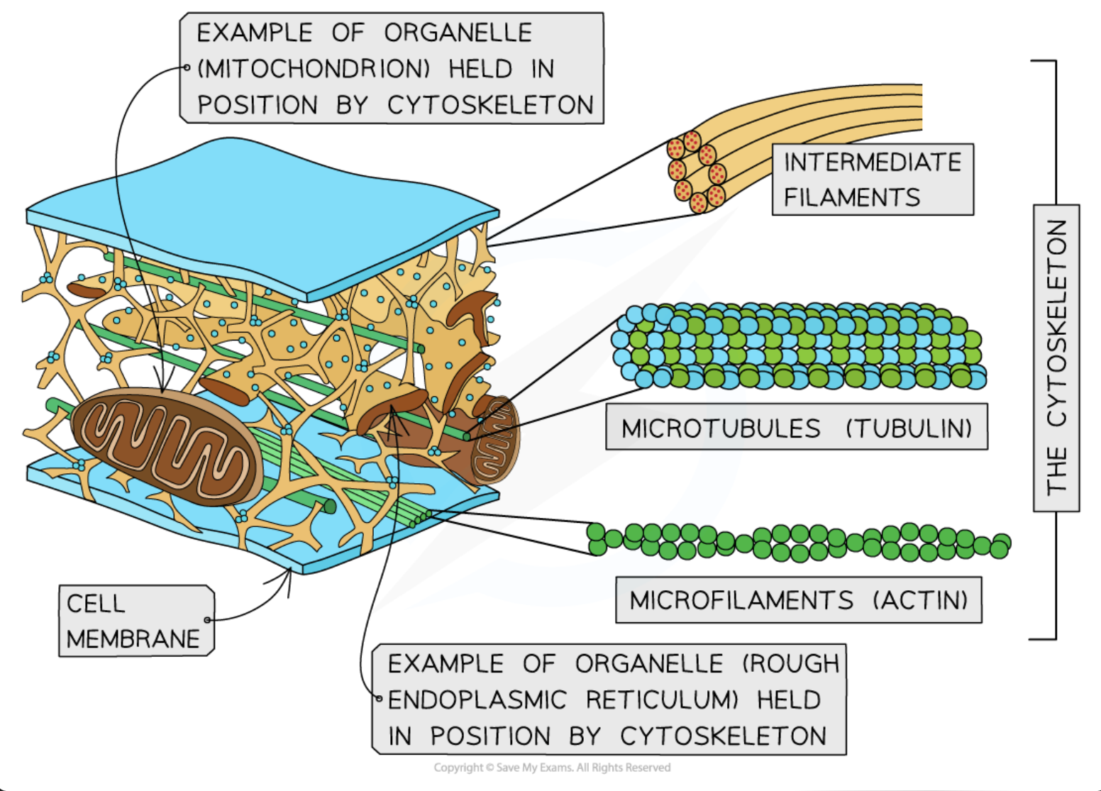
Outline microtubules
hollow tubes made up of α and β tubulin which combine to form dimers, which are then joined to make protofilaments
Thirteen protofilaments in a cylinder make a microtubule
Microtubules make up the cytoskeleton of the cell
providing support and movement of the cell
the assembly of microtubules from tubulin molecules is controlled by the special locations in cells called microtubule organizing centers (MTOCs)
Outline Centrioles (and centrosomes)
one centriole is made up of 9 triplets of microtubules
2 centrioles are present close together at right angles in a region called the centrosome, in animal cells
centrioles are hollow cylinders about 500 nm long
produces spindle fibers
organises microtubules
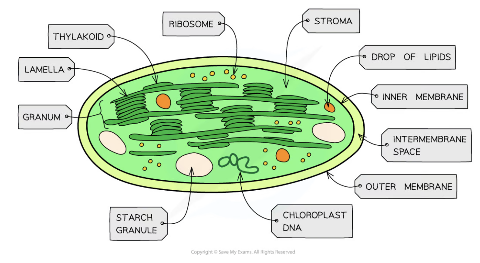
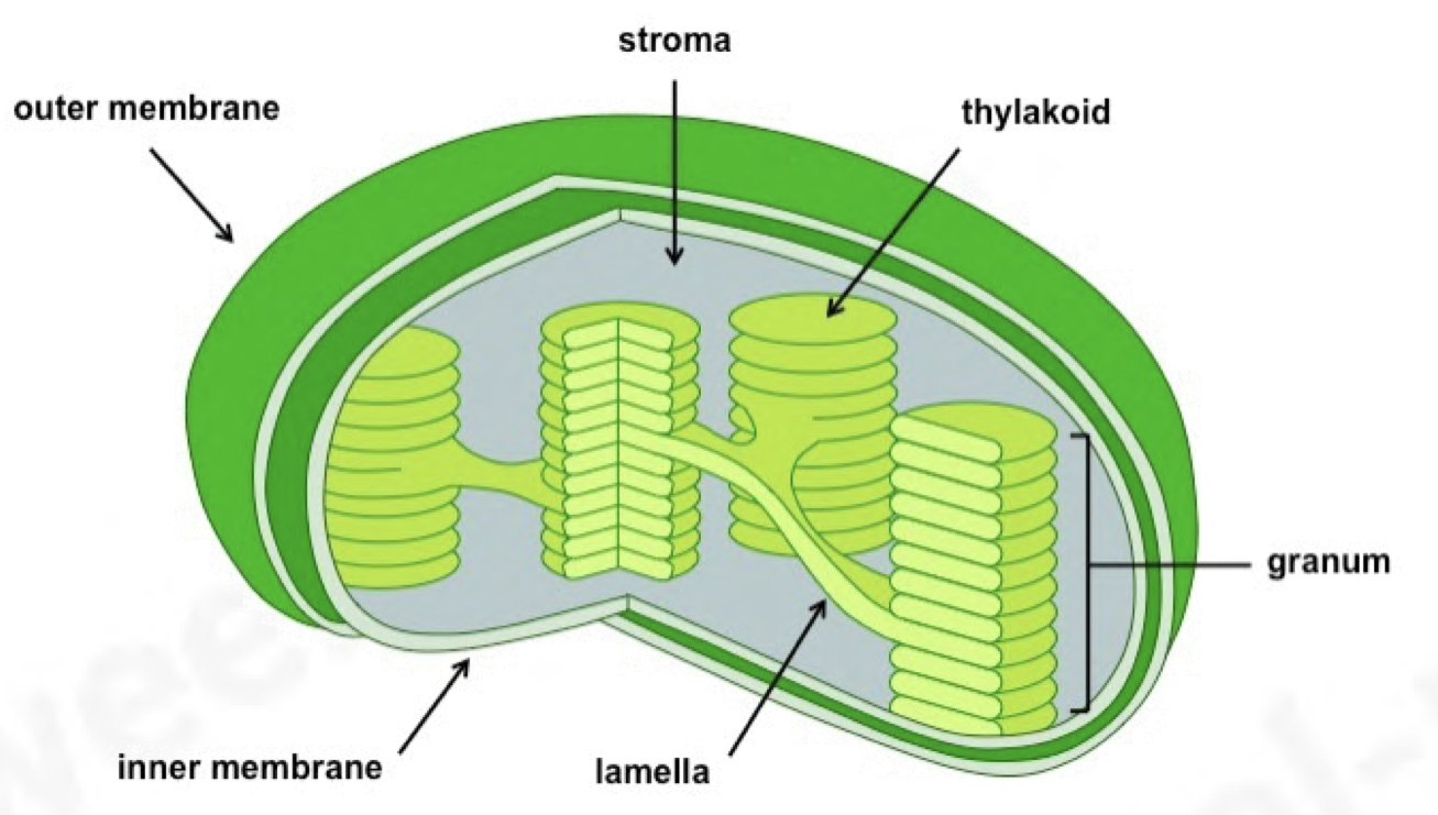
Outline Chloroplasts (3-10μm in diameter)
Chloroplasts are larger than mitochondria, and are also surrounded by a double-membrane
Membrane-bound compartments called thylakoids stack together to form structures called grana
Grana are joined together by lamellae
Photosynthetic pigments such as chlorophyll are found in the membranes of the thylakoids, where their role is to absorb light energy for photosynthesis
contains starch grains
Chloroplasts contain small circular pieces of DNA and 70S ribosomes used to synthesise proteins needed in chloroplast replication and photosynthesis
ATP is also produced here
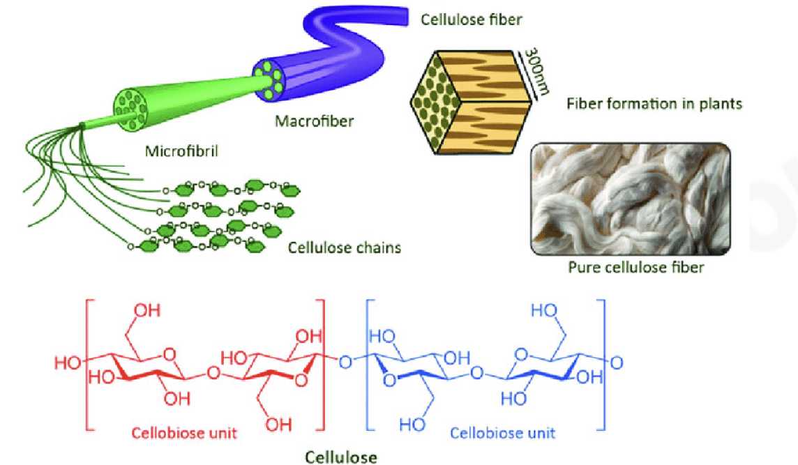
Outline the Cell wall (only present in plants)
gives cell rigidity definite shape as it’s made of cellulose
freely permeable
prevents cell from bursting
Outline the Plasmodesmata
plant cells are linked to neighboring cells by means of fine strands of cytoplasm called plasmodesmata which pass through pore-like structures in their walls
allows the transport of water, sucrose, amino acids, ions, etc., between cells without crossing membranes
this is called movement through the symplastic pathway
allows communication/signaling between cells
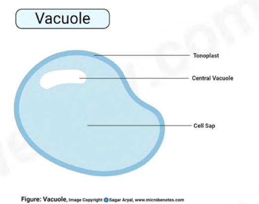
Outline Vacuoles
surrounded by a partially permeable tonoplast which controls exchange between the vacuole and cytoplasm
helps regulate osmotic properties of cells
fluid present in the vacuole consists of:
P igments
E nzymes
S tarch
O rganic molecules
M ineral salts
O xygen
C arbon dioxide
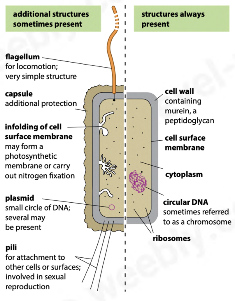
State the Structural features of Prokaryotic cells
organisms that lack nuclei or proper nuclear membranes are called prokaryotes
unicellular
1-5μm in diameter
cell wall made of murein (peptidoglycan = protein + polysaccharides)
no membranes around organelles
70S(smaller) ribosomes
genetic material in the form of circular DNA
have no ER
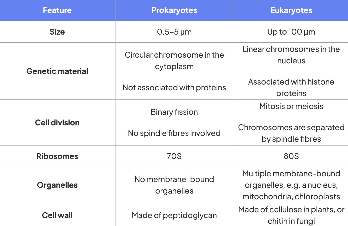
Differences between typical eukaryotic and prokaryotic cells
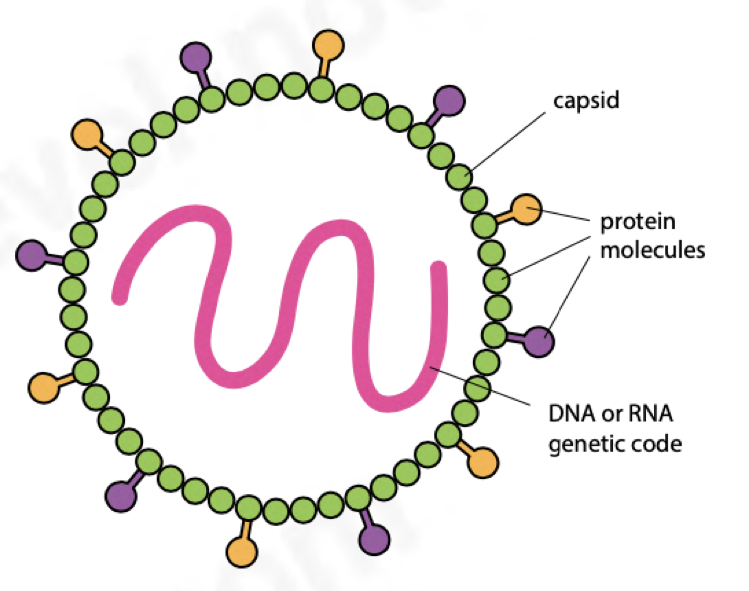
Outline Viruses
noncellular
protein coat called capsid
nucleic acid core; DNA/RNA strand
replicate inside host cells only
show no characteristics of living organism
symmetrical shape
the virus DNA/RNA takes over the protein synthesising machinery of the host cell which helps to make new virus particles
See Chapter 18.2(d) for more details
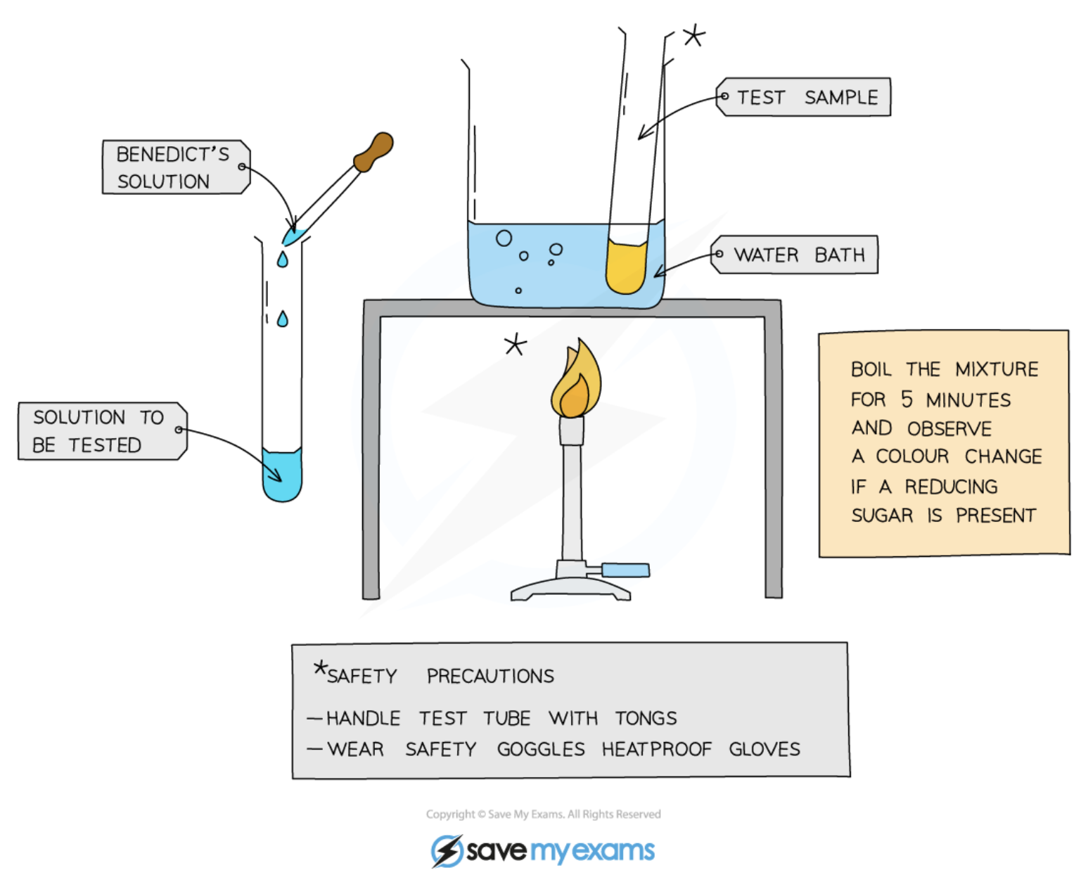
Outline the food test for Reducing sugars
reduce soluble blue copper sulphate containing copper (II) ions into insoluble brick-red copper oxide, containing copper (I) ions
the copper oxide is seen as a brick-red precipitate
add equal volumes of Benedict’s reagent and the food sample to a test tube
heat in a water bath at 80°C
if reducing sugars are present, the following colour
changes are observed:
BLUE → GREEN → YELLOW → ORANGE → BRICK-RED
— CONCENTRATION OF REDUCING SUGARS INCREASING →
Outline the food test for Non-reducing sugars
e.g., sucrose
disaccharide is first broken down into its 2 monosaccharide constituents in a hydrolysis reaction
this is done by adding HCl and then neutralising the acid with an alkali such as sodium bicarbonate
constituent monosaccharides will be reducing sugars and their presence can be tested by Benedict’s test
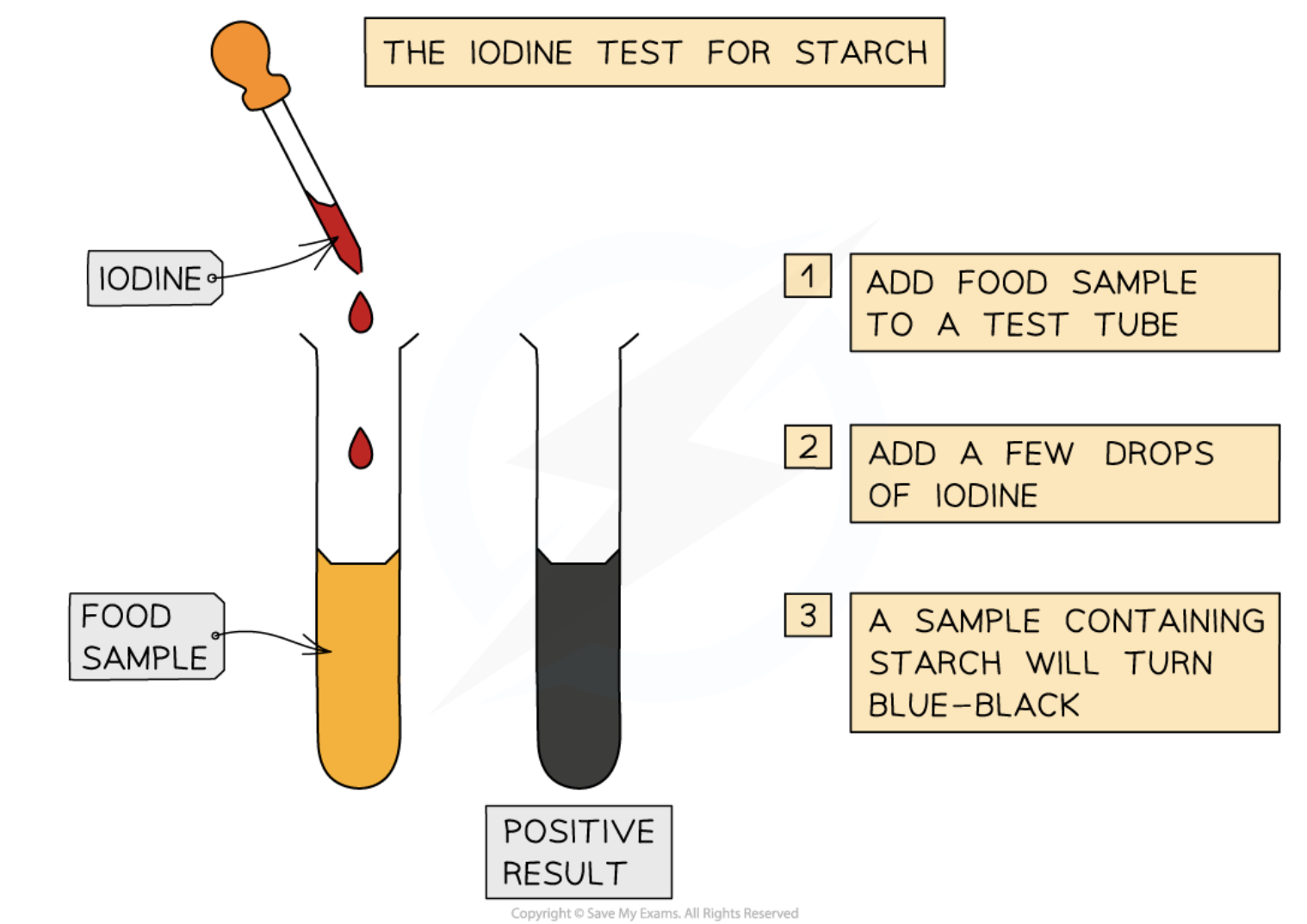
Outline the test for Starch
add drops of iodine solution to the sample
if a blue-black colour is quickly produced, starch is present
iodine solution is yellow brown(not present)
This test is useful in experiments for showing that starch in a sample has been digested by enzymes
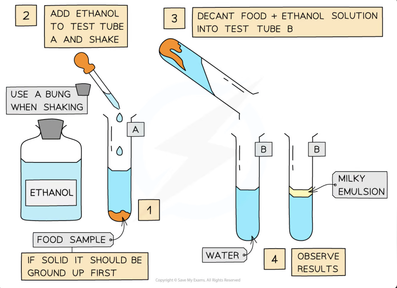
Outline the test for Lipids (ethanol emulsion test)
Lipids are nonpolar molecules that do not dissolve in water but will dissolve in organic solvents such as ethanol
Add ethanol to the sample to be tested, shake to mix and then add the mixture to a test tube of water
If lipids are present, a milky emulsion will form (the solution appears ‘cloudy’); the more lipid present, the more obvious the milky colour of the solution
If no lipid is present, the solution remains clear
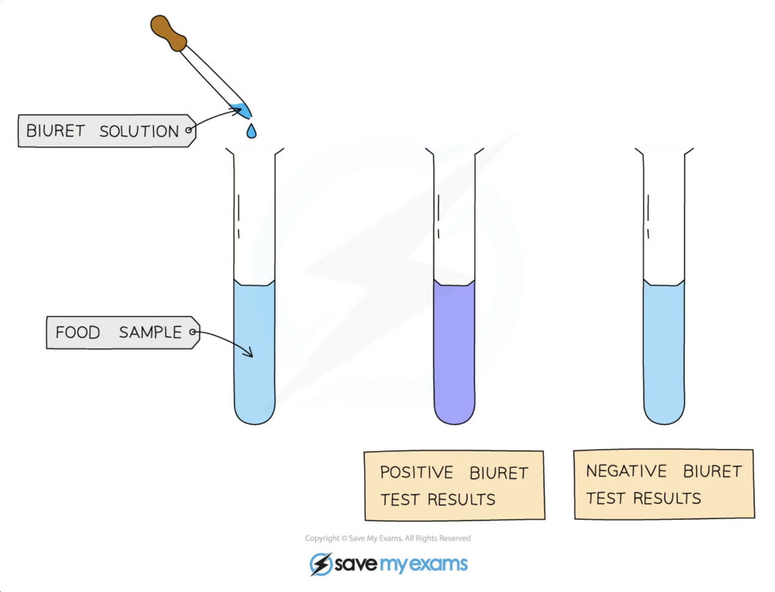
Outline the biuret test for proteins
all proteins have peptide bonds containing nitrogen atoms which form a purple complex with Copper(II) (oxidised carbon) ions
first, equal volumes of the sample and Biuret reagent are mixed
if proteins are present, the colour changes from blue to lilac
instead of biuret reagent, potassium hydroxide and diluted copper (II) sulphate can be used
For this test to work, there must be at least two peptide bonds present in any protein molecules, so if the sample contains amino acids or dipeptides, the result will be negative
What are Carbohydrates
Carbohydrates are one of the main carbon-based compounds in living organisms
composed of C, H, O
As H and O atoms are always present in the ratio of 2:1 (e.g. water H2O, which is where ‘hydrate’ comes from) they can be represented by the formula Cx (H2O)y
divided into monosaccharides, disaccharides,
polysaccharides
What is a Monomer
one of many small molecules that combine to form a polymer, e.g. – monosaccharides, amino acids, nucleotides
What is a Polymer
large molecule made from many similar repeating subunits, e.g. – polysaccharides, proteins, nucleic acids
What is a Macromolecule
large molecule formed due to polymerisation of monomers
e.g. – polysaccharides, proteins (polypeptides), nucleic acids (polynucleotides)
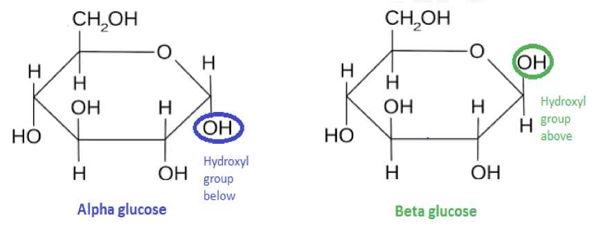
What is a Monosaccharide
A soluble molecule consisting of a single sugar unit all of which are reducing sugars, with the general formula C(H2O)n
the main types of monosaccharides are:
trioses (3C), pentoses (5C), hexoses (6C)
{glucose, fructose galactose}- HEXOSES
{ribose, deoxyribose}- PENTOSES
What are the roles of Monosaccharides
are a source of energy in respiration -
C-H bonds can be broken to release a lot of energy which is transferred to help make ATP from ADP
are the building blocks for larger molecules
glucose is used to make the polysaccharides starch, glycogen, and cellulose; ribose is one of the molecules used to make RNA and ATP, deoxyribose is one of the molecules used to make DNA
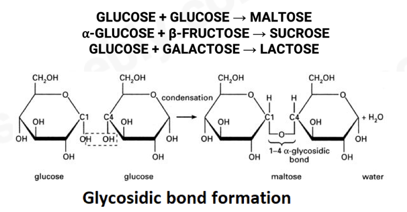
What is a Disaccharide
Sugar molecule, consists of 2 monosaccharides joined by a glycosidic bond.
Examples:
Maltose (α glucose + α glucose)
Sucrose (α glucose + fructose)
Lactose (α glucose + β galactose)
• formed by a condensation reaction where an H2O molecule is removed; the bond formed by condensation is called a glycosidic bond
Functions:
Sugar in germinating seeds (maltose)
Sugar stored in cane sugar (sucrose)
Mammal milk sugar (lactose)
What is a Polysaccharide
A polymer consisting of many subunits which are monosaccharides joined by glycosidic bonds
e.g., starch, glycogen, and cellulose (all polymers of α-glucose)
not sugars
Functions
Energy storage – convenient, compact, inert, insoluble.
In plants - starch
animals - glycogen
Structural cell wall (cellulose)
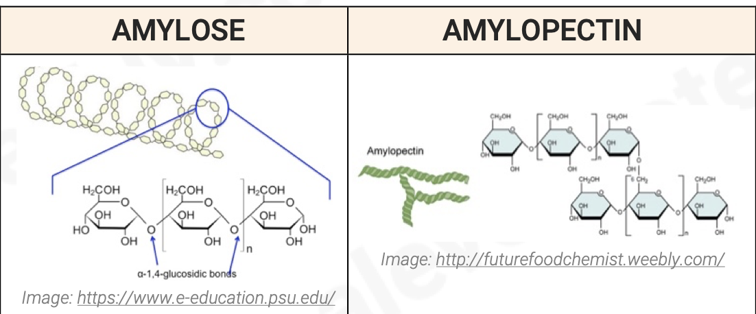
What are the two polysaccharides that make up starch?
AMYLOSE:
made by condensation reactions between 1,4 linked ⍺-glucose molecules
long, unbranching chain
chains are curved and coil into helical structres making the final molecule more compact
AMYLOPECTIN:
also made of 1,4 linked ⍺- glucose molecules
chains are shorter than amylose and branch out to the sides
branches are formed by 1-6 linkages
formed by glycosidic bonds
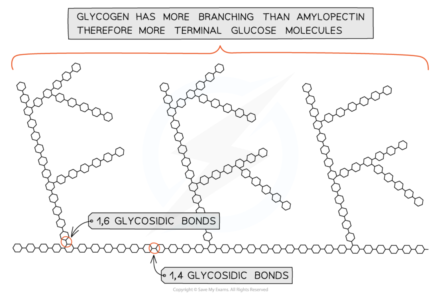
What is Glycogen
A polysaccharide
made of chains of 1-4 linked ⍺-glucose molecules with 1-6 linkages forming branches
tend to be more branched than amylopectin molecules
the many ends due to branching, aid in easy addition and removal of glucose
compact and insoluble, doesn’t affect the water potential (Ψ)
High concentration in liver & muscle cells due to higher cellular respiration
Function: Energy storage polysaccharide in animals and fungi
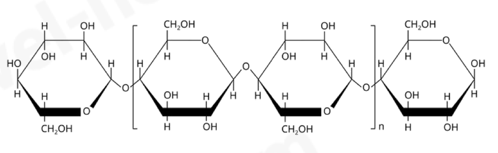
Cellulose → polymer of β-glucose
A polysaccharide
formed by 1-4 beta glucose linkages where every second glucose is rotated 180 degrees so one oxygen is up and the other is down
tightly cross-linked to form bundles which are held together by hydrogen bonds
cellulose fibers have very high tensile strength – making it possible for a cell to withstand high osmotic pressure and are freely permeable
Function: Compose cell wall in plants
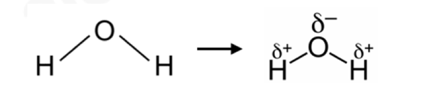
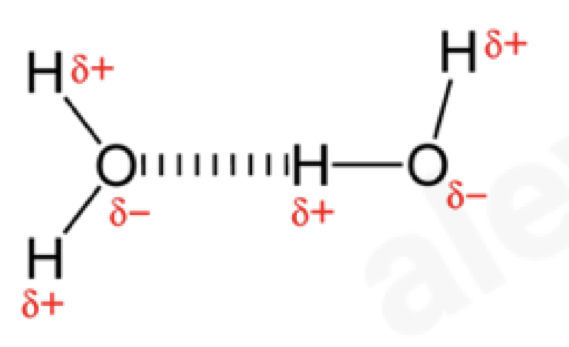
Explain dipoles and hydrogen bonds
an unequal distribution of charges in a covalent bond is called a dipole
molecules which have groups with dipoles are polar
in water, oxygen atoms get more electrons due to them being more electronegative and therefore get a small negative charge denoted by delta (𝛅-)
hydrogen atoms get less electrons and therefore get small positive charges (𝛅+)
negatively charged oxygen of one molecule is attracted to a positively charged hydrogen of another, this attraction is called a hydrogen bond
Are molecules containing groups with dipoles polar or non polar?
Molecules which have groups with dipoles are polar
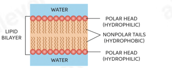
they’re attracted to H2O molecules as they also have dipoles and are considered to be hydrophilic (water-loving)
soluble in water
e.g., glucose, amino acids, NaCl
Molecules which do not have dipoles are non-polar
they’re not attracted to water and hydrophobic (water-hating)
insoluble in water
e.g., oils, cholesterol
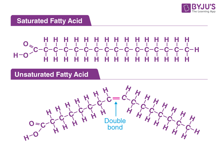
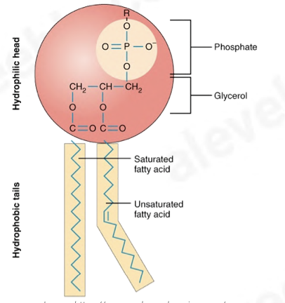
Outline Fatty acids
Fatty acids
contain the acidic (carboxyl) group –COOH
larger molecules in the series have long hydrocarbon tails attached to the acid which are 15- 17 carbon atoms long
two types: saturated and unsaturated
unsaturated fatty acids have C=C double bonds
therefore don’t have maximum amount of hydrogen atoms
form unsaturated lipids
mostly liquid at room temp (unsaturated)
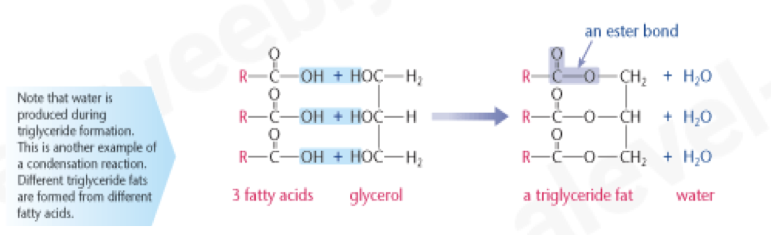
Outline Alcohols & Esters
alcohols contain the hydroxyl group (–OH) attached to C atom
reaction between (fatty) acid (–COOH) and alcohol (– OH) produces an ester
the chemical link between acid and alcohol is called an ester bond and is formed by a condensation reaction
glycerol has 3 hydroxyl groups; each one is able to undergo a condensation reaction with a fatty acid
triglycerides are insoluble in water due to the non- polar nature of hydrocarbon tails – they don’t have uneven distribution of charges and are hydrophobic
What are the roles of Triglycerides?
energy reserves
insulator
protect vital organs
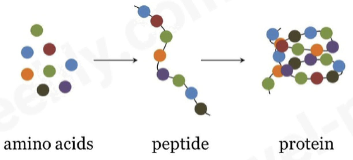
What are Proteins made of?
All proteins are made from the same monomer - amino acids.
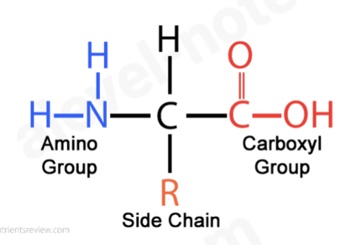
What is the structure of Amino acids?
All have a central carbon atom bonded to –
an amine group (–NH2)
a carboxylic group (–COOH)
a hydrogen
an R-group that determines what type of amino acid it is
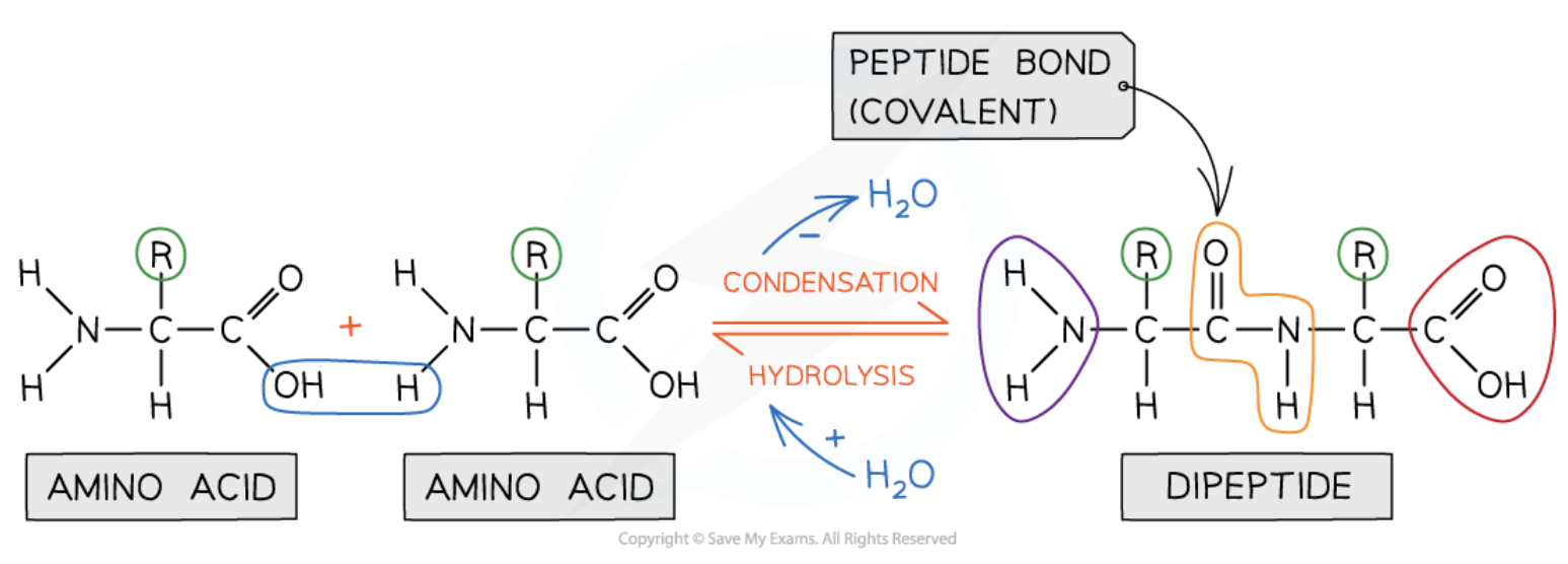
What is a peptide bond?
Forms when the carboxyl group of one amino acid loses an -OH and the amine group of another loses a hydrogen. The carbon of the first amino acid then bonds to the nitrogen of the second, releasing water in a condensation reaction.
a molecule made up of many amino acids linked together by peptide bonds is a polypeptide
polypeptides can be broken down to amino acids by breaking the peptide bonds in a hydrolysis reaction
this happens naturally in the stomach and small intestine during digestion
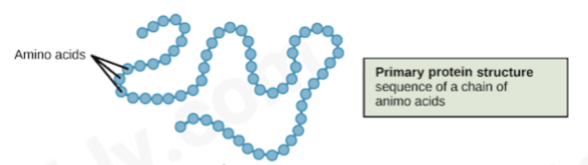
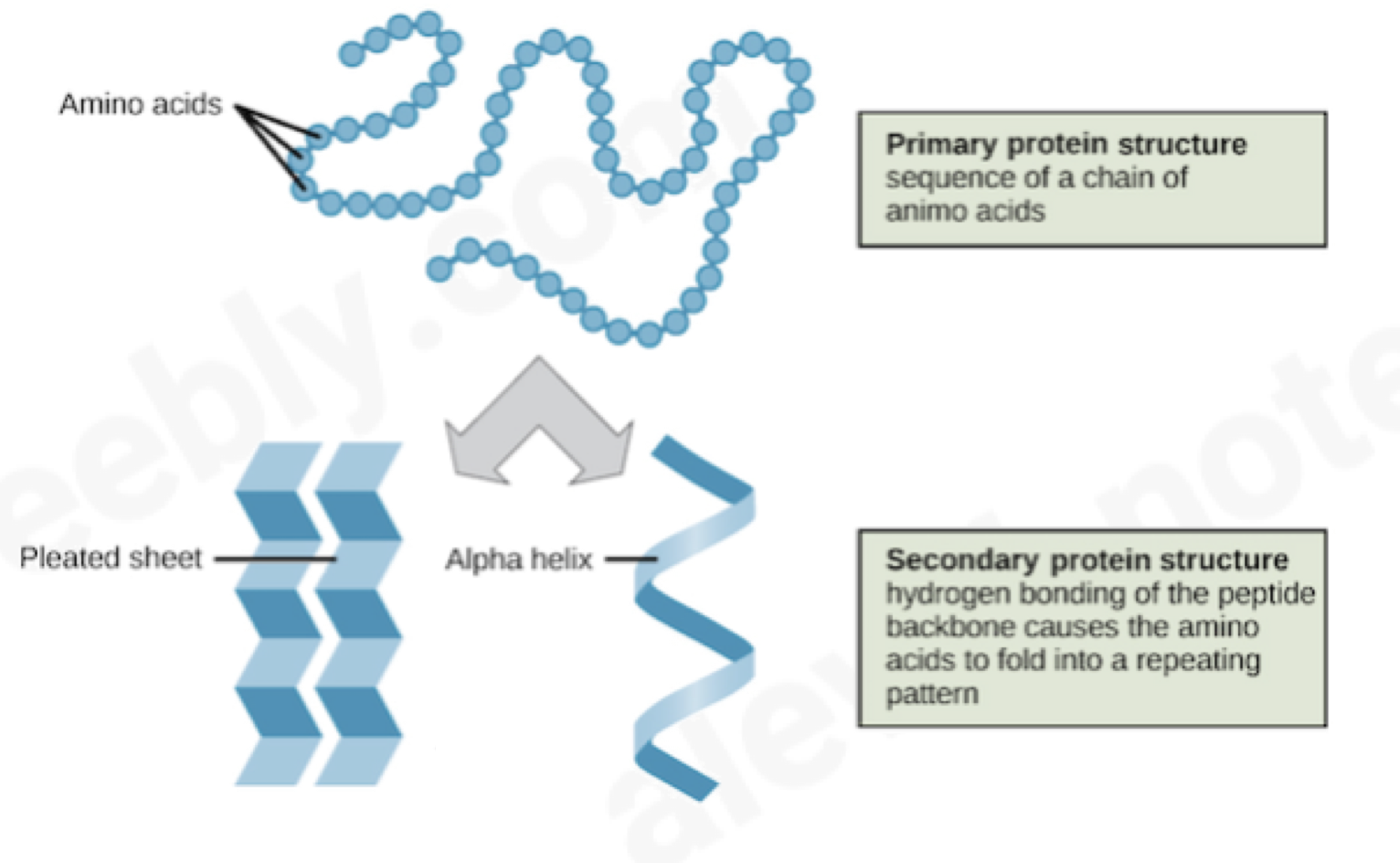
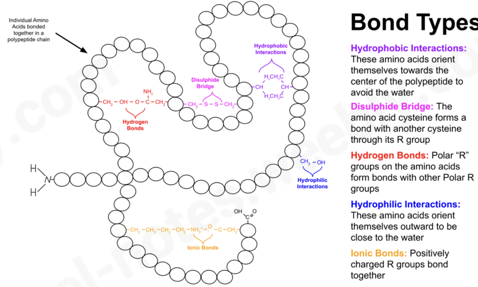
Primary protein structure
sequence of an amino acid chain
Secondary protein structure
hydrogen bonding of the peptide backbone causing amino acids to fold into a repeating pattern
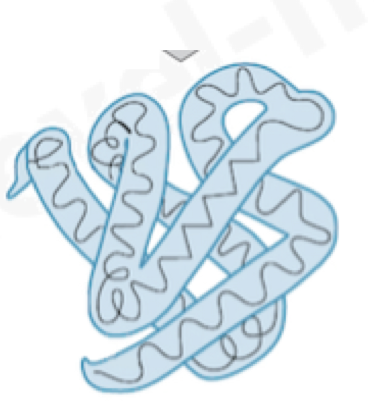
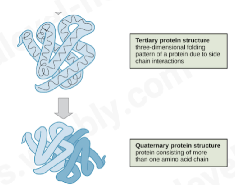
Tertiary protein structure
three-dimensional folding pattern of a protein due to side chain interactions
Quaternary protein structure
protein consisting of more than one amino acid chain
Bonds in the tertiary structure
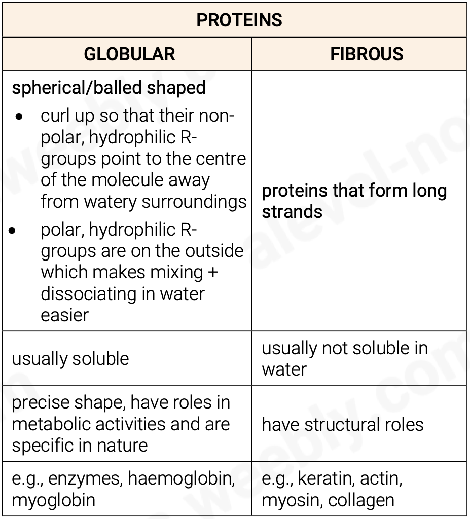
Globular vs Fibrous Proteins
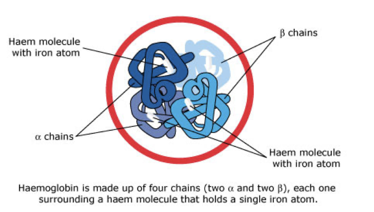
Outline Haemoglobin: a globular protein
made of 4 polypeptide chains therefore they have a quaternary structure
2 of the haemoglobins ⍺-chains, are made of ⍺- globin
the other 2 chains, β-chains, are made of β-globin
each polypeptide chain has a haem group attached
(prosthetic group) to it
haem contains a charged particle of iron
the haem group is also responsible for the colour of haemoglobin
each polypeptide chain can carry one molecule of oxygen
therefore, in total, haemoglobin can carry 4 molecules of oxygen or 8 oxygen atoms
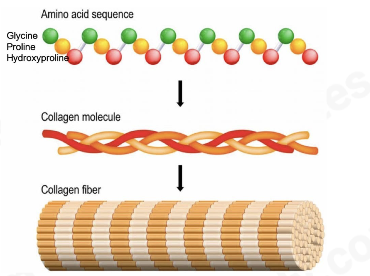
Outline Collagen: a fibrous protein
a structural protein
consisting of 3 helical polypeptide chains wound together into a triple helix and held together by hydrogen and some other covalent bonds formed between R-groups of amino acids where every 3rd amino acid in each chain is glycine
each 3 stranded molecule interacts with other collagen molecules running parallel to it
these cross-links hold many collagen molecules side by side forming fibrils
many fibrils lie alongside each other forming strong bundles called fibres
collagen is flexible but has tremendous tensile strength
collagen fibres line up according to the forces they withstand
found in skin, tendons, cartilage, bone, teeth, etc.
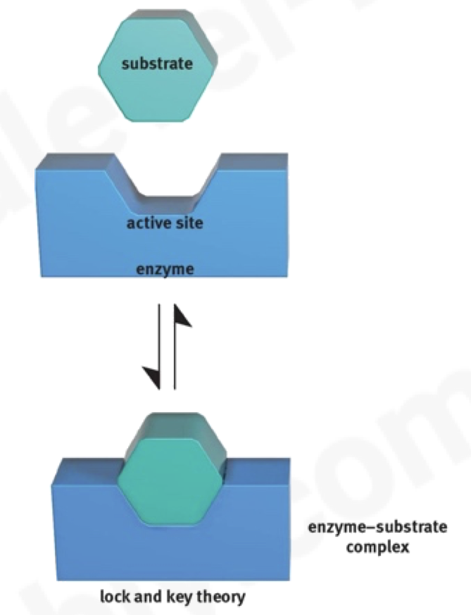
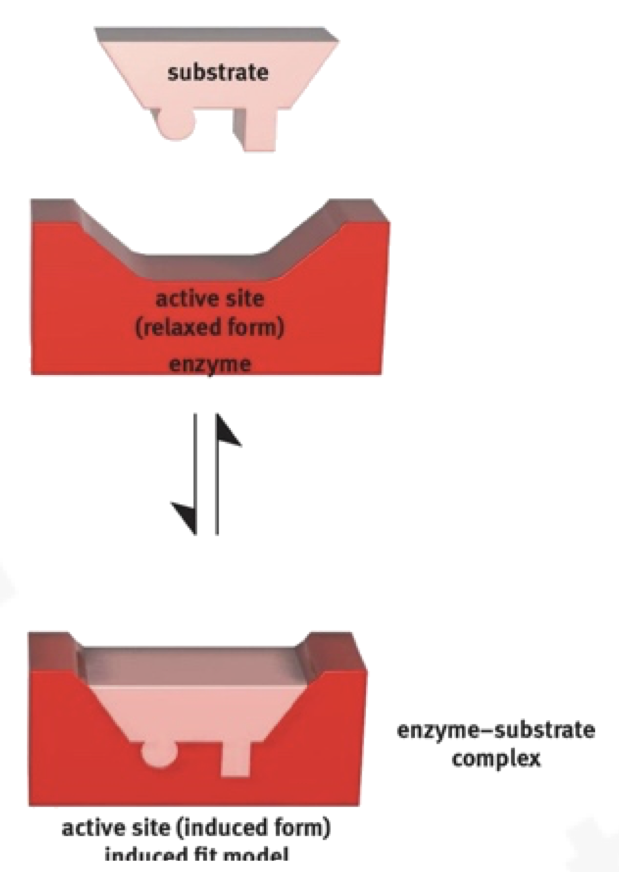
outline the difference between the induced fit mechanism and lock and key mechanism of enzyme action [4]
induced fit
1. shape of substrates not fully complementary to shape of active site
2. active site flexible / moulds around substrates
3. provides better fit / fully complementary
lock & key
1. shape of substrates complementary to shape of active site
2. active site does not change shape
3. substrate fits into active site
What are enzymes?
enzymes are globular proteins that catalyse metabolic reactions
function as biological catalysts
specific in nature
precise 3D shape with hydrophilic R-groups on the outside ensuring they’re soluble
possess active sites which are clefts to which a substrate can bind
Define the Lock and key theory
idea that enzymes have particular shapes into which their substrate fits into exactly
enzyme is said to be specific for a substrate
Define the induced fit hypothesis
substrate is partially complementary to the active site
the active site changes shape slightly to ensure a better fit and stronger binding of substrate, making catalysis even more efficient
Enzymes reducing activation (Ea)
in many chemical reactions, the substrate will not be converted to a product unless it’s temporarily given extra activation energy (Ea)
enzymes do this by holding their substrates in a way that bonds can be broken more easily hence reducing Ea
or the shape is slightly changed, making it easier to change the substrate to a product (induced fit theory)
Outline the course of a reaction
when the enzyme and substrate are first mixed, their reaction rate is initially high as there’s a large number of substrate molecules therefore almost every enzyme has a substrate in its active site.
How does Temperature affect enzyme action
rate of reaction is slow at lower temperatures as molecules are moving slowly which makes collisions happen less frequently
as temperature rises, enzymes and substrates move faster, and collisions happen more frequently
when they collide, they do so with more energy which makes it easier for bonds to be formed and broken
if temperature keeps increasing, bonds holding enzyme in shape (ionic, hydrogen bonds) break and the enzyme is said to be denatured, in humans this is around 40°C
the temperature at which enzymes catalyse a reaction at maximum rate is the ‘optimum temperature’
in humans, this is around 37.5°C
How does pH affect enzyme activity
pH is a measure of the H+ ions in a solution
H+ ions can affect the R-groups of amino acids which affects the ionic bonding between groups which in turn affects the 3D structure of the enzyme
Active site may also be changed, reducing chances of a substrate fitting in
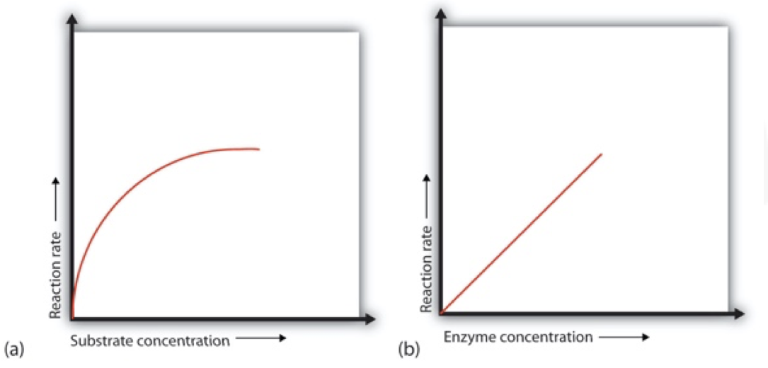
How does enyme concentration affect enzyme activity
the more enzymes present, the more active sites are available for substrates to bind to
as long as there’s plenty of substrate available, initial rate of reaction increases linearly with enzyme concentration
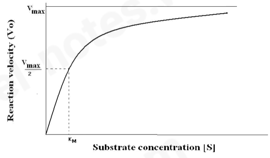
How does substrate concentration affect enzyme activity
as substrate concentration increases, initial rate of reaction also increases
the more substrate molecules there are, the more often an enzyme’s active site can bind with one
saturation point – enzymes working at max (Vmax)
all active sites are filled up
enzyme moves to find substrates as they decrease, collision forces start to decrease
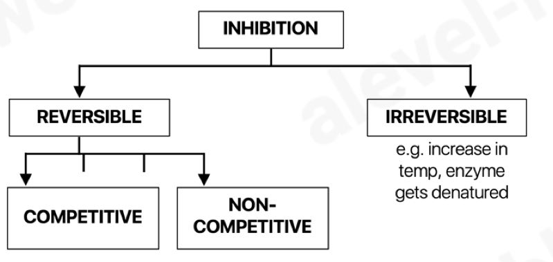
How does the inhibitor concentration affect enzyme activity
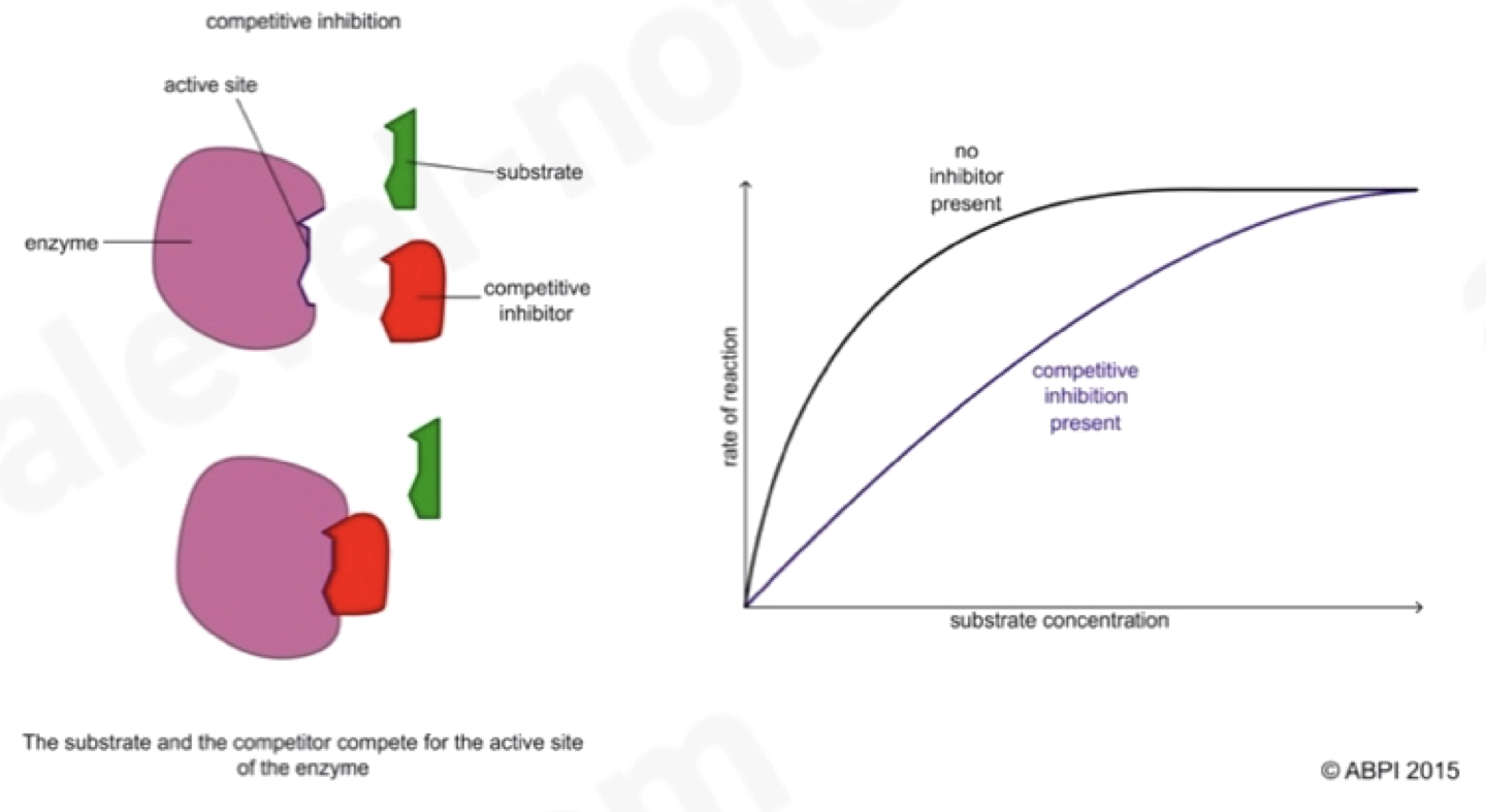
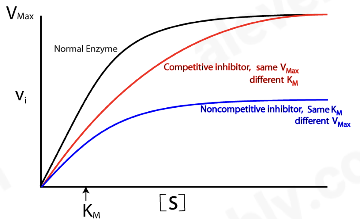
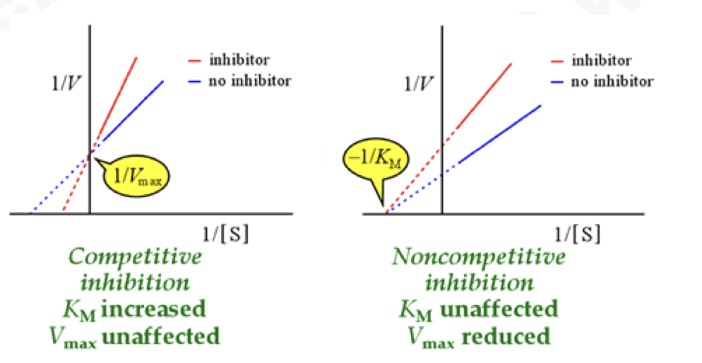
Outline competitive inhibition (enzymes)
As the inhibitor molecule is similar in shape to the enzyme’s substrate, it competes with the substrate for the active site and binds with the active site inhibiting the enzymes function
if the concentration of the inhibitor rises or the substrates falls, it becomes less likely that the substrate will collide with an active site
can be reversed by increasing the concentration of substrate
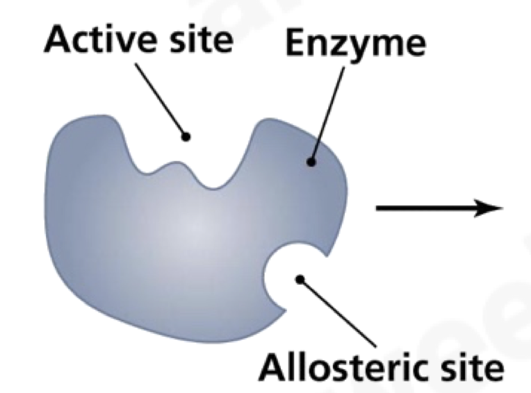
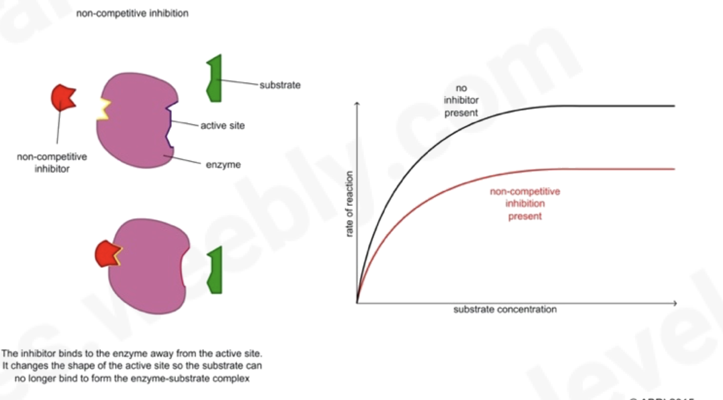
Outline non-competetive inhibitor
Molecule fits into the allosteric site of the enzyme rather than the active site.
disrupts the three-dimensional shape of enzyme preventing the substrate from fitting into the active site as its distorted
increasing the substrate concentration has no change on the rate of reaction here
End product inhibition – as enzyme converts substrate into product, rate is slowed down at the end as the product binds to another part of the enzyme and prevents more substrate binding
Outline enzyme affinity
affinity – enzyme willingness to bind to a substrate
at Vmax, all enzyme molecules are bound to substrate molecules; the enzyme is saturated with substrate, as substrate concentration is increased, reaction rate rises until the max rate i.e., Vmax
what is the Km (Michaelis-Menten constant)?
the substrate concentration at which enzyme works at half its maximum rate
half the active sites of enzymes are occupied by substrate
An enzyme with a lower value of Km has a high affinity to its substrate
Outline the process of immobilising enzymes
enzyme is mixed with a solution of sodium alginate
droplets of this mixture are added to calcium
chloride solution
a reaction occurs forming jelly/beads
enzyme is immobilised in the bead
Advantages of immobilising enzymes
enzyme can be reused
enzyme is easily recovered
product isn’t contaminated with enzymes
reduces product inhibition
enzyme is more stable/less likely to denature
longer shelf-line of enzyme
Exocytosis
Bulk movement of liquids or solids out of a cell by the fusion of vesicles containing the substance with the cell surface membrane
The membrane surrounding the vacuole, called the tonoplast, has a fluid mosaic structure. Describe the structure of this membrane. (4)
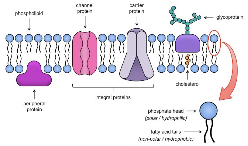
1) phospholipid bilayer
2) phospholipids have hydrophilic heads and hydrophobic tails
3) labile nature of bilayer structure is due to phospholipids moving within their monolayer
4) protein molecules, interspersed
5) many different protein molecules present
6) idea of most proteins moving / not in fixed position
why do cells need cholesterol?
1) for membrane stability
2) regulating fluidity of membrane
3) production of steroid hormones
What is the meaning of “Fluid mosaic” model?
‘fluid’ refers to the movement of phospholipids while ‘mosaic’ refers to the scattered proteins (and glycoproteins) in the phospholipid bilayer
How are phospholipids arranged in the fluid mosaic model?
phospholipids are arranged so that hydrophobic, non- polar tails do not face water. Water is on both the intracellular and extracellular sides
therefore, tails point inwards, and hydrophilic heads face the aqueous medium
What is Membrane fluidity?
The viscosity of the lipid bilayer of a cell membrane.
What factors affect membrane fluidity?
tail length –
longer the tail, the less fluid the membrane
saturation of fatty acid –
the more unsaturated they are, the more fluid the membrane. This is as unsaturated fatty acid tails are bent and fit together more loosely
cholesterol -
regulates the fluidity of membrane
at low temperatures, cholesterol increases the fluidity of the membrane preventing it from being too rigid, this is because it prevents close packing of phospholipid tails
at high temperatures, cholesterol decreases the fluidity of membrane and stabilises the cell
Outline Glycolipids and glycoproteins
Lipid and protein molecules on the outer surfaces of cell membrane have carbohydrate chains attached to them forming glycolipids and glycoproteins
These carbohydrate chains projecting out like antennae:
stabilise the membrane structure by forming hydrogen bonds with water molecules surrounding the cell
glycocalyx – sugary cell coating formed by carbohydrate chains
act as receptor molecules:
→ signalling receptors – recognise messenger
molecules like hormones and neurotransmitters
→ endocytosis – bind to molecule to be engulfed by membrane
act as cell markers/antigens allowing cell-cell recognition
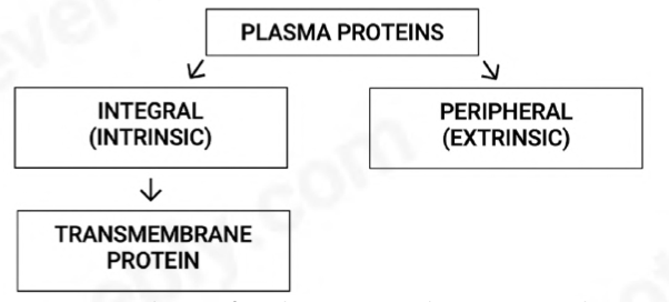
What are integral(intrinsic) - transmembrane proteins
proteins that are found embedded within the membrane
may be found in inner layer, outer layer or spanning the whole membrane (these are transmembrane proteins)
helps in movement in and out of cell
What are peripheral(extrinsic) - proteins
can be present inside or outside of the cell membrane i.e., intracellular, and extracellular
extracellular peripheral proteins –
communication, receptors, and recognition proteins
intracellular peripheral proteins- structural support, attached to the cytoskeleton of the cell
What is the function of transmembrane proteins
act as gateways and can transform, helping in facilitated diffusion and active transport
Outline Channel proteins
do not require energy
transport substances through membrane passively,
along their concentration gradient
used for both active transport and facilitated diffusion
Outline Carrier proteins
require energy
go against the concentration gradient
take substances from outside and pumps it inside or vice versa
used for active transport
Outline the Cell surface receptors
present in membranes and bind with particular substances
used for signalling, endocytosis, cell adhesion, cell markers
Outline Cell surface antigens
act as cell identifying markers
each type of cell has its own antigen
this enables cells to recognise other cells and behave in an organised way
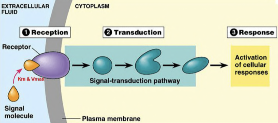
What is Cell signnalling
method in which cells detect signals with cell receptors, i.e., glycoproteins and glycolipids, present on their membrane
the signalling molecule binds to the receptor as their shapes are complementary to each other
this creates a chain of reactions in the cell, leading to a response
What if the signalling molecules are hydrophobic(.e.g., steroid hormones such as oestrogen)?
they can diffuse directly across the cell membrane and bind to receptors in the cytoplasm or nucleus.
What if the signalling molecule is water-soluble
signal arrives at protein receptor in cell membrane
the receptor’s shape is complementary to the ligand
the signal brings about a change in the receptor’s shape
changing the shape of the receptor allows it to interact with the next component of the pathway so the message gets transmitted
binding triggers/stimulates reactions within the cell
cell signalling results in a response which may be intracellular or extracellular
Define Diffusion
> Net movement of molecules or ions from a region of higher concentration to a region of lower concentration down a gradient, as the result of the random movement of particles.
passive process
molecules tend to reach an equilibrium situation
What factors affect diffusion?
as steepness of gradient increases, diffusion increases
as temperature increases, diffusion increases
as surface area increases, diffusion increases
as diffusion distance increases, diffusion decreases
smaller and non-polar molecules like fats diffuse much easily across the cell surface membrane as they’re soluble in phospholipid tails