Ruminant Skull and Eye
1/56
There's no tags or description
Looks like no tags are added yet.
Name | Mastery | Learn | Test | Matching | Spaced |
|---|
No study sessions yet.
57 Terms
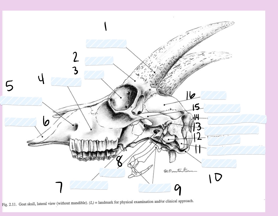
label the left lateral view of the caprine skull.
cornual process
frontal bone
orbita
facial tubercle
infraorbital foramen
incisive bone
pterygoid hamulus
oval foramen
hyoid apparatus
occipital condyle
paracondylar process
tympanic bulla
external acoustic meatus
external occipital protuberance
zygomatic arch
parietal bone
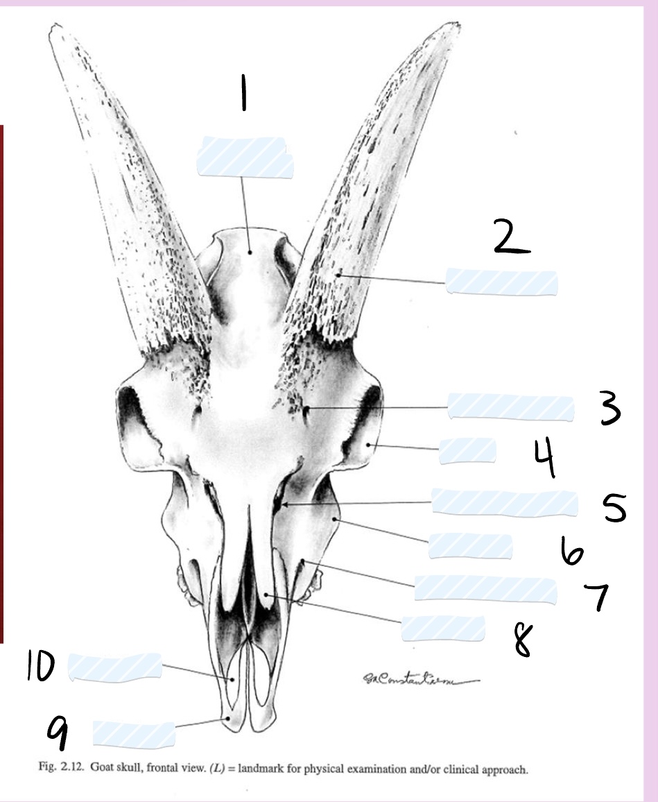
label the dorsal view of the caprine skull.
external occipital protuberance
cornual process
supraorbital foramen
orbita
frontomaxillolacrimal notch
facial tubercle
infraoribital foramen
nasal bone
incisive bone
palatine fissure
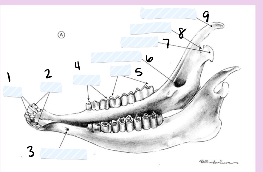
label the caprine mandible.
incisors
canines
mental foramen
premolars
molars
mandibular foramen
condylar process
mandibular notch
coronoid process
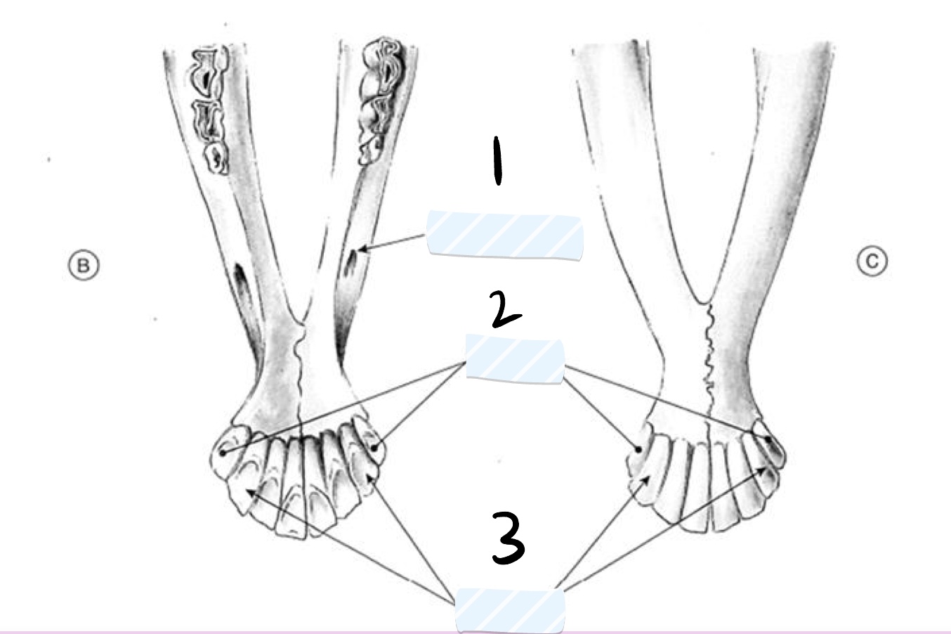
label the dorsal and ventral views of the caprine mandible.
mental foramen
canines
incisors
caprine/ovine permanent incisors
incisor 1 → 12-18 months
incisor 2 → 18-24 months
incisor 3 → 30-36 months
incisor 4 → 36-48 months
caprine/ovine permanent premolars
premolars 2, 3 and 4 are permanent at 18-24 months
caprine/ovine permanent molars
molar 1 → 3 months
molar 2 → 9 months
molar 3 → 18 months
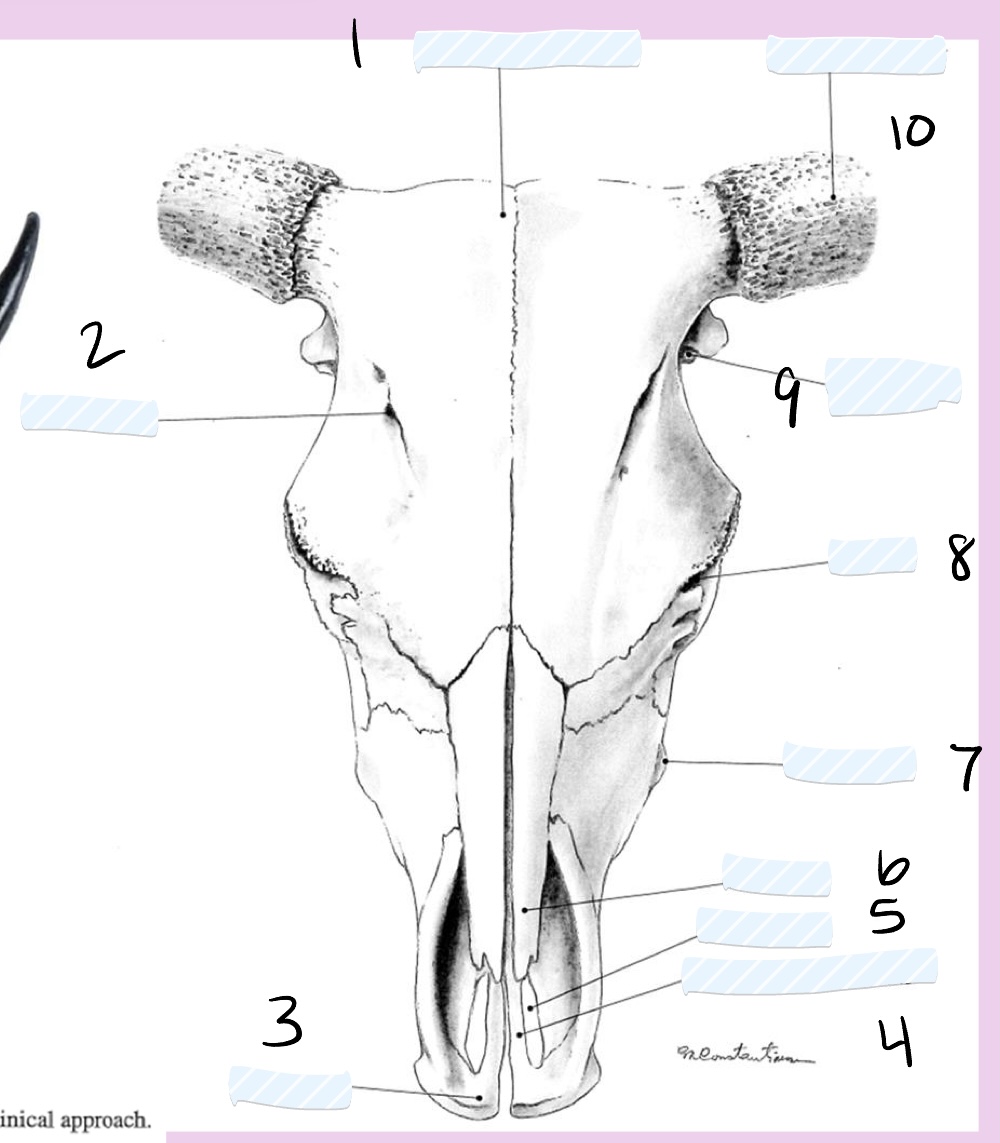
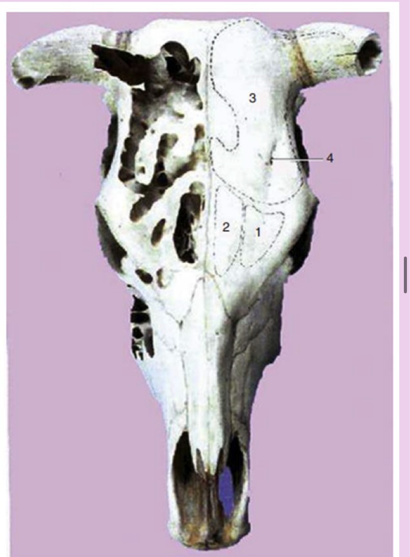
label the dorsal view of the bovine skull.
intercornual protuberance
supraorbital foramen
incisive bone
palatine process of incisive bone
palatine fissure
nasal bone
facial tubercle
orbita
external acoustic meatus
cornual process
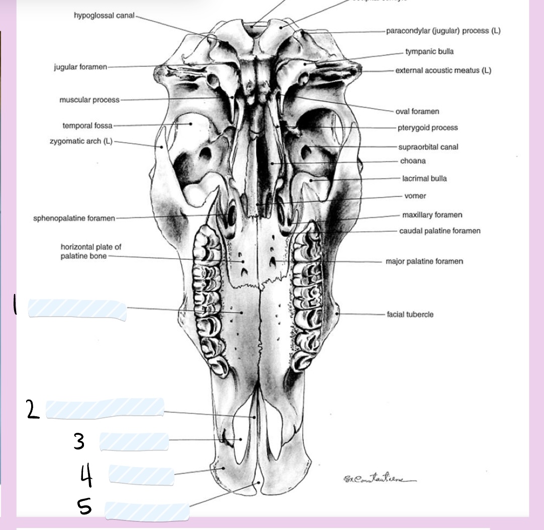
label these parts of the ventral bovine skull.
palatine process of maxilla
palatine process of incisive bone
palatine fissure
incisive bone
interincisive fissure
bovine permanent incisor eruption
incisor 1 → 1.5-2 years
incisor 2 → 2-2.5 years
incisor 3 → 3 years
incisor 4 → 3.5-4 years
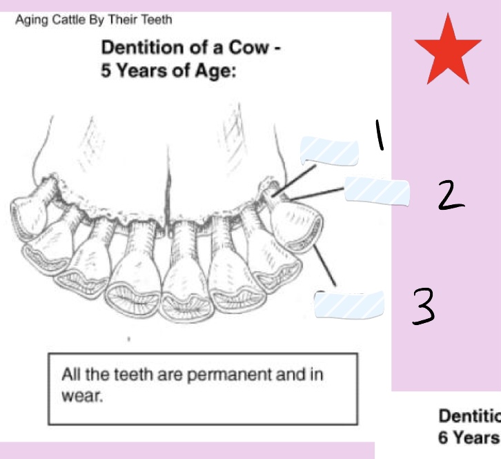
when are all bovine incisors in wear?
5 years
bovine central incisors
erupted permanent teeth
larger than the deciduous teeth on either side
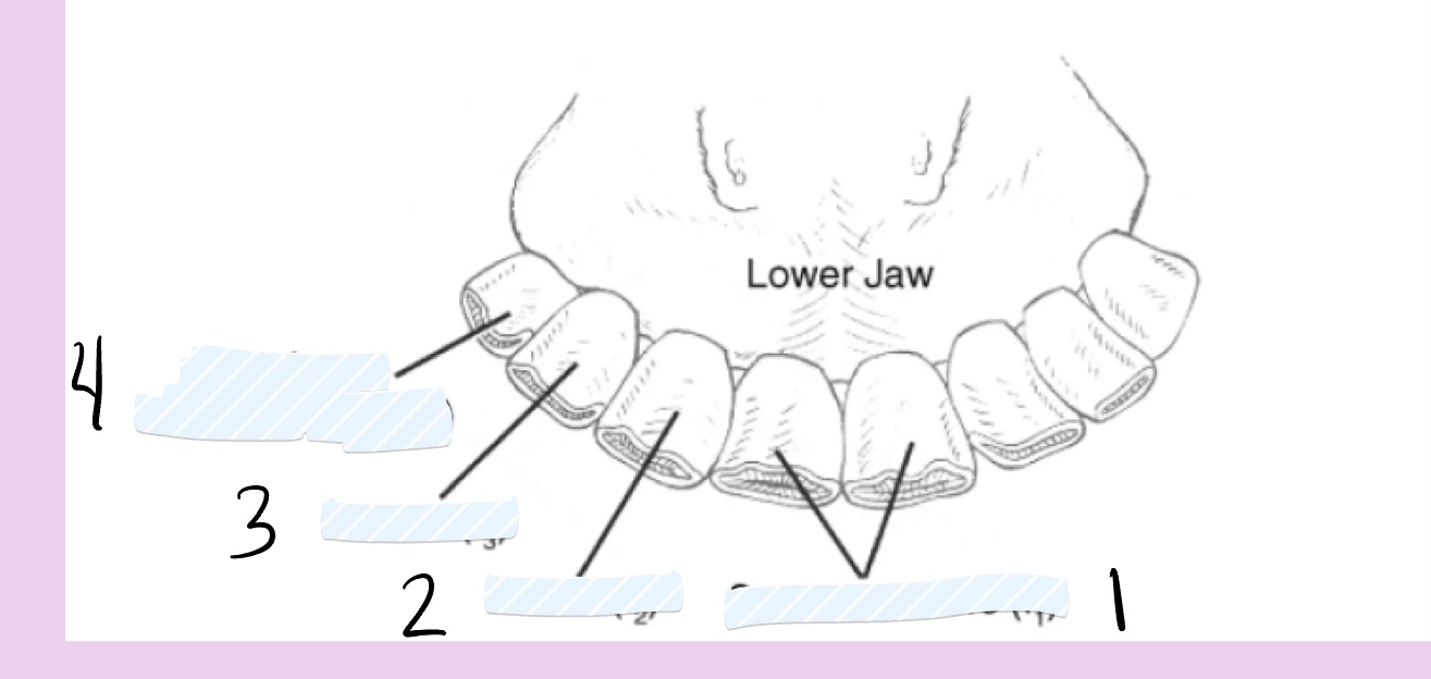
label the teeth of the bovine lower jaw.
central incisors
incisor 2
incisor 3
canine or corner incisor
label the parts of the bovine permanent teeth in wear.
root
neck
crown
by 9 years of age, the bovine incisors are
level and the necks have emerged from the gums
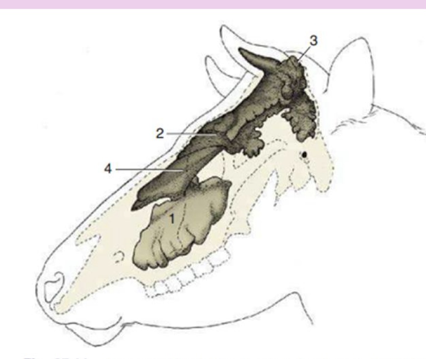
label the lateral view of the bovine skull sinuses.
maxillary sinus
rostral frontal sinuses
caudal frontal sinus
dorsal conchal sinus
label the dorsal view of the bovine skull sinuses.
lateral rostral frontal sinus
medial rostral frontal sinus
caudal frontal sinus with cornual diverticulum
supraorbital foramen
what does the dehorning procedure do?
often exposes cornual sinuses
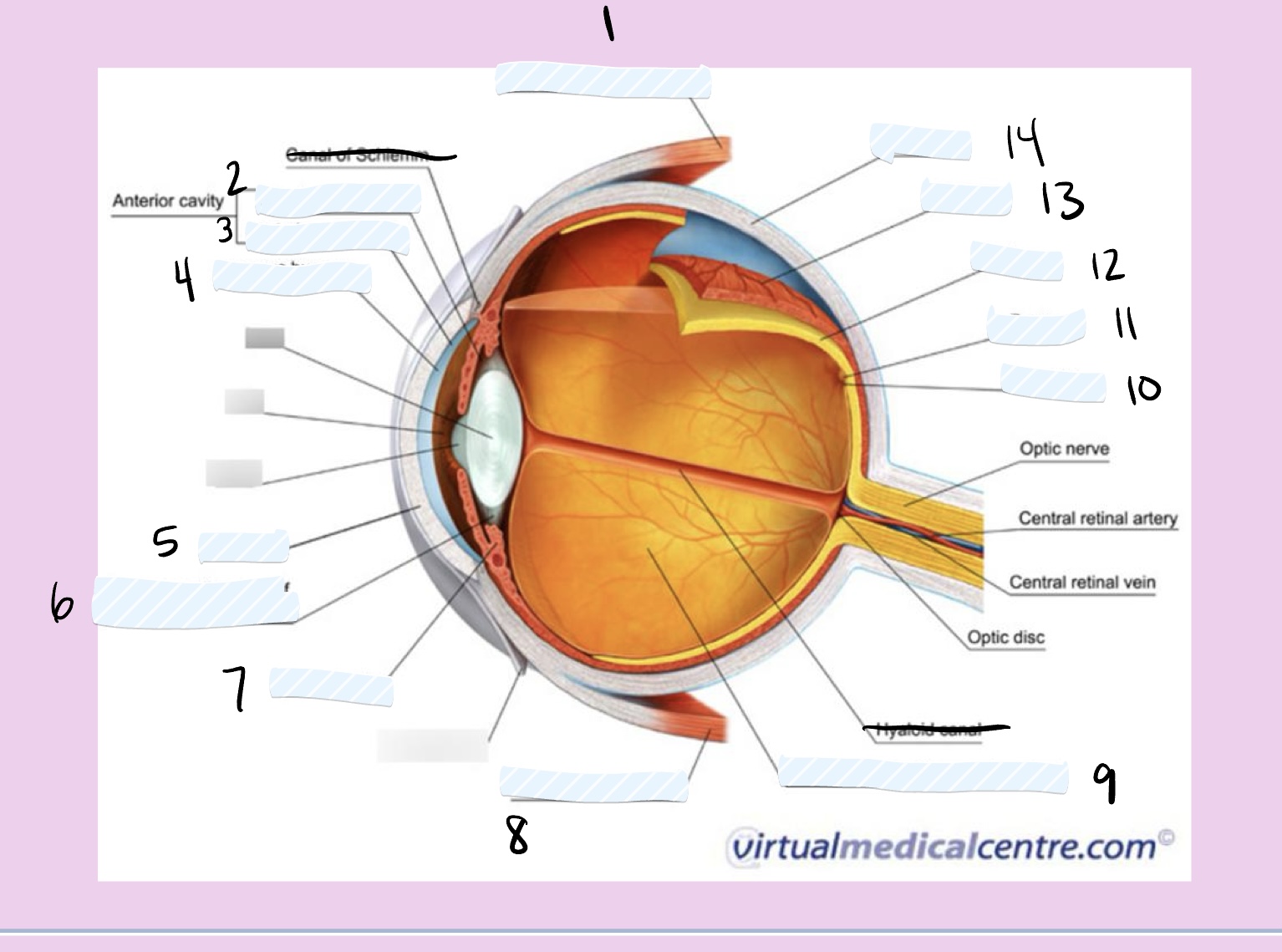
label the parts of the eye.
superior rectus muscle
posterior chamber
anterior chamber
aqueous humor
cornea
suspensory ligament of the lens
ciliary body
inferior rectus muscle
posterior cavity (vitreous chamber)
macula
fovea
retina
choroid
sclera
what is the movement of fluid in the eye?
aqueous humor produced by ciliary body in the posterior chamber of the eye
flows from posterior chamber through pupil to anterior chamber
fluid drains from anterior chamber
if fluid drainage is blocked in the eye, what can happen?
glaucoma
inferior oblique muscle
innervated by CN III
elevates and turns eye laterally
inferior rectus muscle
innervated by CN III
pulls eye inferiorly
superior rectus muscle
innervated by CN III
pulls eye superiorly
medial rectus muscle
innervated by CN III
pulls eye medially
lateral rectus muscle
innervated by CN VI
pulls eye laterally
superior oblique muscle
innervated by CN IV
depresses and turns eye laterally
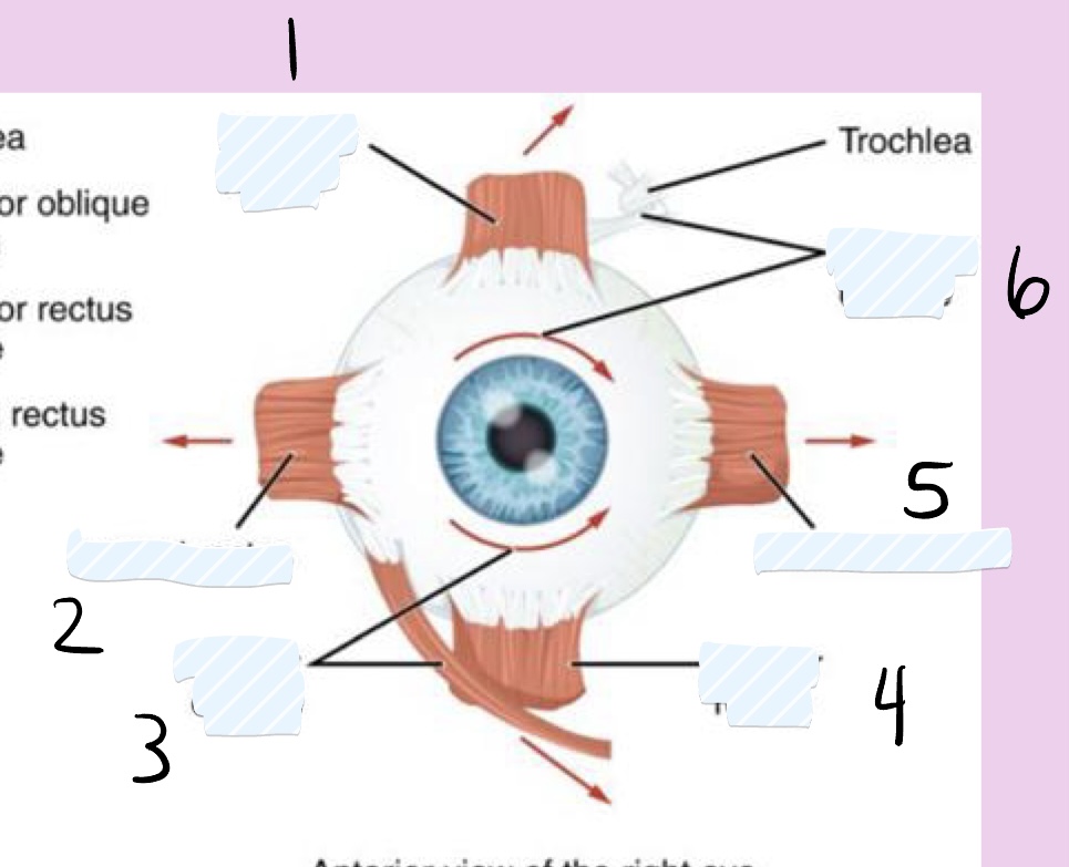
label the eye muscles.
superior rectus
lateral rectus
inferior oblique
inferior rectus
medial rectus
superior oblique
label the hemi-section of the eye.
conjunctiva
lens
cornea
pupil
iris
ciliary body
retina
tapetum lucidum
optic nerve
optic disc
sclera
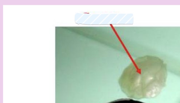
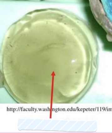
what is this?
cornea
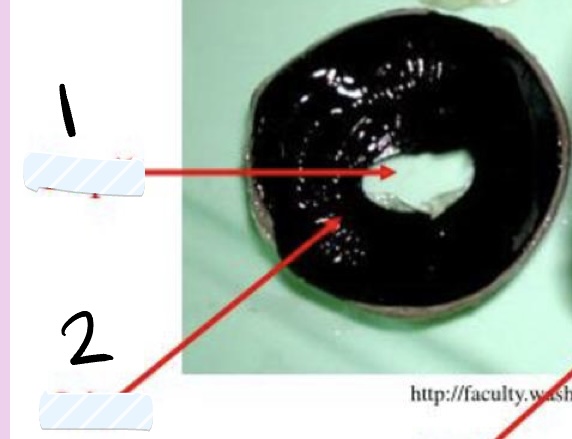
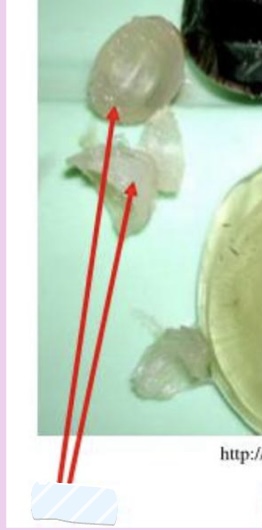
what are these?
pupil
lens
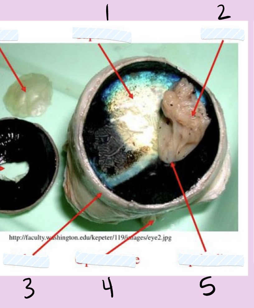
label the internal tissue components of the eye.
tapetum
retina
sclera
optic nerve
optic disc
what is this?
lens
what are these?
cornea
iris
what is the arrow pointing to?
vitreous humor
what are the 3 layers of the eye?
fibrous tunic, vascular tunic, nervous tunic
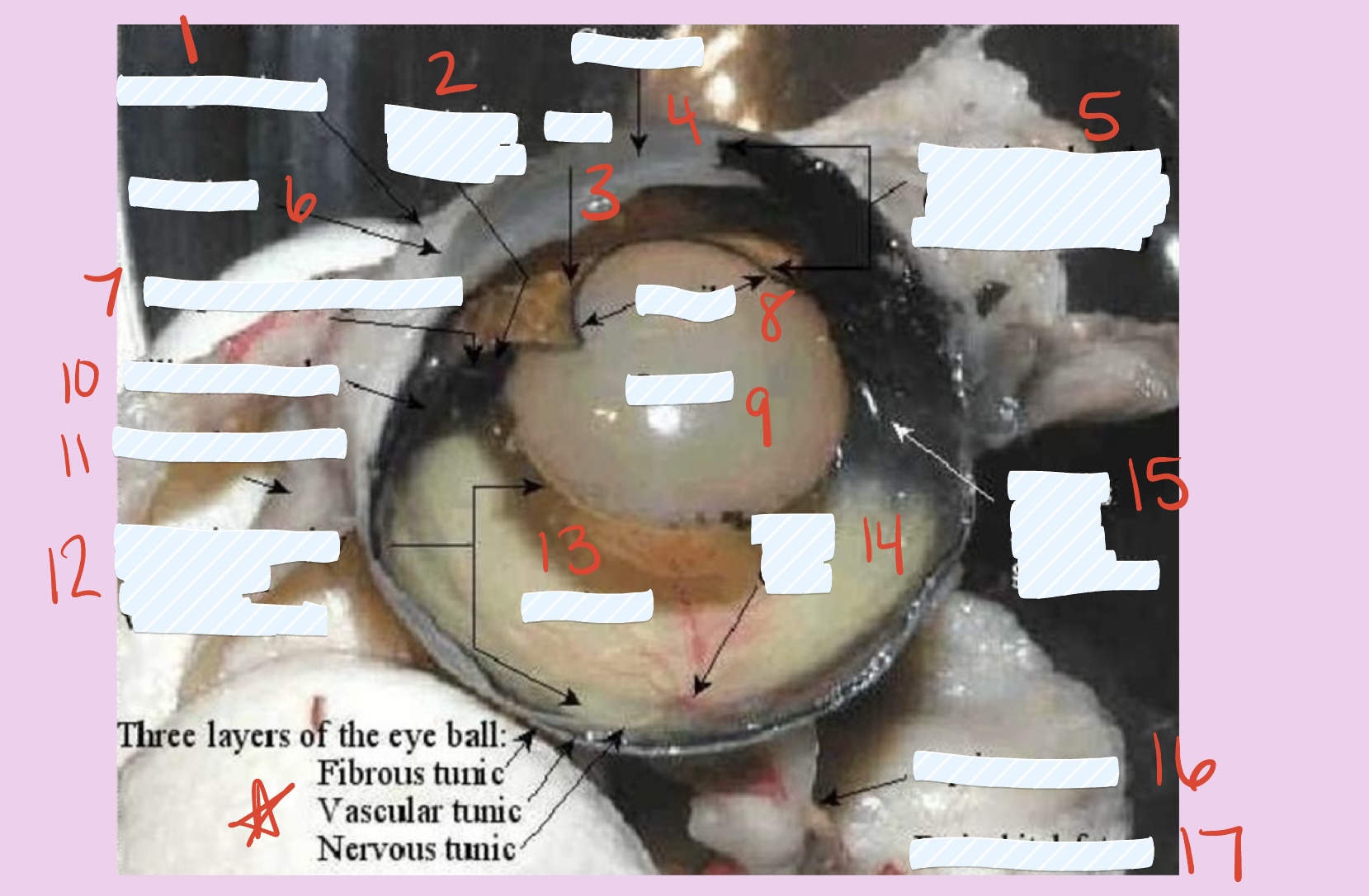
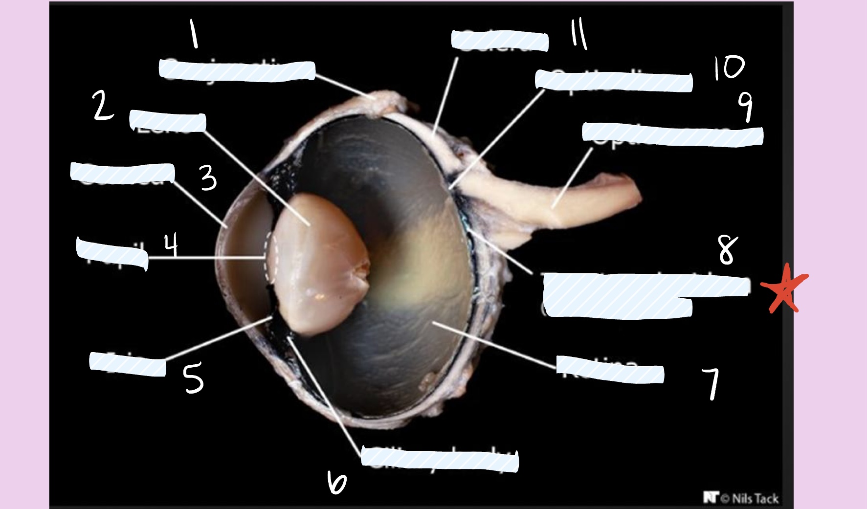
label this bovine eye dissection.
conjunctiva
posterior chamber
iris
cornea
anterior chamber
limbus
suspensory ligaments
pupil
lens
ciliary muscle
lateral rectus
posterior cavity filled with vitreous humor
retina
optic disc
vitreous humor glittering
optic nerve
periorbital fat
where would you give injections in the eye?
the limbus
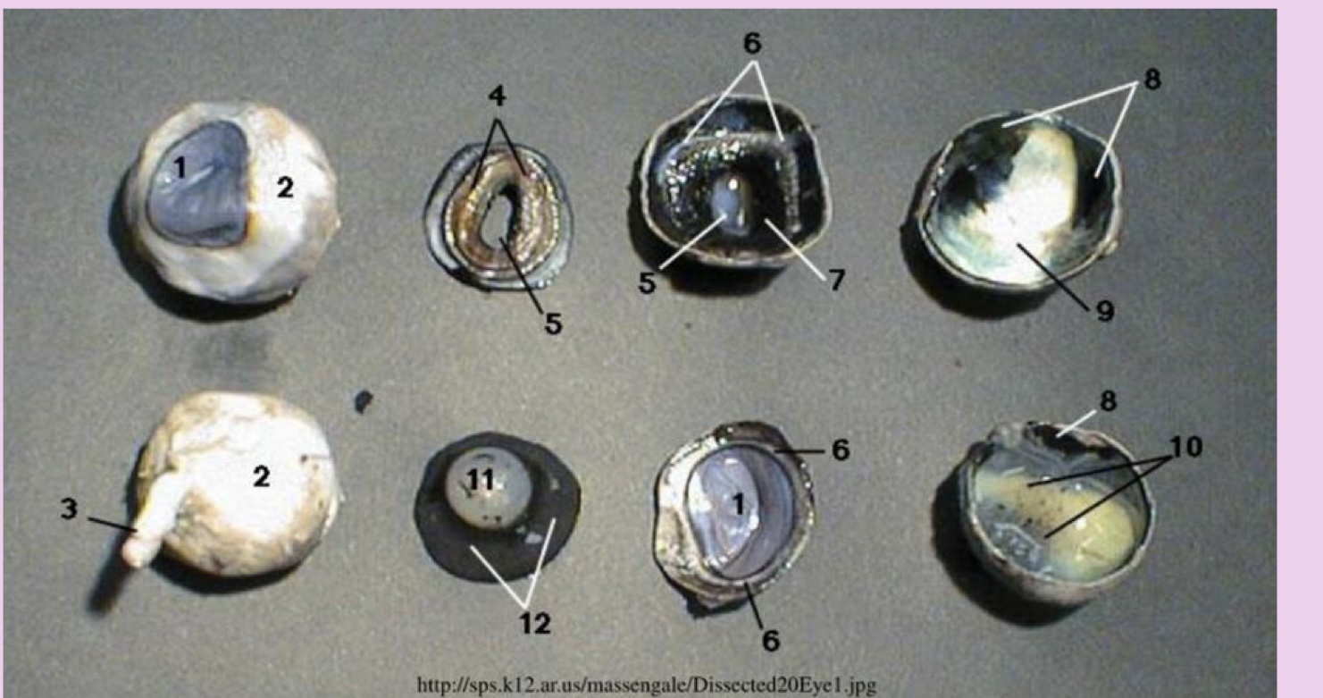
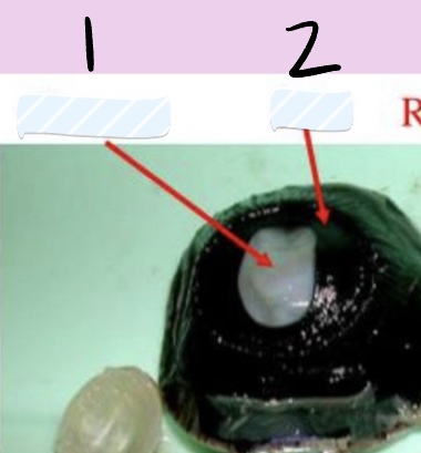
what is 1
cornea
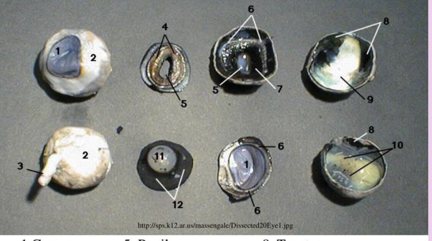
what is 2
sclera
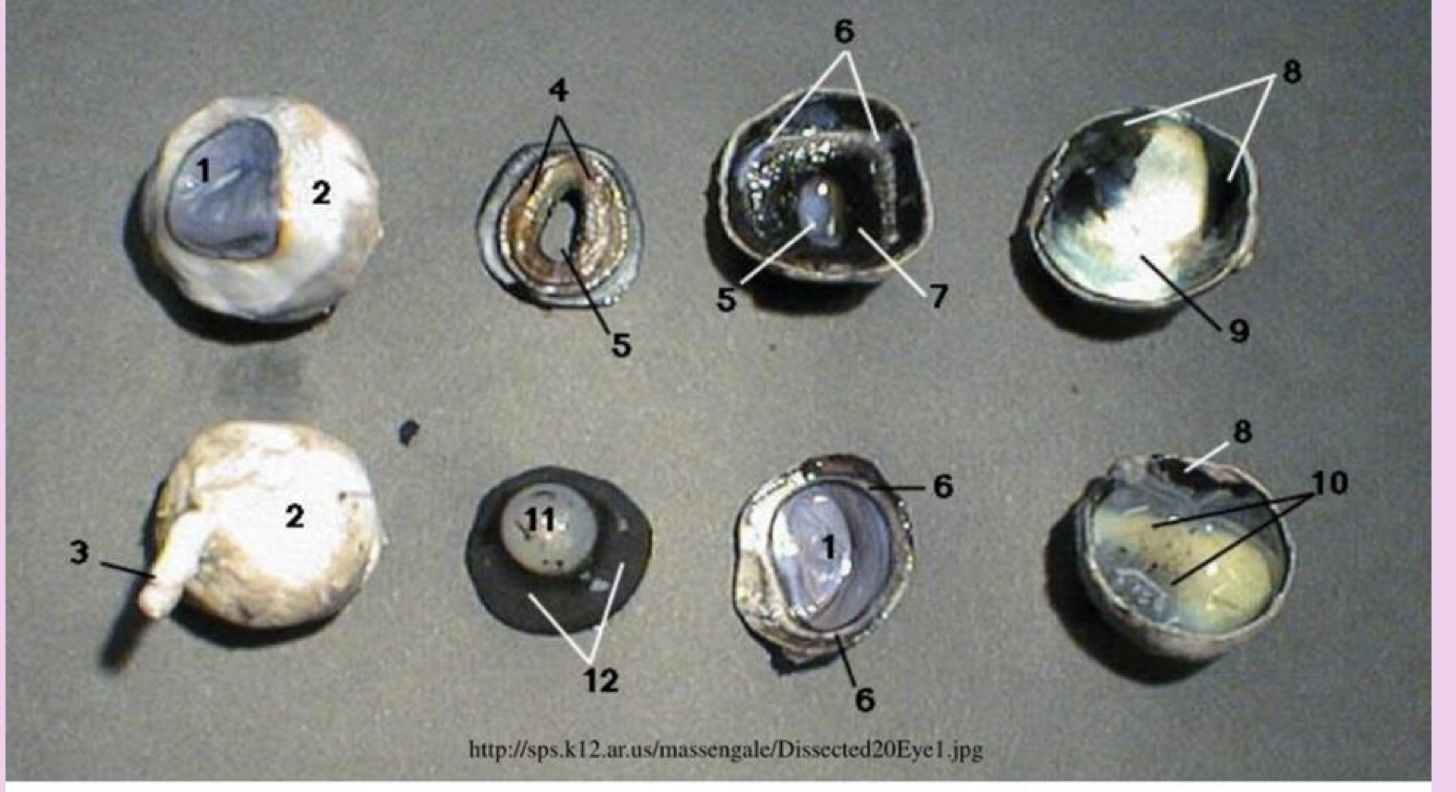
what is 3
optic nerve
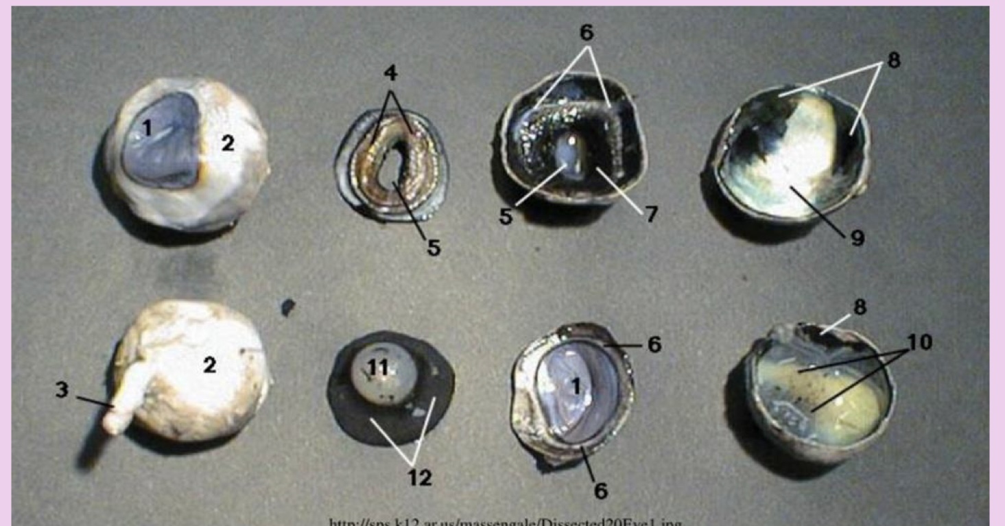
what is 4
ciliary bodies
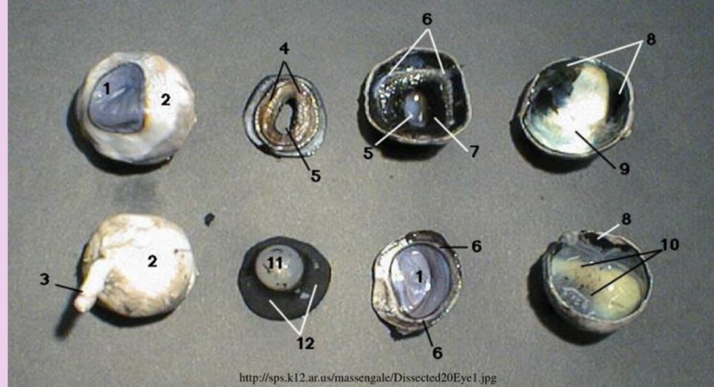
what is 5
pupil
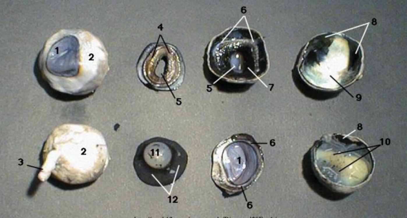
what is 6
limbus
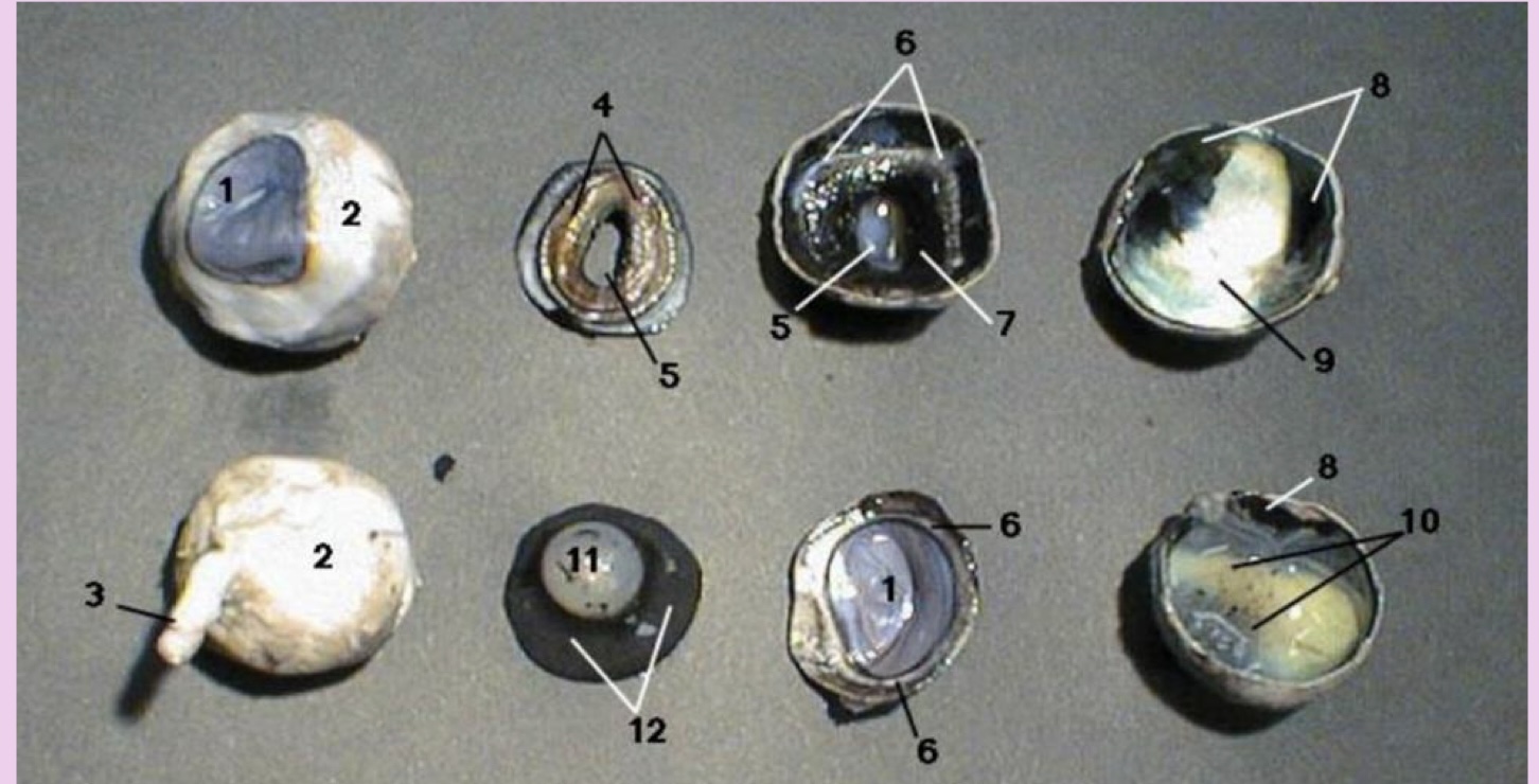
what is 7
iris
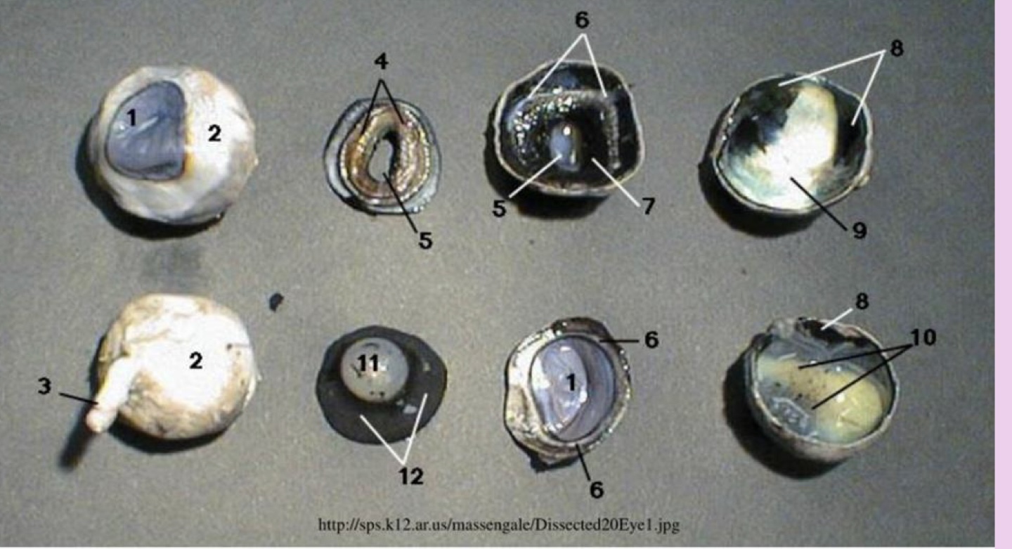
what is 8
choroid coat
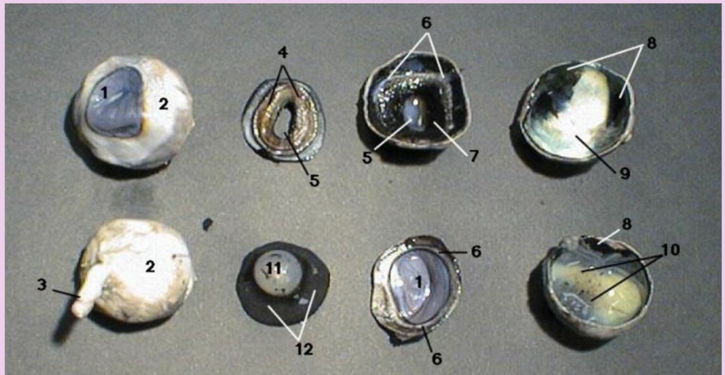
what is 9
tapetum lucidum
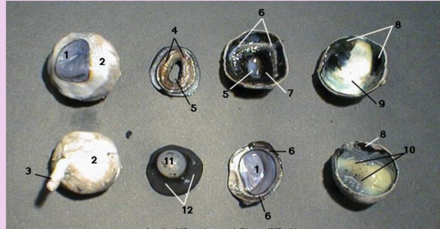
what is 10
retina
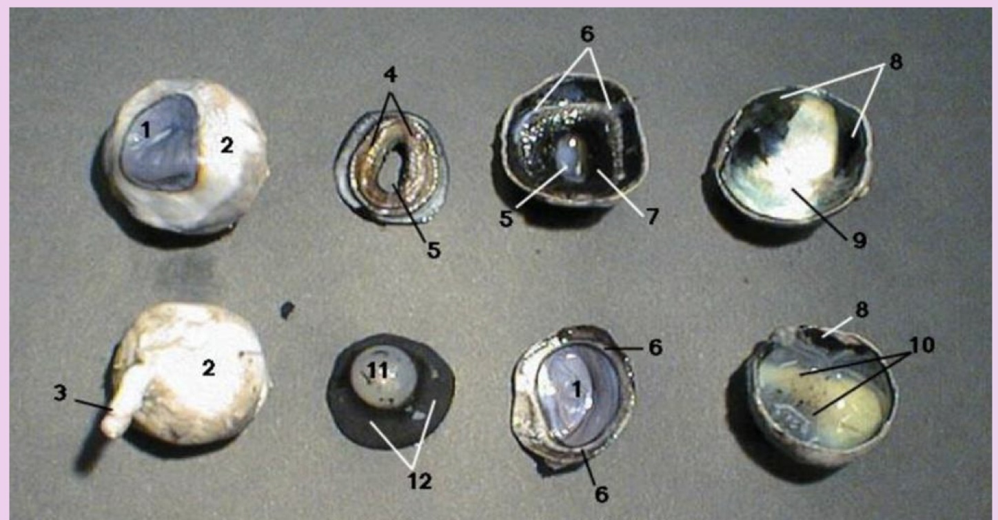
what is 11
lens
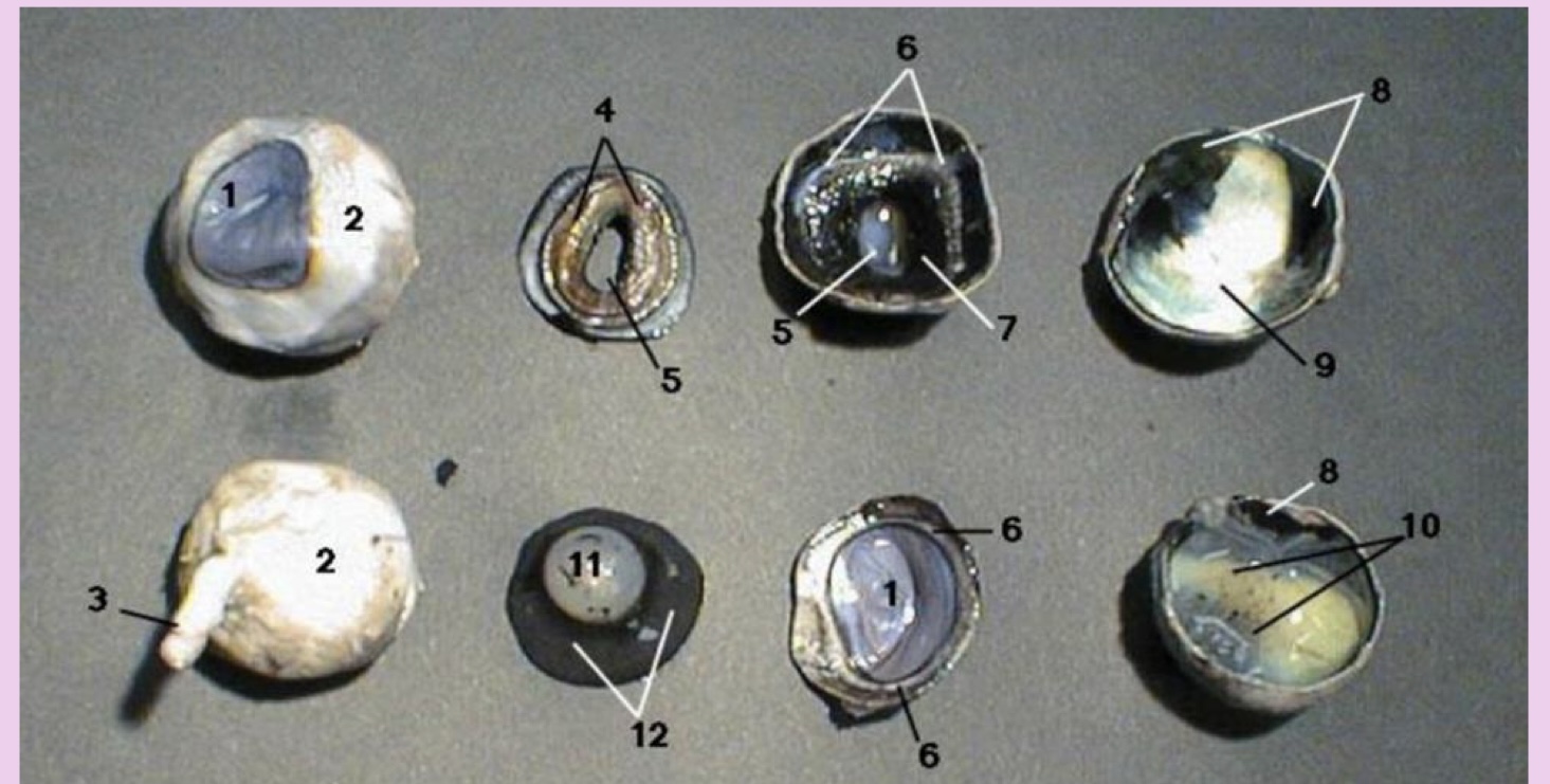
what is 12
vitreous humor

what is 1
iris

what is 2
pupil
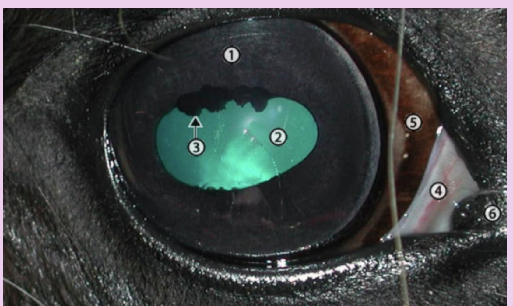
what is 3
iridic granule

what is 4
nictitating membrane

what is 5
conjunctiva

what is 6
puncta
what are iridic granules?
ruffles in the eye are that are used to shade the eye from bright sunlight