3.6 Organisms respond to changes in their internal and external environments
1/260
There's no tags or description
Looks like no tags are added yet.
Name | Mastery | Learn | Test | Matching | Spaced |
|---|
No study sessions yet.
261 Terms
Define stimulus
A detectable change in the internal or external environment of an organism that leads to a response in the organism.
Define response
a reaction to a stimulus
Define receptor
a cell adapted to detect changes in the environment
How does response help organisms pass on alleles?
There is always a selection pressure which favours the organisms with more appropriate responses. These organisms survive longer, reproduce more and are therefore more likely to pass on their alleles to the next generation.
What are the two sorts of response?
Hormonal and nervous
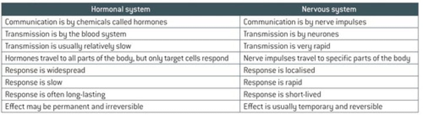
Describe hormonal control.
- slower, longer lasting, transport in bloodstream, plants and animals, menstrual cycle in females
Describe nervous control.
faster, rapid communication, transport through nerve cells, synapses, animals only, catching prey uses nervous control
Define taxis
a simple response whose direction is determined by the direction of the stimulus
What are the two types of taxes?
- positive: movement towards a favourable stimulus
- negative: movement away from an unfavourable stimulus
Give examples of taxis.
- single celled algae move towards light; positive phototaxis
- bacteria move towards higher glucose conc: positive chemotaxis
What is a kinesis?
A non-directional response to a stimulus. Speed and the rate of direction changes are changed.
What is a tropism?
the growth of a part of a plant in response to a directional stimulus,
What are the three types of tropism we need to know in plants?
phototropism
gravitropism
hydrotropism
How does a kinesis increase survival chances?
They turn faster in favourable environments and move more slowly so they stay in the favourable environment.
The opposite occurs in unfavourable environments.
What are plant growth factors?
"PGFs"
hormone-like substances which allow the plant to respond to external stimuli
How are PGFs different to animal hormones?
They exert their influence by affecting growth. They are made by cells located throughout the plant, not by particular organs.
Some PGFs affect the tissues that release them rather than acting on a distant region.
What is an example of a PGF?
indoleacetic acid (IAA), an example of an auxin
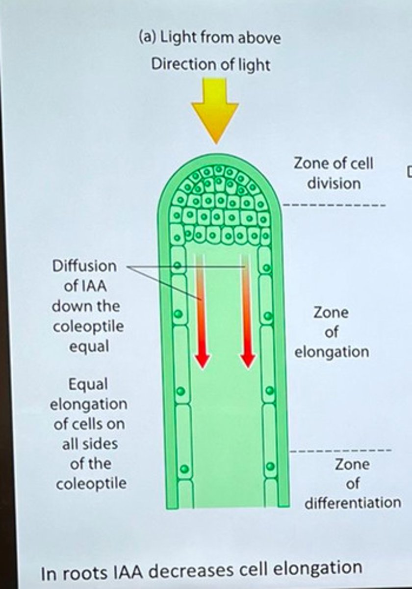
Explain phototropism in flowering plants?
1. Cells in tip of shoot / root produce IAA
2. IAA diffuses down shoot / root (evenly initially)
3. IAA moves to shaded side of shoot / root (so conc. ↑)
4. In shoots this stimulates cell elongation whereas in
roots this inhibits cell elongation
5. So shoots bend towards light
whereas roots bend away from light
What are auxins?
the main PGFs responsible for cell elongation in phototropism and gravitropism.
Define positive phototropism.
growth of a coleoptile (tip of a shoot) towards the direction of a light source.
What are the three zones in a coleoptile when you're considering the effect of IAA?
- zone of cell division
- zone of elongation
- zone of differentiation
Describe gravitropism in flowering plants.
1. cells in the tip of the root produce IAA, which is then transported along the root
2. the IAA is initially transported to all sides of the root
3. gravity influences the movement of IAA from the upper side to the lower side of the root
4. a greater concentration of IAA builds up on the lower side
5. As IAA inhibits elongation in root cells, the cells on this side elongate less on the lower side
6. the relatively greater elongation of cells on the upper side compared to the lower side causes the root to bend downwards towards the force of gravity
Describe one difference and one similarity between a taxis and a stimulus.
- both are a directional response to a stimulus
- a taxis moves a whole organism, but a tropism causes growth
What are the two major divisions of the nervous system?
- the central nervous system (CNS), the brain and spinal cord
- the peripheral nervous system (PNS) (made up of pairs of nerves that originate from either the brain or the spinal cord)
What is the peripheral nervous system divided into?
- sensory neurones which carry nerve impulses from receptors towards the CNS
- motor neurones which carry nerve impulses away from the CNS to effectors
What is the motor nervous system divided into?
- voluntary nervous system, which carries nerve impulses to body muscles and is under conscious control
- autonomic nervous system which carries nerve impulses to glands, smooth muscle and cardiac muscle and is not under voluntary control
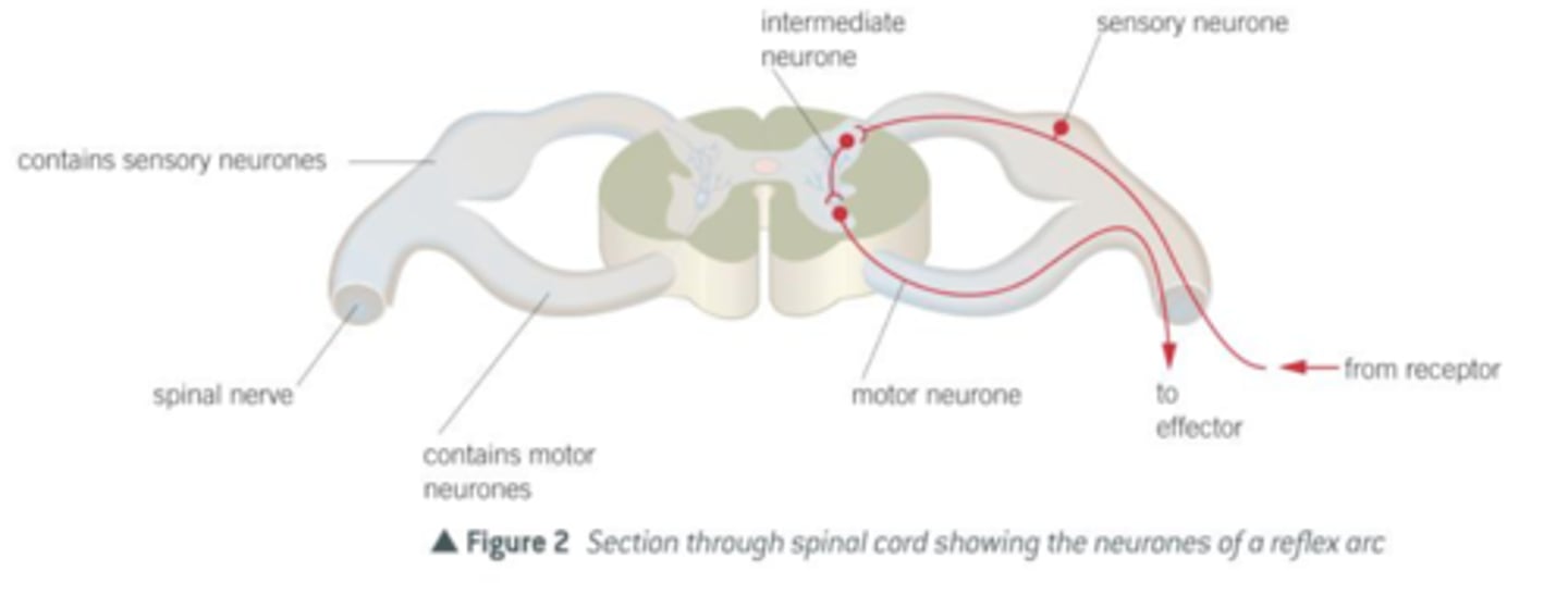
Describe the spinal cord.
A column of nervous tissue that runs along the back and lies inside the vertebral column for protection. Emerging at intervals along the spinal cord are pairs of nerves.
Define reflex
rapid, involuntary response to a stimulus
Define reflex arc
pathway of neurones involved in a reflex arc
How many neurones does a reflex arc involve?
3
Describe a reflex arc.
stimulus → receptor → sensory neurone → intermediate neurone passes impulses across the spinal cord → motor neurone passes impulse to the muscle → effector contracts → response
Why are reflex arcs important?
1) Since it is an involuntary response it doesn't require any decision-making powers of the brain. The brain is also not overloaded with situations in which the response is always the same.
2) They protect the body from harmful stimuli.
3) They are fast since the neurone pathways are short with only one or two synapses.
this is important in withdrawal reflexes
4) they are effective from birth so dont have to be learnt
What is the Pacinian corpuscle?
They are receptors responsible for responding to mechanical pressure. They respond to large pressure changes and vibrations, but they stop responding if the stimulus remains constant.
Give some examples of reflexes?
- patellar: knee jerk reflex
- corneal: blink reflex
- grasp reflex in newborns
- pupillary light reflex in response to light intensity falling on the retina
What are receptors?
Specialised cellls that repsond to various mechanical, thermal or chemical stimuli. They are specific and only respond to one type of stimulus. Receptors are energy transducers.
What are energy transducers?
they convert the energy of the stimulus into a frequency of impulses or generator potentials
What is the structure of the pacinian corpuscle?
the sensory neurone is located at the centre of layers of tissue, each separated by a gel.
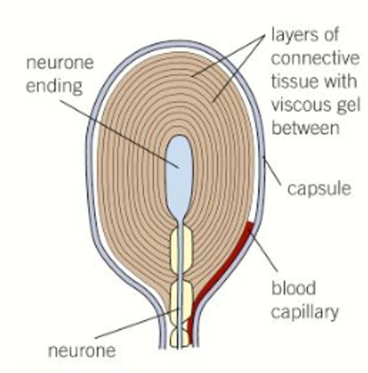
Where are pacinian corpuscles found?
deep in the skin. They are most abundant on the fingers, soles of the feet and the external genitalia. They also occur in joints, legaments and tendons, where they enable the organism to know which joints are changing direction.
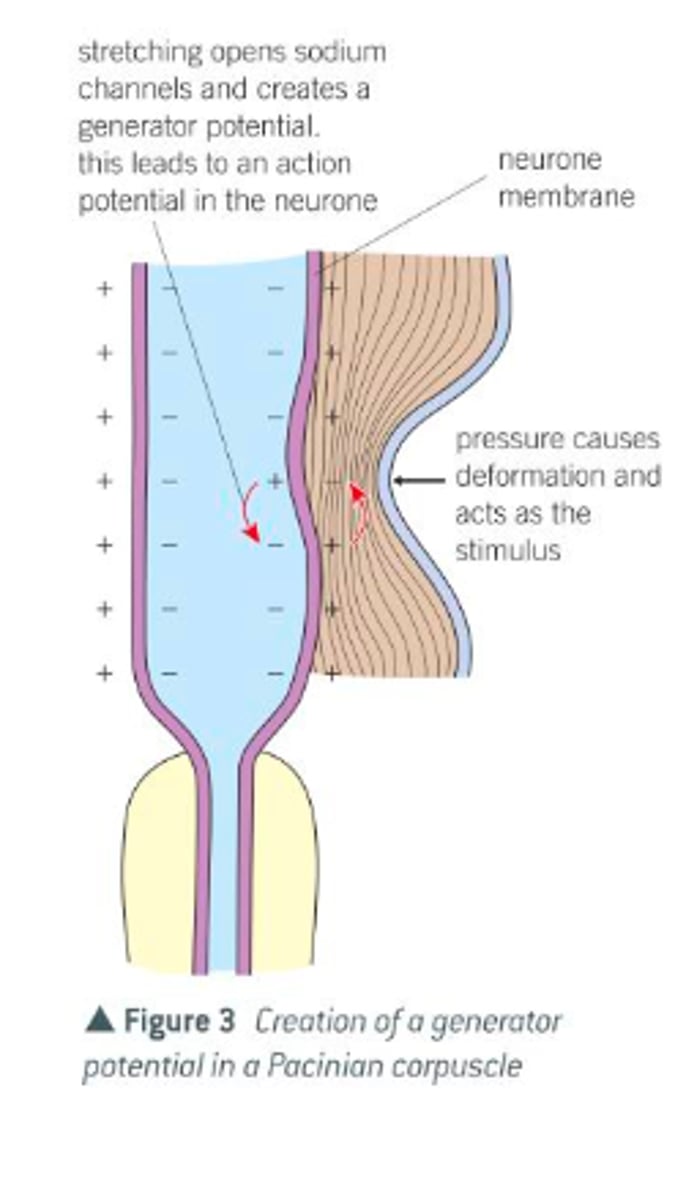
What is the important structure in the plasma membrane of a pacinian corpuscle called and why?
stretch-mediated sodium channel
their permiability to sodium changed when they are deformed, for example, by stretching.
Describe how the pacinian corpuscle functions.
1) the the normal resting state, the stretch-mediated sodium channels of the membrane around the neurone of a Pacinian corpuscle are too narrow to allow sodium ions to pass along them. In this state, the neurone of the Pacinian corpuscle has a resting potential.
2) when pressure is applied to the pacinian corpuscle, it is deformed and the membrane around its neurone becomes stretched
3) this stretching widens the sodium channel in the membrane and sodium ions diffuse into the neurone
4) the influx of sodium ions changes the potential of the membrane (i.e. it becomes depolarised) , thereby producing a generator potential.
5) the generator potential in turn creates an action potential that passes along the neurone, and then, via other neurones to the central nervous system
What are the types of receptor?
photoreceptor
chemoreceptor
thermoreceptor
mechanoreceptor (pacinian corpuscle)
proprioreceptor
What is a generator potential?
localised depolarisation in a Pacinian corpuscle and other receptor cells. The more intense the stimulus, the greater the generator potential.
Why is it that the more intense the stimulus, the greater the generator potential?
Because the more intense the stimulus, the more stretch-mediated channels will open, so the influx fo Na+ ions is faster and so the generator potential is greater.
What happens when the generator potential reaches or exceeds a threshold value?
it triggers an action potential in the sensory neurone attached to the Pacinian corpuscle.
What are the two types of receptors in in the retina?
rod and cone
Why do rod cells lead to images only being seen in black and white?
they cannot distinguish different wavelengths.
Which are more numerous; rod or cone cells?
rod cells; there are around 120 million in each eye
What are rod cells used for?
to detect light of a very low intensity
Why are many rod cells connected to a single sensory neurone in the optic nerve?
A certain threshold of light has to be exceeded before a generator potential is created in the bipolar cells to which they are connected. As a number of rod cells are connected to a single bipolar cell, there is a much greater chance that the threshold value will be exceeded than if only a single rod cell were connected to each bipolar cell. This is due to summation. As a result, rod cells allow us to see in low light intensity.
Why do rod cells give low visual acuity?
Many rod cells are linked to a single bipolar cell so light received by rod cells sharing the same neurone will only generate a single impulse travelling to the brain regardless of how many neurones are stimulated. This means that, in perception, the brain cannot distinguish between the separate sources of light that stimulated them.
How is a generator potential created in rod cells?
The pigment rhodopsin must be broken down. There is enough energy from low-intensity lifht to cause this breakdown.
This is why is takes time to adjust your eyes to the dark because if all the rhodopsin is broken down, you have to wait for it to be reformed.
Where are cone cells found in the highest density?
the region of the retina called the fovea. When you look directly at something, the image is focused on the fovea.
How many types of cone cells are there?
3
they detect red, blue or green light.
What is bleaching?
Rhodopsin molecule breaks down into retinal and opsin at a faster rate than it can be re-formed at.
What is acuity?
sharpness of vision; ability to distinguish fine detail
What are the differences between rod and cone cells?
- one type of rod cells v 3 types of cone cell
- rod cells are rod-shaped but cone cells are cone-shaped
- rod cells are found in greater distribution outside the fovea, but cone cells are most concentrated at the fovea
- cone cells give good visual acuity, but rod cells give poor visual acuity
- cone cells are not sensitive to low-intensity light, unlike rod cells
- there are a greater number of cone cells
What is the pigment in cone cells?
iodopsin
How is a generator potential created in cone cells?
photodecomposition of iodopsin produces a generator potential, but in cones the iodopsin quickly reforms.
Define transducer cell.
a cell that converts a non-electrical signal into an electrical signal and vice-versa.
Explain why rod cells rather than cone cells enable vision at night.
Rhodopsin is more sensitive to light than iodopsin, so rod cells respond to lower light intensities.
A person looked at several small white dots clustered close together on a sheet of black paper. Explain why cone cells would be able to distinguish between the separate dots, but rod cells may not.
Light from different dots absorbed by different rod cells but responses to stimuli may merge along a single neural pathway, because multiple rod cells can be connected to the same bipolar cell. This is why rod cells have lower visual acuity.
What is the autonomic nervous system?
the part of the nervous system responsible for control of the bodily functions not consciously directed, such as breathing, the heartbeat, and digestive processes. It carries impulses to glands, smooth muscle and is not under voluntary control.
What are the two divisions of the autonomic nervous system?
sympathetic and parasympathetic
Describe the sympathetic nervous system.
In general, this stimulates effectors and so speeds up any activity. It acts rather like an emergency controller; it controls effectors when we exercise strenuously or experience powerful emotions. In other words, it helps us cope with stressful situations by heightening our awareness and preparing us for activity.
Describe the parasympathetic nervous system.
This inhibits effectors and so slows down any activity. It controls activities under normal resting conditions. It is concerned with conserving energy and replenishing the body's reserves.
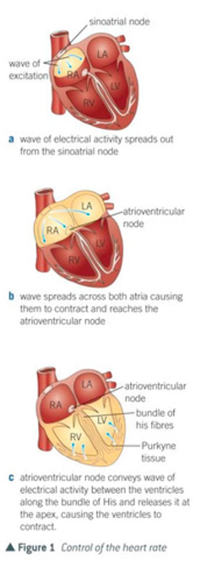
What are where is the sinoatrial nerve (SAN)?
A distinct group of cells within the wall of the right atrium
What does it mean to say that the cardiac muscle is myogenic?
its contraction is initiated from within the muscle itself rather that by nervous impulses from outside (neurogenic), as is the case with other muscles.
Why is the SAN referred to as the pacemaker?
it has a basic rhythm of stimulation that determines the beat of the heart
Describe the sequence of events that controls the basic heart rate?
- a wave of electrical excitation spreads out from the SAN across both atria, causing them to contract
- a layer of non-conductive tissue (atrioventricular septum) prevents the wave crossing to the ventricles
- the wave of excitation enters a second group of cells called the atrioventricular node (AVN) which lies between the atria.
- the AVN, after a short delay, conveys a wave of electrical excitation between the ventricles along a series of specialised muscle fibres called Purkyne tissue which collectively make up a structure called. the bundle of His.
- the bundle of His conducts the wave through the atrioventricular septum to the base of the ventricles, where the bundle branches into smaller fibres of Purkyne tissue
- the wave of excitation is released from the Purkyne tissue, causing the ventricles to contract quickly at the same time, from the bottom of the heart upwards.
Which region of the brain controls the heart rate?
medulla oblongata
What are the two centres of the medulla oblongata concerned with heart rate?
- a centre that increases heart rate, which is linked to the SAN by the sympathetic nervous system
- a centre that decreases heart rate, which is linked to the sinoatrial node by the parasympathetic nervous system
What is the AVN?
a group of cells called the atrioventricular node which lies between the atria
Where are chemoreceptors found?
the wall of the carotid arteries
What are chemoreceptors sensitive to?
blood pH, CO2, and oxygen concentration
Describe the process of control of heart rate by chemoreceptors when CO2 conc is high?
- when the blood has a higher than normal CO2 conc, pH decreases
- chemoreceptors detect this and increase frequency of nervous impulses to the centre in the medulla oblongata that increases heart rate
- centre increases the frequency of impulses via the sympathetic nervous system to the sinoatrial node; this increases the rate of production of electrical waves by the sinoatrial node and therefore increases heart rate
Where are pressure receptors found?
In the walls of the carotid arteries and the aorta
How do pressure receptors operate when blood pressure is higher than normal?
pressure receptors transmit more nervous impulses to the centre in the medulla oblongata that decreases heart rate. this centre sends impulses via the parasympathetic nervous system to the sinoatrial node of the heart, which leads to a decrease in the rate at which the heart beats.
How do pressure receptors operate when blood pressure is lower than normal?
pressure receptors transmit more nervous impulses to the centre in the medulla oblongata that increases heart rate. this centre sends impulses via the sympathetic nervous system to the sinoatrial node, which increases the rate at which the heart beats,
The cardiac cycle is controlled by the sinoatrial node (SAN) and the atrioventricular node (AVN). Describe how. (5)
SAN initiates heartbeat/acts as a pacemaker
SAN sends wave of electrical activity
AVN delays electrical activity
allowing atria to empty before ventricles control act
AVN sends wave of electrical activity down Purkyne fibres
causing ventricles to contract
When a wave of electrical activity reaches the AVN, there is a short delay before a new wave leaves the AVN. Explain the importance of this short delay. (2)
Allow atria to empty
Before ventricles contract
Which hormones stimulate the sympathetic nervous system to increase heart rate?
adrenaline and noradrenaline
Which hormone stimulate the parasympathetic nervous system to decrease heart rate?
acetylcholine
Describe how the regular contraction of the atria and ventricles is initiated and coordinated by the heart itself. (5)
1. (cardiac) muscle is myogenic;
2. sinoatrial node/SAN;
3. wave of depolarisation/ impulses /electrical activity (across atria);
4. initiates contraction of atria
atrioventricular node/AVN;
5. bundle of His/purkyne tissue spreads impulse across ventricles;
6. ventricles contract after atria/time delay enables ventricles to fill;
Describe the role of the nervous system in modifying the heart rate in response to an increase in blood pressure (5)
pressure receptors
in aorta/carotid artery/sinus
send impulses
to medulla
send impulses along parasympathetic nerve
slows heart rate
Describe how an increase in exercise will result in the heart rate increasing (6).
- increased exercise leads to increased rate of aerobic respiration
- increased concentration of CO2 released into the blood
- decrease in pH in blood
- detected by chemoreceptors in the wall of the carotid arteries and aorta
- increased nerve impulses to the centre in the medulla oblongata that increases heart rate
- impulses via the sympathetic nervous system
- heart rate increases
What are the two main forms of coordination in animals?
nervous system and hormonal system
What are the differences between the hormonal system and nervous system?
What is the role of the nissl granule?
they are the site for protein synthesis
they are made of rough ER with rosettes of free ribosomes.
in order to generate an action potential, cells need lots of channel proteins in their membranes.
What are the main parts of a mammalian motor neurone?
- cell body
- nissl granule
- dendrons
- axon
- schwann cells
- myelin sheath
- nodes of Ranvier
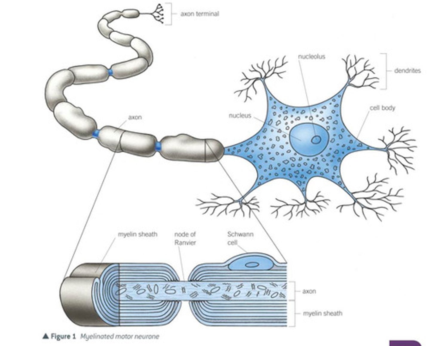
What is the role of the cell body in a motor neurone?
It contains all the usual cell organelles, including a nucleus and large amounts of rough endoplasmic reticulum. This is associated with the production of proteins and neurotransmitters.
What is the role of the dendron?
dendrons are extensions of the cell body which subdivide into smaller branched fibres called dendrites, that carry nerve impulses towards the body
What is the role of the axon?
a single long fibre that carries nerve impulses away from the cell body
What are Schwann cells? What do they do?
They surround the axon, protecting it and providing electrical insulation. They also carry out phagocytosis (removal of cell debris) and play a part in nerve regeneration. Schwann cells wrap themselves around the axon many times, so that layers of their membranes build up around it.
What is the role of the Myelin sheath?
This forms a converging to the axon and is made up of the membranes of the Schwann cells. These membranes are rich in a lipid known as Myelin. Neurones with a myelin sheath are called myelinated neurones.
What is the role of the nodes of Ranvier?
Constrictions between adjacent Schwann cells where there is no myelin sheath.
Define nerve impulse.
A self-propagating wave of electrical activity that travels along the axon membrane. It is a temporary reversal of the electrical potential difference across the axon membrane. This reversal is between two states, called the resting potential and the action potential.
How is the movement of ions such as sodium and potassium ions across the axon membrane controlled?
- phospholipid bilayer prevents the ions diffusing across it
- channel proteins can be opened or closed, and some remain open all the time
- some carriers, like the sodium-potassium pump actively transport potassium ions into the axon and sodium ions out of the axon.
What causes the establishment of the resting potential in the axon?
- sodium ions are actively transported out of the axon by the sodium-potassium pumps, and potassium ions are actively transported in.
- 3 sodium ions move out for every 2 potassium ions that move in
- They are both positively charged. So, as there are more sodium ions in the tissue fluid surrounding the axon than in the cytoplasm and more potassium ions in the cytoplasm than the tissue fluid, there is an electrochemical gradient
- sodium ions begin to diffuse naturally back into the axon, but only very few, whilst many of the potassium ions begin to diffuse back out
- so the potassium can leave, meaning there is a more negative charge in the axon than outside.
- the proteins in the axon are negatively charged
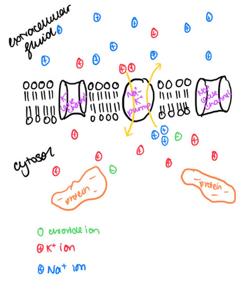
What is the action potential?
+40mV
the axon membrane is said to be depolarised at this point
Describe the formation of an action potential.
- at a resting potential some potassium voltage-gated channels open (namely those that are permanently open) but the sodium voltage-gated are closed
- the energy of the stimulus (see generator potential) causes some sodium voltage-gated channels in the axon membrane to open and therefore sodium ions diffuse into the axon through these channels along their electrochemical gradient. Being positively charged, they trigger a reversal in the pd across the membrane.
- as sodium ions diffuse into the axon, so more sodium channels open, causing an even greater influx of sodium ions by diffusion
- this forms an action potential of around 40mV, and once formed, the voltage gates on the sodium channels close, and those on the potassium ion channels begin to open