2nd Plant Exam
1/125
There's no tags or description
Looks like no tags are added yet.
Name | Mastery | Learn | Test | Matching | Spaced | Call with Kai |
|---|
No analytics yet
Send a link to your students to track their progress
126 Terms
What is the Lateral Meristem and what is it responsible for?
Occur on axis of the plant, responsible for increases in width/circumference in plant.
two types are Vascular Cambium and Cork Cambium
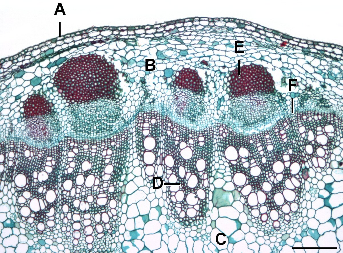
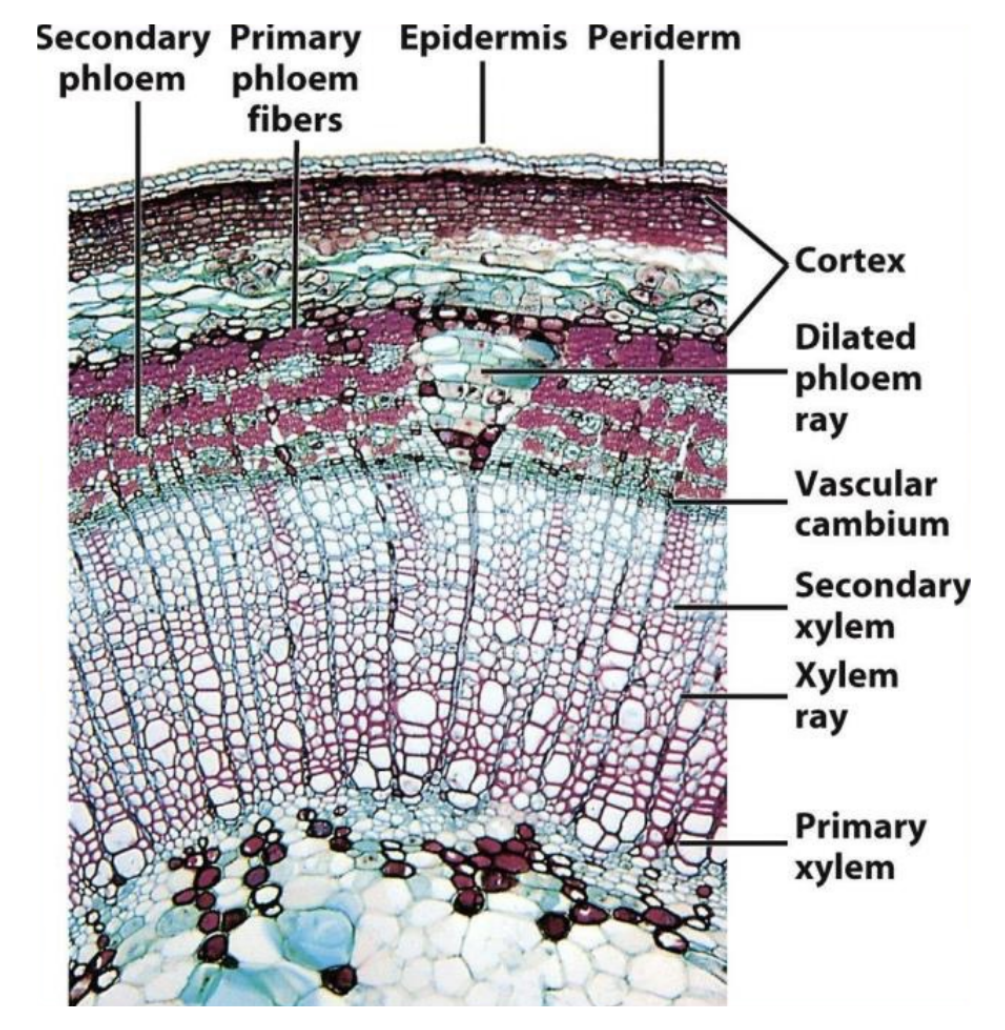
What meristem is in F and what does it do?
arises between and
within the primary phloem/xylem
• produces the secondary vascular tissues
• secondary xylem tissue to inside & secondary phloem
tissue to outside
(see F) Vascular Cambium
What meristem is the needle pointing to and what does it do?
cork cambium
arises from parenchyma in
cortex (just under epidermis)
• produces periderm (secondary epidermal tissues)
• Cork cells to outside
• Parenchyma cells to inside
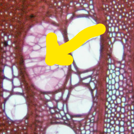
What is this tissue (Assuming it’s secondary), what system is it apart of, and what meristem? How can you tell and what is the function?
Tracheid, part of Axial system, Vascular Cambium meristem. Longer, thinner, ends taper. Function is water conduction
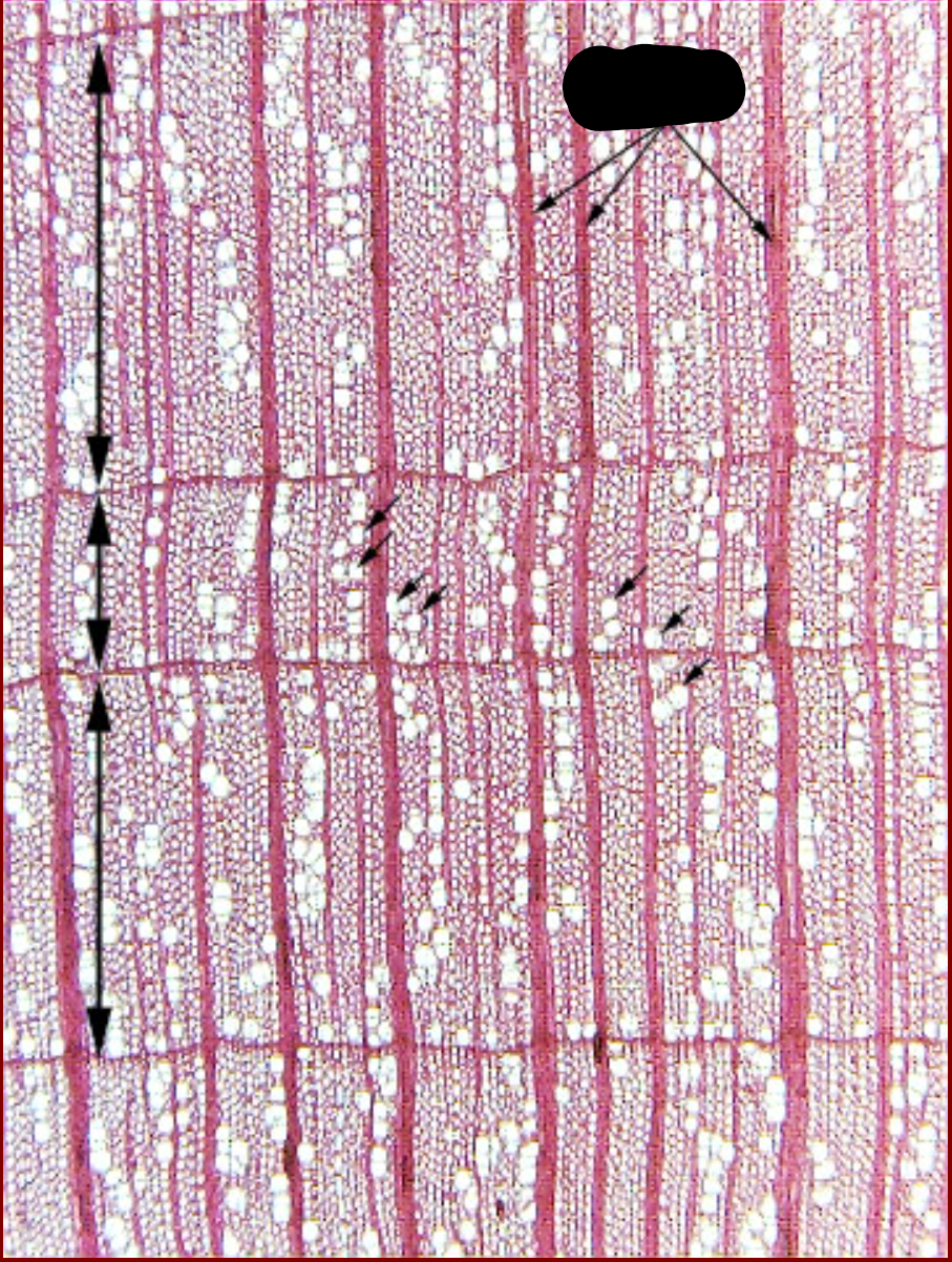
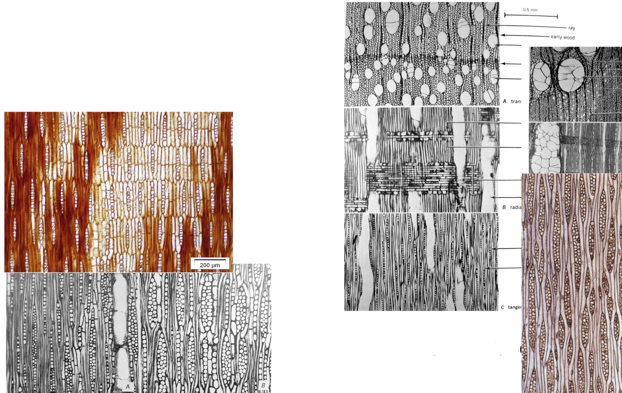
Tell-Tale Signs of Secondary Growth
1: Vascular Cambium
2: Rays and Ray Cells
3: Growth Rings
Identify all this shiz
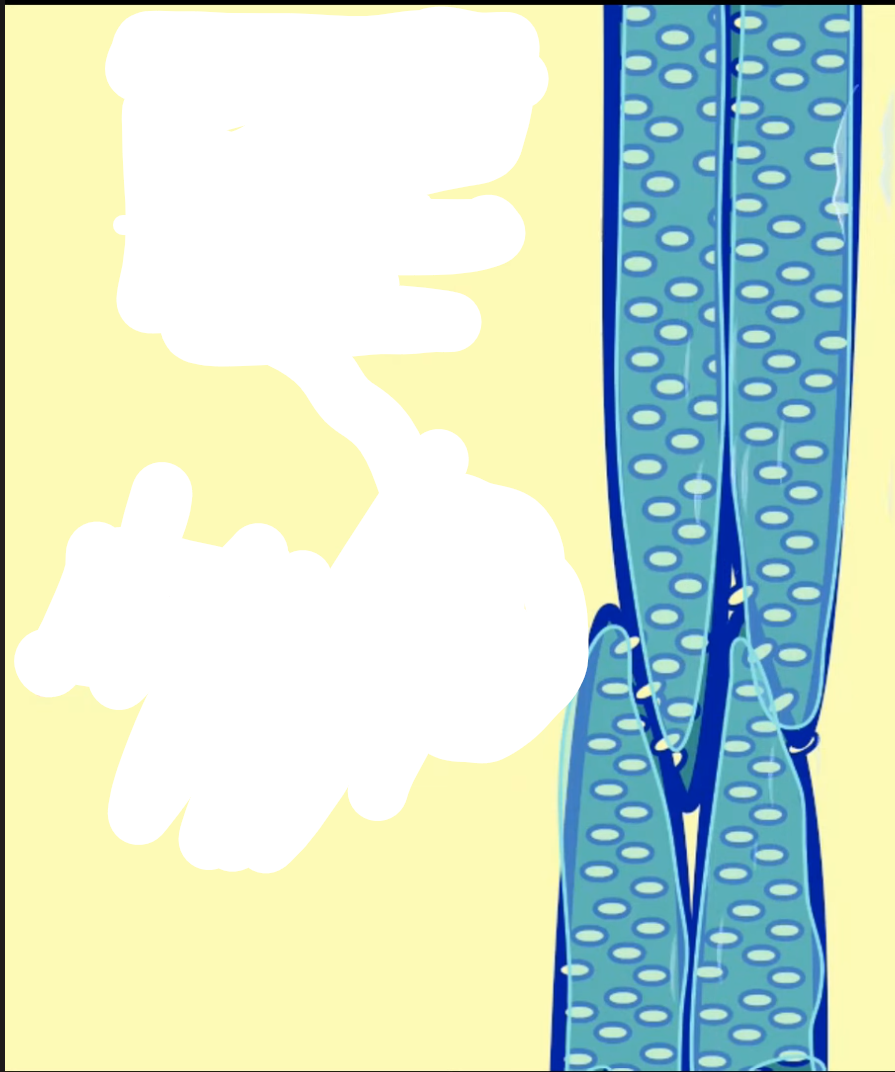
What cell is this? Trace it back to its meristem and give the function
ray parenchyma → Ray System → Axial System → Secondary Xylem → Vascular Cambium Meristem
function: store and transport water, nutrients, and carbohydrates (NSCs) between the xylem and phloem
Lateral Meristems
Responsible for large increases in
width/circumference of plant
• Occur along the axis of the plant
Vascular Cambium:
arises between and
within the primary phloem/xylem
• produces the secondary vascular tissues
• 20 xylem tissue to inside & 20 phloem
tissue to outside2
Cork Cambium:
arises from parenchyma in
cortex (just under epidermis)
• produces periderm (20 epidermal tissues)
• Cork cells to outside
• Parenchyma cells to inside
Give me the Flow Chart for Lateral Meristems and the functions
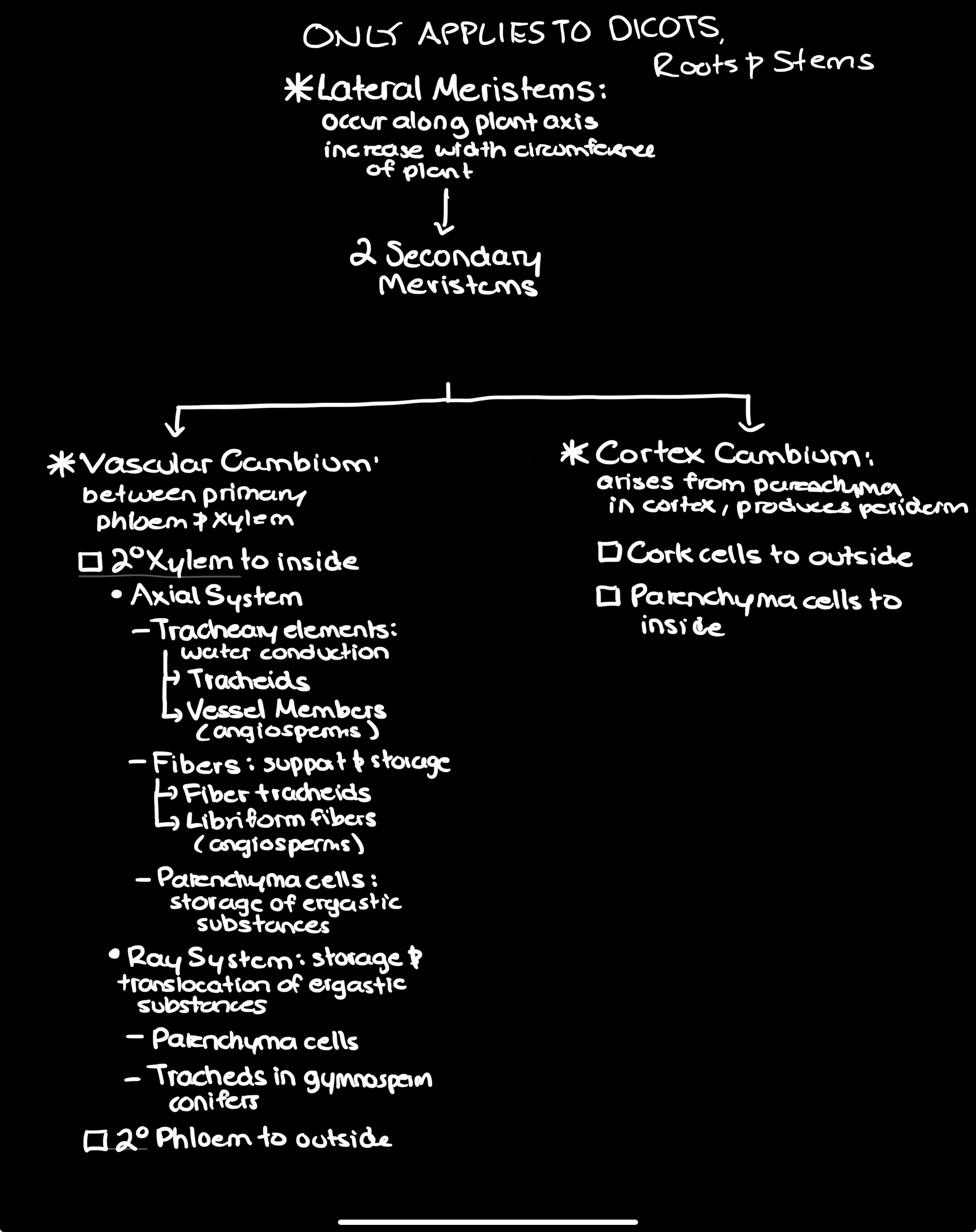
Fiber tracheids:
, small pits small and thin shape
•
• Present in both angiosperms and
gymnosperms
Libriform fibers –
very elongated;
thick secondary walls with lignin,
small cell lumen
• have no pit cavity and the pit pair is
simple (no border). “slit like”
• Fiber tracheid walls become thicker
at end of season and libriform fibers
formed in early season can have
thinner walls
• Sometimes no sharp boundary
between fiber tracheids and
libriform fibers.
Septate fibers with ray function
These store materials (like
xylem parenchyma in function and
difficult to distinguish between them).
What are the two kinds of parenchyma and what is their funciton?
axial and ray
• Function: store starch, oils, ergastic substances such as
tannins, crystals.
• Walls may develop secondary thickenings and become
lignified.
• They can develop into sclereids.
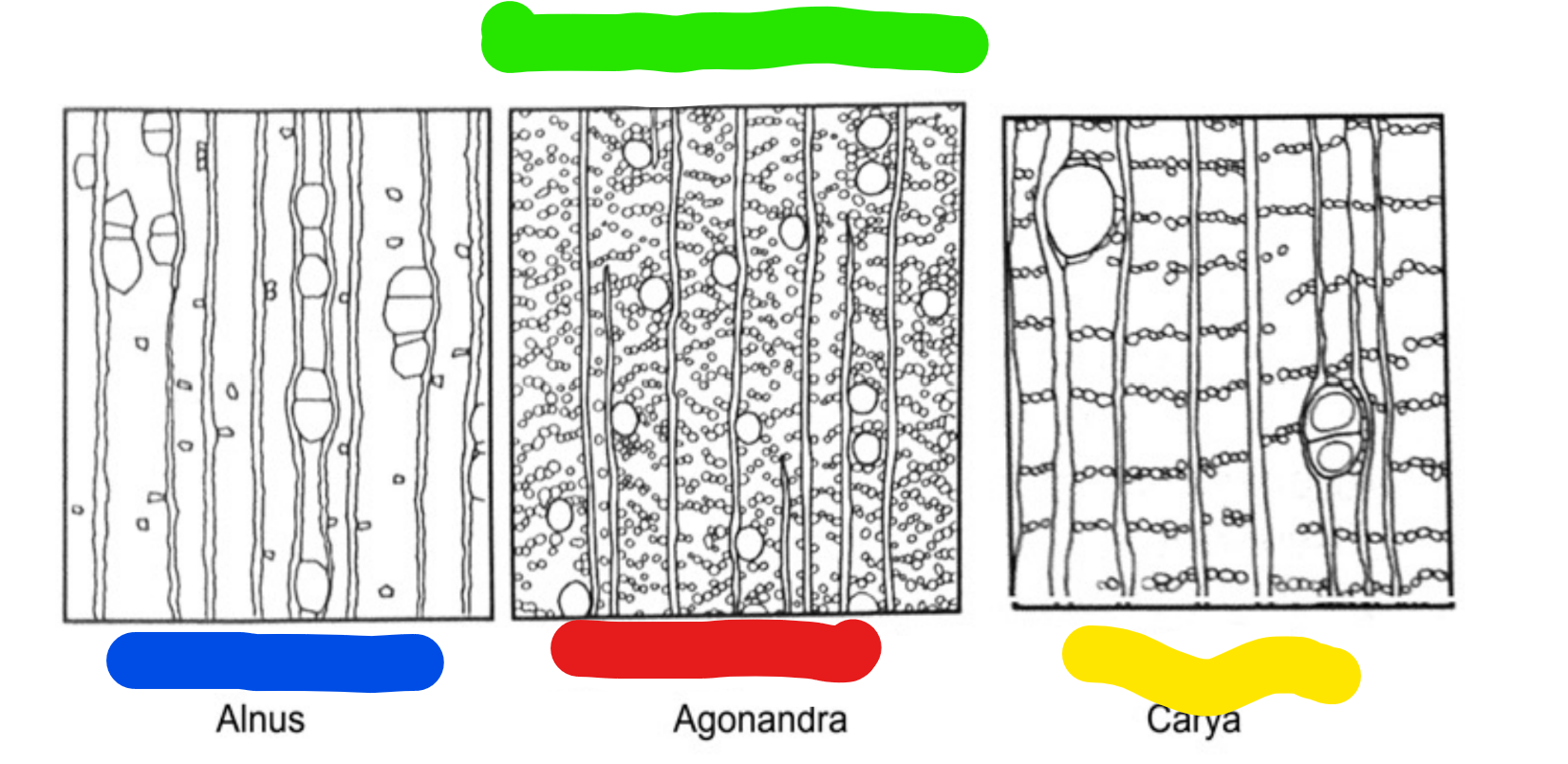
Name all this, and give the signs
Diffuse singly. Example: Alnus [the parenchyma shows up as
dark dots in the wood]
b. Diffuse in aggregation.
c. In bands – may or may not be at margin of growth ring.
Marginal may be in early wood (initial parenchyma) or late
wood (terminal parenchyma).
![<p>Diffuse singly. Example: Alnus [the parenchyma shows up as</p><p>dark dots in the wood]</p><p>b. Diffuse in aggregation.</p><p>c. In bands – may or may not be at margin of growth ring.</p><p>Marginal may be in early wood (initial parenchyma) or late</p><p>wood (terminal parenchyma).</p>](https://assets.knowt.com/user-attachments/f3d84416-6b1f-40dd-accd-165e8c0508aa.png)
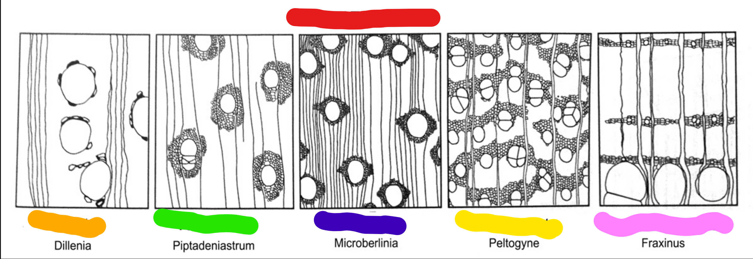
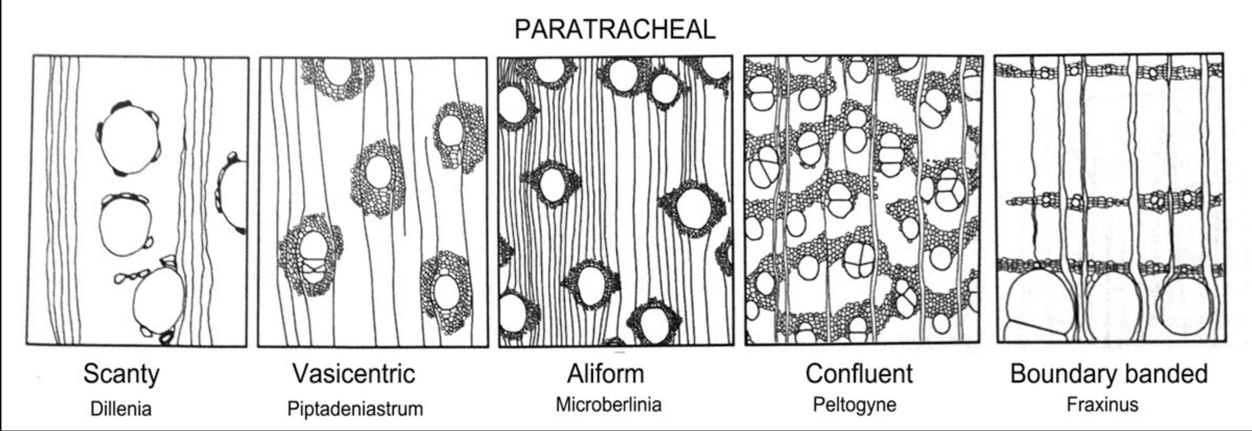
Name all this shiz and tell me what the signs are
associated with vessels
a. Scanty – scattered, usually solitary, next to vessels.
b. Vasicentric – surround the vessel.
c. Aliform (with wings) – AP surrounds the vessel and extends
to either side in wings.
d. Confluent – wings connect adjacent vessels.
e. Boundary banded – in growth ring, forms sheath around
vessels and narrow bands that connect with other vessels.
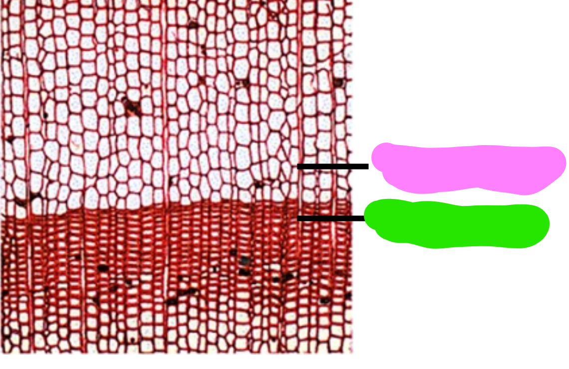
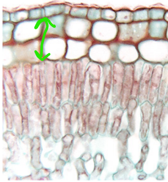
Which is Spring Wood which one is Late Wood, how can you tell?
Early in growing season (pink) - lots of water,
light, cells have thinner walls larger
lumens
Late in growing season (green) – decreased
water availability, leaves are being
shed, cooler temps, cells produced
smaller cells with increased cell wall
thickness
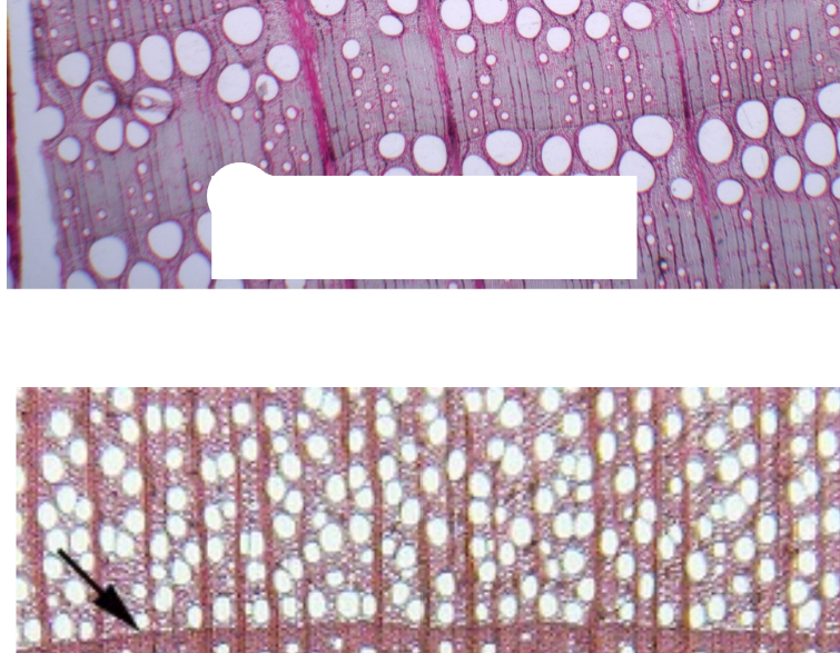
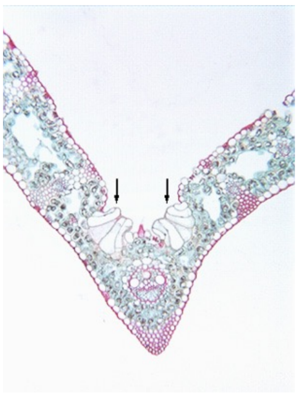
What are these types of Porous arrangements? What are there differences?
Ring Porous vs. Diffuse porous:
arrangement of vessel members
(pores)
Ring Porous wood: large diameter
vessel members produced early in
spring wood (top)
Diffuse porous wood: diameter of
vessel members doesn’t change
and uniformly distributed thru-out
the growing season. (bottom)
Diffuse porous
Ring porous
Gymnosperms don’t produce vessels:
Nonporous Wood
What is this and what is the function?
Tyloses: defense mechanism against pathogens and stress
Storied vs Non Storied
Storied on the left, Non storied on the right
Hardwood vs. Softwood
Hardwood – angiosperms due to abundant presence of fibers
Softwood – gymnosperm wood due to absence of numerous fibers
What system is leaves part of?
Shoot System
Plants adapted to dry environments
Xerophytes.
Plants adapted to habitats that are neither too wet or too dry
Mesophytes.
Floating, emergent, or submergent plants
Hydrophytes.
Typical Mesophyte Characteristics
Bundle sheath extensions may be present (parenchyma or sclerenchyma)
• Bundle Sheath cells surround vascular bundle – composed of parenchyma
or sclerenchyma
• Most guard cells on abaxial (lower) surface of leaf
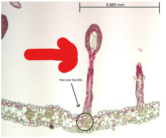
What is this and what is it generally a sign of and what’s the function?
bulliform cells, large, colorless cells that cause
the leaf to roll or fold to reduce water loss, found in mesophytes
Leaf with hydrophytic features characteristics
1. Well-developed aerenchyma (large air spaces) to facilitate gas exchange and
buoyancy.
2. Submerged leaves often lack stomata entirely & floating leaves are typically
epistomatic, with stomata confined to the upper epidermis.
Cross sections through a hydrophytic leaf from Nymphaea (water lily)
4. Veins run parallel and are connected by small veins. Vascular bundles are
generally small.
5. Possess poorly developed or reduced vascular bundles, particularly the
xylem, as water is readily available
Leaves with Xeromorphic features
1. Leaves with high volume / surface ratio (i.e. small and compact)
2. Palisade mesophyll strongly developed
3. Small intercellular spaces in mesophyll
What is this? What’s the function? What is it a sign of?
Hypodermis, made of collenchyma cells, Xeromorphic
Leaf structure in Pinus
1. Needles from short shoots in groups (fascicles); may be one or
several per fascicle.
2. Shape is either round or triangular.
3. Epidermis thick-walled, with a heavy cuticle, sunken stomata,
overarching subsidiary cells (Figure 7.8 in Esau). Stomata on all sides,
in rows.
4. Sclerified hypodermis below the epidermis
6. No palisade parenchyma - only spongy parenchyma.
7. Resin ducts (or canals). secreting antiseptic, aromatic, and sticky
resin to seal wounds, prevent fungal infections, and deter insects.
prevents freezing, and helps absorb heat.
8. Vascular bundles may be one or two side by side. Xylem consists
of protoxylem and metaxylem as well as xylem parenchyma
alternating with tracheids. The phloem appears in regular vertical
stacks.
9. Vascular bundles surrounded by transfusion tissue composed of
tracheids and parenchyma.
10. Vascular bundle and transfusion tissue surrounded by a thick-
walled endodermis with Casparian strip
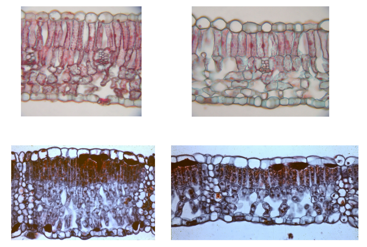
Difference Between Sun and Shade leaves
Sun leaves (left) are often thicker, with a smaller blade
area and more developed palisade layer (shown left)
• Shade leaves (right) are generally larger, longer, wider with
a thinner palisade layer. They increase surface area to
capture light.
What is guttation?
At night-in some small plants roots continue to
take up water; as the water accumulates in the
leaf pressure builds and forces water out of
wide stomata called water pores (arrows). These
stomata are open-don’t close.
The process of secreting excess water is called
guttation. Typically see it in the morning and
only in small plants
All the Leaf Modifications
1. Asexual reproduction – produce clonal “plantlets”; genetically identical to
parent
2. Spines - Leaves are reduced to sharp structures to reduce water loss and defend
against herbivores, common in cacti
3. Tendrils - Thread-like structures that allow weak-stemmed plants to climb and
gain support, found in peas
Leaf Modification
4. Storage Organs - Water and Food Storage storage. Fleshy, succulent leaves that
store water or food, seen in onion, garlic, and Aloe.
Leaf Modification
5. Insect Traps - Specialized traps for capturing insects to overcome nitrogen
deficiency in soil, such as the pitcher plant, bladderwort, Venus flytrap.
How do Bladderworts work?
possess highly specialized leaves that function as
sophisticated, vacuum-powered, aquatic traps (bladders) to catch, digest, and absorb
nutrients from tiny prey.
How does Drosera (sundew) leaf work?
sticky trichomes that release volatiles to attract and
trap insects
• Once trapped leaves release digestive enzymes to absorb nutrients
What the shiz is this?
Glandular hair, traps insects
How does Darlingtonia (pitcher plant) pitcher work?
pitcher plant is lined with a slippery wax.
Insects crawling on the lip slip and fall into a pool of water in the bottom of the
pitcher, where they are digested by bacteria. The plant then absorbs the smaller
molecules.
Two Processes of Photosynthesis
1) Light reactions:
• Light absorption by chlorophyll is able to
hydrolyze water into protons, electrons &
oxygen (2H20 → 4e- + 4H+ + 02)
• Electron transport thru a series of distinct
carriers results in the synthesis of ATP and
NADPH
2) Calvin Cycle (aka dark reactions)
• ATP and NADPH is used to reduce C02 to
CH20 (carbon dioxide is converted into
sugars/food for the plant)
Light Reactions
1) light absorption by chlorophyll is able to hydrolyze water & 2)
electron transport of the electrons results in the synthesis of NADPH & ATP
The Calvin Cycle (aka the dark reactions):
Uses the ATP and NADPH made in light reactions to reduce CO2 to
carbohydrate (CH20)
• Occurs primarily within chlorenchyma tissue in the stroma of chloroplasts
• The C-H bonds produced provides almost all of the energy for life on earth
Calvin Cycle: three distinct processes
Carboxylation, Reduction, Regeneration
Carboxylation
1. Carboxylation (“fixation”) Phase:
• CO2 is added to a 5-carbon sugar called RuBP (ribulose 1,5-bisphosphate)
• RuBisCO (RuBP carboxylase/oxygenase) catalyzes the reaction.
• The 6-carbon molecule is unstable and quickly splits into two 3-carbon
molecules: 3-phosphoglycerate (3PG)
In C3 plants the first stable product produced is this 3-carbon substrate
(3-phosphoglycerate).
Reduction Phase:
the products of the light rxs are used to reduce C02 to carbohydrate
• ATP phosphorylates 3-phosphoglycerate (3PG) to form 1,3 bisphosphoglycerate (1,3 bPG)
• NADPH will reduce 1,3 bPG to Glyceraldehyde 3-phosphate (G3P)
• Glyceraldehyde 3-phosphate is THE SUGAR that is made during the Calvin Cycle
RuBisCO can bind to
O2 as well of CO2
• RuBisCO can bind CO2 (functions as a carboyxylase)
• RuBisCO can also bind O2 (functions as an oxygenase)
• RuBisCO binding to oxygen is known as photorespiration
Unstable 5C 2C + 3C (glyceraldehyde 3-phosphate)
re-used in Calvin Cycle – but it will take
some extra energy to return it to the
Calvin Cycle
photorespiration)
Two 2C molecules combine to reform a 3C sugar while one C is oxidized
to CO2 (i.e. photorespiration)
Under normal spring temp, RubisCo will
bind to 1 02 for
every 2 C02 bound
Under warm, dry conditions – the pore of the stomatal complex
becomes
smaller to limit water loss-but it also doesn’t allow C02 to enter the leaf
• In addition 02 levels increase inside the leaf due to photosynthesis
H20
Guard cell closure leads to
increased oxygen levels within leaf
and decreased CO2 concentrations.
This leads to increased
photorespiration
Conifer phloem
Simple, less variable than dicot secondary phloem.
Variety of cells including: Sieve Cells with associated Rays
Parenchyma cells singly or in strands
Albuminous cells at ends of rays
Fibers and sclereids may be present
Stems are extremely varied in plants, with many modifications.
Aspects that affect this variation include:
1. leaf arrangement (around the stem)
2. leaf insertion (angle, position, attachment)
3. presence of axillary buds
4. where (at what level) does branching occur
5. is the shoot vertical or horizontal, free-standing or climbing,
etc.
6. where does the shoot system grow? (above ground,
underground, in water)
two leaves arise at a single node in the same vertical
plane but in opposite directions.
Phyllotaxy: • If successive pairs of leaves are parallel it is termed Superposed,
• if successive pairs of leaves are at right angles it is termed
Decussate.
single leaves arise at each node in an
alternating manner.
• Alternate: A single plane where they appear to go from side
to side (Distichous), or a
• Spiral where the leaves are arranged around the stem. Leaves
arise at repetitions of an angles until one eventually arises over
the first.
three or more leaves arise at a node and radiate in
different directions
Whorled
Vegetative SAM produces
stems (nodes, internodes), leaves,
axillary buds
Reproductive SAM produces
flowers
SAM locations:
1. Terminal bud-end of primary shoot
2. Axillary buds in the axils of the leaf
petiole/stem
• Will produce lateral branches with
their own terminal bud.
• Will produce leaves with their
own apical meristems
Shoot apical meristems: are protected by the newest formed leaves
(leaf primordia)
Arise initially as small “bumps” (leaf buttresses) along the meristem
and then grow into leaf primordia that surround the meristem
(leaf buttresses), Their development will reflect the phyllotaxy you see in the plant
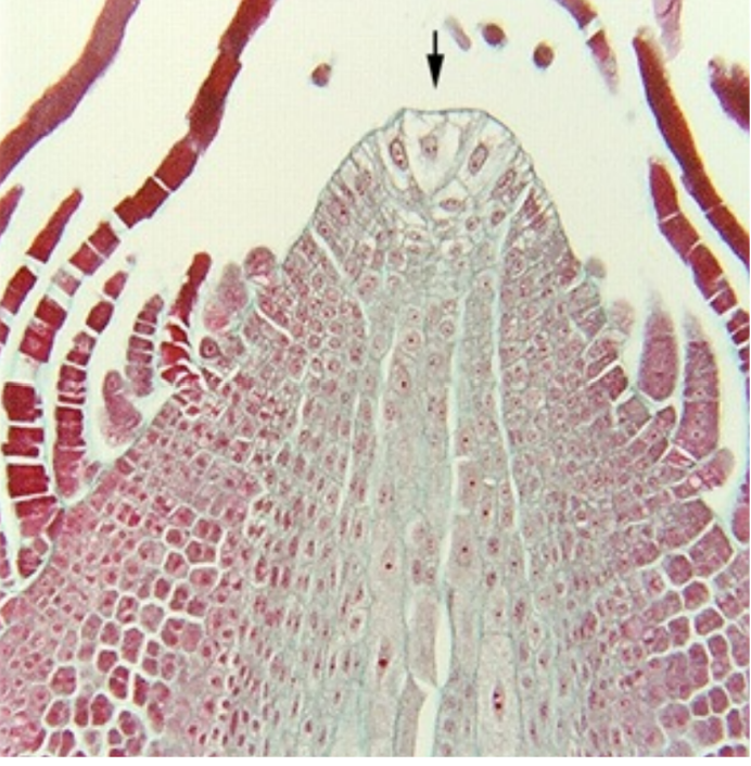
What is this and what type of organization is it and what does it do?
1. Apical Meristem with a single Apical Cell (ferns, mosses)
• a prominent, single tetrahedral (four-sided) pyramidal cell
located at the shoot apex
• serves as the main site of cell division
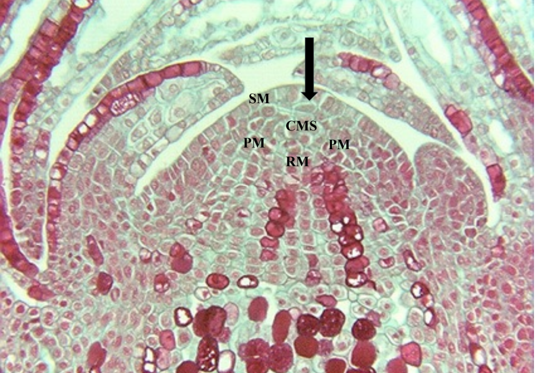
What is this and what type of organization is it and what does it do?
2. Apical Meristem with a Conspicuous Cyto-Histological Zonation
• Produce zones rather of cell division (rather than layers)
• Surface meristem has occasional periclinal divisions and
contribute cells to the central mother cells (arrow). Since the
Tunica does not-this is one way to tell the two meristems apart
CMC: central mother cells. Don’t
divide as often as other cells in apical
meristem. Often larger and more
vacuolate.
RM: Pith (rib) Meristem: produces pith
PM: Peripheral Meristem:
produces cortex, leaf primordia,
vascular tissues, sometimes the outer
part of the pith
SAM in Pine
SM: Surface Meristem
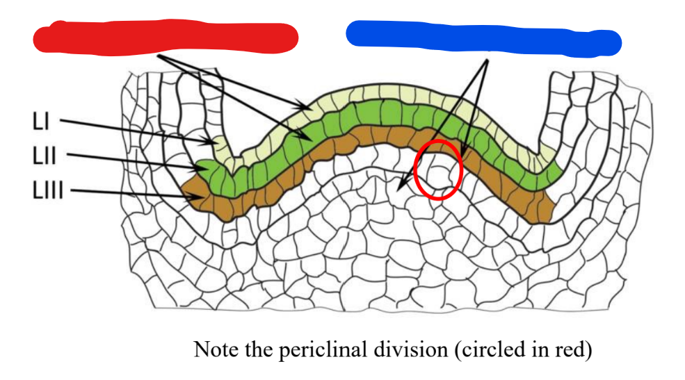
What is this, what type of organization is it, what is the function
Tunica (L1, L2): Comprises the
peripheral outer layer(s) that divide
perpendicular to the meristem
surface (anticlinal divisions). This
increases surface area, allowing for
surface expansion. L1 typically
forms the epidermis, while L2
contributes to subepidermal tissues.
Corpus (L3 and below): Located
beneath the tunica, this mass of cells
divides in multiple planes (periclinal,
anticlinal), increasing the volume
and bulk of the shoot. Look for the
periclinal divisions to help you
identify the corpus
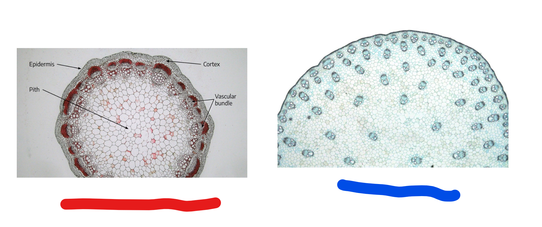
This slide is looking at which part of the plant? Which belongs to which class of plant?
red is dicot, blue is monocot
Phloem to the outside of the xylem (“typical”)
Collateral:
phloem to the inside and to the outside of xylem
• Result of two cambia!
Bicollateral:
xylem is concentric and surrounds phloem
Concentric, Amphivasal
phloem is concentric and surrounds xylem
Concentric, Amhicribral:
Anomalous Growth
Anomalous Growth are deviations from the normal, predictable
pattern of development in plants
• often resulting from irregular cambial activity, accessory cambia,
or unusual vascular bundle arrangements
Example: Cucurbita:
• No interfasicular cambium
produced
• Isolated phloem strands inside
of the perivascular fibers
• Bicollatoral bundles
Example of anomalous growth:
Aristolochia & Clemaatis produce
medullary rays (during secondary growth-the bundles look like they
are being maintained)
• secondary parenchyma is produced from the vascular cambium that
formed between the original bundles.
Stems as storage organs:
2. Storage Organ: store excess carbohydrate-numerous kinds
a. Rhizomes: horizontal stem just under soil
• Daffodils, lillies, ginger
Stems as tubers
enlarged end of a rhizome
Stems as bulbs
vertical underground shoot
• lots of enlarged leaves that
store carbohydrates/food;
• i.e. onions, garlic
Stems as water storage
Water Storage-organs are often adapted for
dry habitat (xerophytic)
• Adaptions include: photosynthetic stem with
chlorenchyma, often with sunken stomata,
thick cuticle, cells that store water, Reduced
Surface Area to minimize transpiration,
stems may be cylindrical, spherical, or
reduced in length
• Leaves are modified (spines)
What are the adaptations seen in aquatic
plant stems (hydrophytic environment)?
Adaptations provide buoyancy,
flexibility, gas exchange, direct nutrient
absorption
• aerenchyma (spongy, air-filled tissue)
for flotation, light weight
• reduced, structures for direct nutrient
absorption-structure maximizes contact
with water
• lack the need for rigid support or
extensive, woody vascular systems.
Monocots do not have true ”secondary growth” since they don’t have
vascular or cork cambia.
They do however unique meristems that increase their width
1. Primary Thickening Meristem – occurs close to shoot apex
2. Secondary Thickening Meristem – occurs in area of plant
where cell elongation has stopped.
• It can be continuous with primary thickening meristem
Primary Thickening Meristem
occurs close to shoot apex
• Occurs primarily in short compact plants; often immediately &
greatly increases the width of the apex
• Diffuse primary meristem, narrow zone of meristematic cells with
radial derivatives
• Produces lots of parenchyma towards the inside; may produce
vascular bundles
Secondary Thickening Meristem
occurs in area of
plant where cell elongation has stopped.
• Arises in the cortex-produces radial derivatives
• Produces “secondary” vascular bundles that are
often amphivasal (xylem surrounds phloem) to
the inside and parenchyma to the outside
Intercalary meristems:
Specialized region in monocot stems
• Small undifferentiated cells at the base of some internodes (or
above a node)
• Cells retain capacity for cell elongation
• Particularly important when plant is dislodged (falls over)
• Stem senses gravity and elongates the cells on bottom side of
stem so shoot rights itself.
Fusiform initials
Elongated to isodiametric in shape, fusiform = spindle shaped, really
prismatic, wedge-shaped ends.
• rectangular in radial section, fusiform shape in tangential section,
smaller rectangle in transverse (cross) section.
Cell divisions
1) Ain angiosperms, cambial initials are usually bifacial, which means they
produce cells off of two sides.
• They divide periclinally to produce xylem to the inside and phloem to
the outside, sometimes alternating.
• Also called additive divisions
Cell divisions
2) To keep up with the increasing girth of the plant a fusiform initial must
produce more of themselves
• Divisions that add to the cambial cells are called multiplicative
divisions
3. Radial anticlinal divisions gives more vascular cambium cells that are
arranged in stacks, gives rise to storied wood (considered an advanced
condition). The oblique divisions result in non-storied wood, no tiers of
cells.
Robinia- storied vascular cambium
Will produces a storied wood Pyrus- non storied vascular cambium
produces non storied wood
4. After the anticlinal cell divisions, the derivatives can enlarge tangentially,
elongate via apical intrusive growth (remember this type of growth with
fibers). Intrusive growth also causes forking and invasion of rays (Figure
10.5 H-L, Figure 10.6 C) which may result in the ray splitting.
Ray initials
Transverse divisions of the fusiform
initial results in several cells, only some
of which may survive and become ray
initials.
• Rays begin as a group of only 1-2 cells
but they increase in height through later
transverse divisions and by fusion with
other rays.
• To become multiseriate, radial anticlinal
divisions and fusions occur.
Robinia Vascular cambium
Initials may divide again before their derivative cell has matured, so a zone of
undifferentiated cells accumulates.
• Sometimes hard to distinguish between the fusiform initial and its
derivative, which is why some call the entire cambial zone the
"cambium".
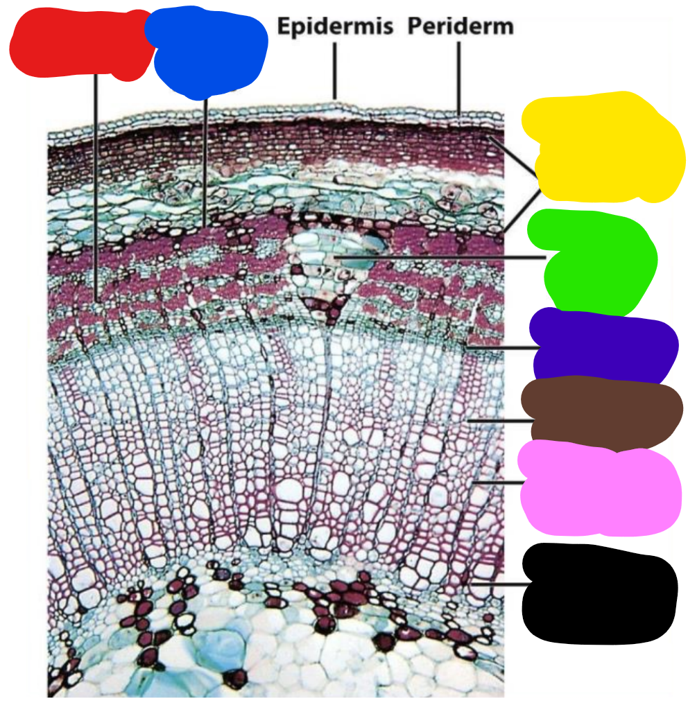
produced by the cork cambium (phellogen)
Periderm
Relatively simple, rectangular, radially
flattened cells. Produce derivatives in radial files.
Phellogen = cork cambium.
Phellem
cork layer. Tissue that differentiates to the outside of
cork cambium variable in in shape. Lack intercellular spaces.
Nonliving but may have solid or liquid contents. In these photos,
tannins were deposited into the phellem cell vacuoles of Quercus
and Clusia.
a. Walls are suberized. Suberin and wax forms a lamellar layer on
top of primary wall (that may or may not be lignified).
b. Walls of Quercus suber (cork oak) phellem are thin, cells filled with
air, thus lightweight and insulating. Form a barrier that is
impervious to water and resistent to oil.
Cork cells
Quercus suber
c. May also have phelloid cells – non-suberized, may become
sclereids.
• Layering of cork and sclerified cells can also occur in seasonal
increments (layers). Similar phenomenon in this cactus
(Oreocereus celsianus) root.
Phelloderm
living parenchyma that differentiates to the
inside of the cork cambium. Usually 1-4 cell layers thick, if
present at all
Cork cambium (phellogen) typically produces less
phelloderm (parenchyma) than phellum (cork)
Development of Periderm
A. Typically - the first periderm develops in the outside portion of the
cortex in stems and roots.
• Often directly under the epidermis but sometimes a bit deeper.
• The phellogen can derive from various different living, potentially
meristematic cells, e.g. epidermis, subepidermal parenchyma or
collenchyma, pericycle (roots), phloem and phloem rays.
B. The first cork cambium often forms at the same time or after the
vascular cambium (first year).
C. Later (sequent) periderms are initiated deeper, either the same
year or many years later
Sequential periderms
1. Forms each year and in successively deeper
layers beneath the first.
2. Originate from parenchyma cells of secondary
phloem and ray cells, which on the outside is
no longer functional.
3. The first phellogen may form in isolated areas
that later become contiguous by lateral
spread and meristematic activity.
• Most cell divisions are periclinal but
anticlinal divisions are involved in the
phellogen keeping pace with the stem’s
increase in circumference.
4. Sequent periderms appear as discontinuous
but overlapping layers (Figure 12.4, 12.7B for
Quercus).
Rhytidome
is the technical term for the outer, dead layer of
bark on trees and woody plants.
• It is primarily composed of dead, suberized cork cells,
cortical tissue, and phloem formed by successive
periderms.
• This protective, often cracked outer layer insulates the tree,
prevents water loss, and shields it from insects and disease
Rhytidome
The outermost bark. Layers of tissue isolated by the
periderm and layers of the non growing periderm.
2. When periderm arises in successively deeper tissue,
accumulation of old periderms have a variety of tissues (cortex,
1 ̊ phloem, 2 ̊ phloem). All of these tissues are no longer
functional – just a barrier to movement of water and gases
3. Eventually the new periderms will arise from living cells within
the secondary phloem
Key Aspects of Rhytidome:
Structure: It consists of multiple layers of dead tissue that
accumulate over time.
• Formation: It is produced by the phellogen (cork cambium).
• Function: It protects the inner, living bark (phloem).
• Appearance: Often rough and fissured, it varies in thickness
from thin (e.g., birch) to very thick (e.g., redwoods).
• Separation: Unlike the inner, living bark, the rhytidome can be
shed, as seen in trees like sycamores or birches.
What is Bark and what is it composed of?
all the tissues outside the
vascular cambium, i.e. secondary
phloem, any primary tissues that are
still there, periderm, and dead tissue
outside the periderm.
Composed of an inner living part and
an outer dead part (rhytidome)
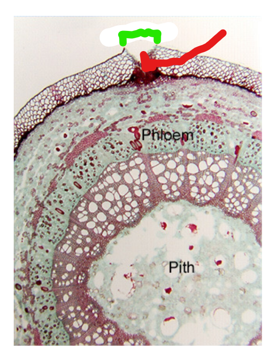
What the shiz is the green and red? What are they doing?
Lenticels in green,
companion cells in red
First appear under stomata (in the stem) Nwhere phellogen is more
active.
• Phellogen is bent downward, and in this “cup” is produced loose
cells called filling (complementary) tissue.
• The outer layers of the lenticel are pushed and the whole structure
ruptures the surface.
As subsequent cork cambia form deeper inside the stem,
they will
form lenticels in the same region as the pre-existing lenticels –
there will be an oxygen diffusion pathway through all layers.
Crassulacian Acid Metabolism:
in cacti, • Primary function of CAM metabolism
is to limit water loss and survive!
• Characterized by an inverted guard
cell cycle (open at night/closed during
the day
• Make 4-carbon acids (with PEP
carboxylase) at night and store them
in vacuole
• IN the morning; Decarboxylate the 4-
carbon acid and use C02 for
photosynthesis (when the light
reactions can make ATP/NADPH)
• GC don’t need to be open-ready
supply of C02
General Functions of Root System
Absorption of water and minerals from
substrate.
• Storage of photosynthates.
• Anchorage of the plant to the soil.
• Control and distribute the the flow of
water throughout the plant.
Tap Roots
The primary root originates from the radicle (embryonic
root) of an embryo and gives rise to the taproot.
• One central root with smaller lateral roots branching off of the main
root.
• Generally, penetrate deeper in the soil for anchorage
• Common in Dicots and Gymnosperms.
Fibrous Roots
Embryonic root dies and all roots arise from base of shoot
• A type of Adventitious root
• Adventitious-arise in unusual place
• What makes them a root and not a leaf is their anatomy!
• Anchorage is shallow but more laterally extensive than tap roots
• Common root system in Monocots
The root cap
protects the meristem and is the site for gravity perception
• Statoliths (starch dense amyloplasts) sediment to new lower wall when
plant is reoriented-this change in pressure redirects growth
• Remove the root cap-roots unable to perceive gravity
stele)
Vascular tissues occur as a solid cylinder in the center (aka stele)
• xylem is “armed” – phloem between arms
Vascular Tissues in Dicots vs Monocots
“many arms in monocots
Central core of parenchyma is
also present in the center of the
stele