DNA packaging
1/99
There's no tags or description
Looks like no tags are added yet.
Name | Mastery | Learn | Test | Matching | Spaced |
|---|
No study sessions yet.
100 Terms
chromatin
in non-dividing cells, a complex of DNA and histones that compresses to form chromosomes
histones
small, positively charged proteins that compact chromosomal DNA into the microscopic space of the eukaryotic nucleus, 8 subunits make up a histone
nucleosomes
structural unit of a chromosome that consists of a length of DNA coiled around histones
30 nm fiber
chromatin that is coiled into a short, thick fiber that is 30 nanometers in diameter so it can fit in the nucleus
chromatid
the two halves of a duplicated chromosome (each one is a "sister chromatid")
centromere
holds the sister chromatids together
chromosome
in dividing cells, the thread-like structure in which DNA is packaged into, each one carries a specific gene
linker DNA
the DNA that joins nucleosomes to one another in chromosomes
diploid
having two complete sets of chromosomes, one from each parent
eukaryotic DNA
shape: linear
proteins to help pack? yes (histones)
# chromosomes: varies (2+)
extra genetic material? yes (introns)
prokaryotic DNA
shape: circular
proteins to help pack? no
# chromosomes: 1
extra genetic material? no
how many chromosomes do humans have?
46
how does the size of the genome relate to the complexity of the organism?
the larger the genome, the more complex the organism
genome
all of the genetic material in an organism
before a cell can divide, what must happen?
-DNA must be duplicated
-organelles must be duplicated
-cell must grow
-cytoplasm must be made
interphase
-before mitotic (M) phase
-consists of the steps G1, S and G2
-G1 phase = gap 1 phase, where the cell grows
-S phase = synthesis phase, where DNA is replicated
-G2 phase = gap 2 phase, cell grows again
mitotic (M) phase
-after interphase
-consists of the steps mitosis and cytokinesis
-mitosis = division of the nucleus
-cytokinesis = cell splits in two
G1 phase (gap 1 phase)
the first stage of the cell cycle (part of interphase) where the cell grows
S phase (synthesis phase)
the second stage of the cell cycle (part of interphase) where the DNA is replicated
G2 phase (gap 2 phase)
the third stage of the cell cycle (part of interphase) where the cell grows again
M phase (mitosis)
the fourth step of the cell cycle (part of the mitotic phase) where the nucleus divides
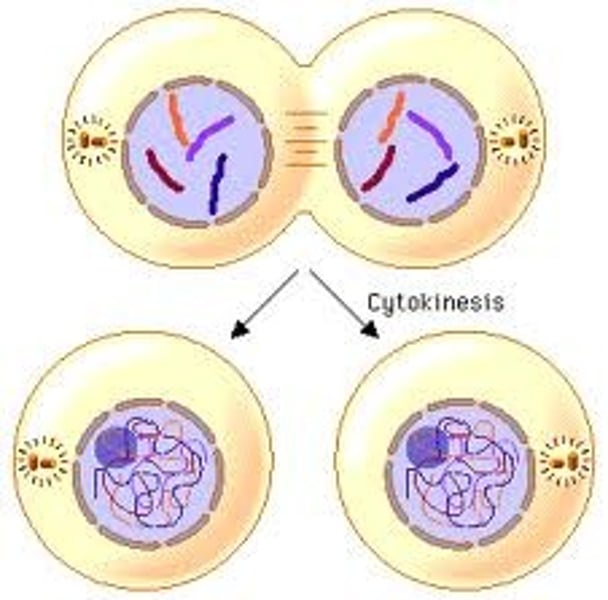
cytokinesis
-the fifth step of the cell cycle (part of the mitotic phase) where the cell splits into two
-division of the cytoplasm
-at the same time as telophase
-in animals, a cleavage furrow is formed which pinches the cell into two daughter cells
-in plant cells, a cell plate is formed and grows outward to create a new cell wall
the cell cycle
the continuous cycle of how cells divide, as soon as cells exit the cycle all cells go back in. includes the steps (in this order) G1, S, G2, M, cytokinesis
can prokaryotes preform mitosis?
no, because they do not have a nucleus
growth factor
increases rate of the cell cycle. it is a protein secreted by certain body cells that simulates other cells to divide. different cells have different growth factors.
external regulator
proteins that respond to the environment outside the cell
density dependent inhibition
decreases rate of the cell cycle. it is a phenomenon where crowded cells stop dividing once the place they operate in is dense enough. cancerous cells do not have a DDI
cell cycle control system
-a cyclically operating set of molecules in the cell that triggers and coordinates key events in the cell
-checkpoints are like stoplights
G1 checkpoint
-determines whether a cell will divide or go to G zero
-if a cell receives a go signal, it may continue in the cycle
-if it receives a stop signal, it enters G zero, a non-dividing stage
-most cells actually exist in G zero
P53
-"guardian on the genome"
-regulates cell cycle, also a tumor suppressor gene
does this in two ways:
-activate the DNA repair pathways
-stop signal at G1 checkpoint for DNA repair
-initiate apoptosis (programmed cell death if DNA damage is too extensive)
cell cycle regulatory proteins
-cyclins
-cyclin dependent kinases
-activate when a cyclin binds to them
-always present but not always on
MPF
-maturation promoting factor
-cyclin-CDK complex that triggers cells' passage past the G2 checkpoint into the M phase
-promotes mitosis
G2 checkpoint
-chromosomes must be replicated successfully
-DNA must be undamaged
-activated MPF must be present
-is there two of everything so the daughter cells will both have everything?
M checkpoint
-ensures that all chromosomes have lined up on the metaphase plate before the sister chromatids
-this makes sure that each daughter cell will have a copy of each chromosome
oncogene
-a gene that has the potential to cause cancer
-it helps cancer escape death (apoptosis)
tumor
abnormally growing mass of cells. 2 types:
benign: harmless, stays in same location (but CAN become malignant
malignant: harmful, spreads into neighboring tissues
metastasis
when a tumor spreads (from the primary tumor) through the circulatory system to another body location (secondary tumor)
naming cancers
-cancers are named based on their site of origin
carcinomas: external/internal body covering
sarcomas: tissues that support body
leukemias: blood forming tissues
lymphomas: lymph nodes, immune system tissues
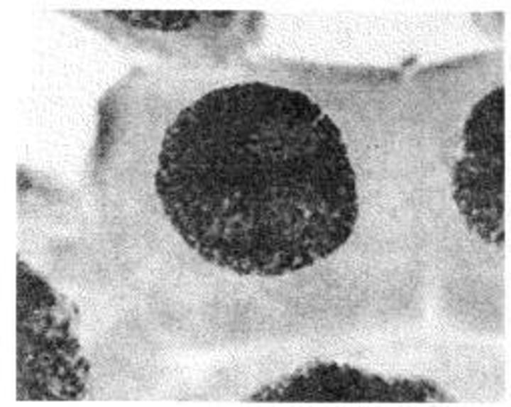
what does a cell in interphase look like under a microscope?
black blob, individual chromosomes not visible
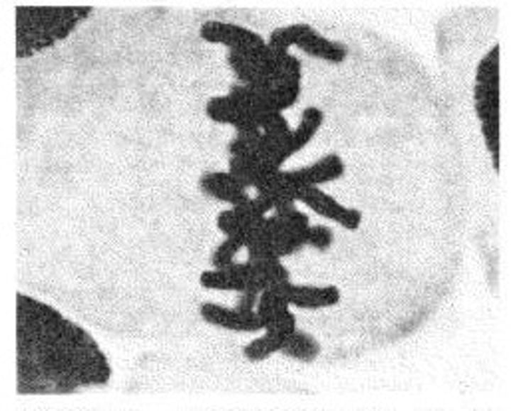
phase 1 of mitosis
PROPHASE
-sister chromatids (duplicated chromosomes) are condensed
-mitotic spindle (microtubules) begin to form in the cytoplasm, begin to grow out of the centrosomes (these make sure each cell gets the right DNA)
-centrosomes move away from each other
-in the last part of this stage, the nuclear envelope begins to break down
what does a cell in prophase look like under a microscope?
black blob with more individual chromosomes
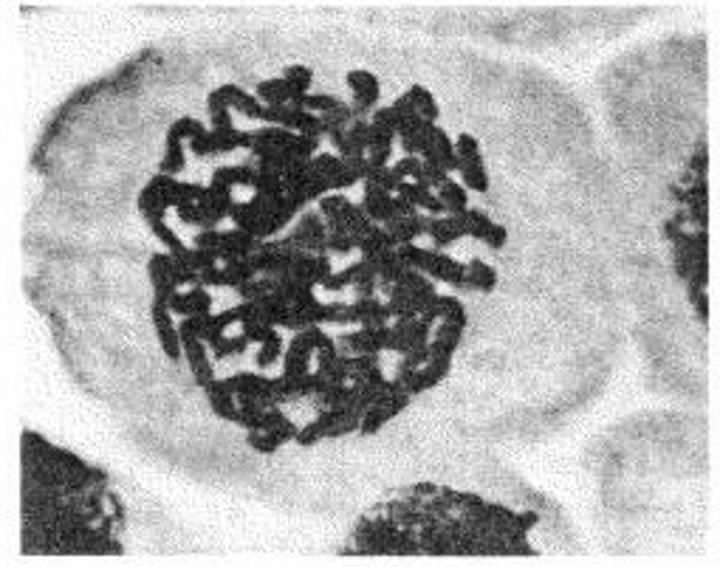
phase 2 of mitosis
METAPHASE
-sister chromatids line up along the metaphase plate (the process may not proceed until all of them line up)
-mitotic spindle is fully formed
-centrosomes are now at opposite poles
-nuclear envelope is gone
what does a cell in metaphase look like under a microscope?
all of the chromosomes in the middle
centriole
an organelle near the nucleus in animal cells, occurring in pairs and involved in the development of spindle fibers in cell division
centrosome
organelle near the nucleus of a cell that contains the centrioles (in animal cells) and from which the spindle fibers develop in cell division
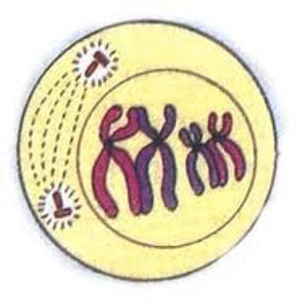
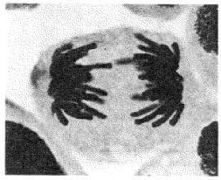
phase 3 of mitosis
ANAPHASE
-sister chromatids pulled apart by motor proteins (they move to opposite poles)
-they are now daughter chromosomes
-spindle apparatus: fibers push apart to opposite sides
-still no nuclear envelope
what does a cell in anaphase look like under a microscope?
chromosomes on either pole
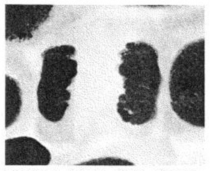
phase 4 of mitosis
TELOPHASE
-chromosomes begins to de-condense to form chromatin
-spindle apparatus disintegrates
-nuclear envelopes appear around chromosomes at the poles
-cell elongation continues
what does a cell in telophase look like under a microscope?
2 dark, dense blobs on either pole
mitotic spindle
-contains two types of microtubules
POLAR: extend from each spindle and overlap through the cell
KINETOCHORE: attach to chromosomes
epigenetics
chemical modifications due to the environment
impact gene expression
-methylation of cytosine (addition of methyl group)
-acetylation of histones (addition of acetyl group)
unmethylated cytosine, acetylated histones:
-switch goes ON
-DNA loosely packed ("active/open chromatin")
-transcription possible
methylated cytosine, deaceylated histones:
-switch goes OFF
-DNA tightly packed ("silent/condensed chromatin")
-prevents transcription
calculating a mitotic index
(# of cells in mitosis) divided by (total # of cells) = percentage mitotic index
mutagen
agent that causes changes in the genetic material of an organism, can be physical, biological or chemical
carcinogen
a mutagen that leads to the formation of cancer
primary tumor
original location of the tumor
secondary tumor
the place to which the tumor spread
asexual reproduction
reproduction that creates genetically identical offspring (mitosis)
sexual reproduction
2 gamete unite to form genetically different offspring (meiosis)
meiosis 1
first cell division in which synapsis and crossing over occur, 2 daughter cells are produced that have half as many chromosomes as the parent
crossing over
occurs in prophase 1. paternal and maternal chromatids break and rejoin at chiasmata, then each chromatid has maternal and paternal DNA
gametes
sex cells (egg and sperm)
somatic cells
all other body cells (non-sex cells)
meiosis 2
second cell division in meiosis in which sister chromatids separate, similar to mitosis
non-sister chromatids
chromosomes that aren't connected but are in the same homologous pair
haploid
having one copy of each chromosome
homologous chromosome
the paternal and maternal pair chromosome (i.e. #23 from both mom and dad)
synapsis
the pairing of homologous chromosomes
prophase 1 (meiosis)
chromosomes condense, nuclear envelope breaks, spindle apparatus forms, synapsis of homologous chromosomes, crossing over of non-sister chromatids
metaphase 1 (meiosis)
migration of tetrads to metaphase plate is complete
anaphase 1 (meiosis)
homologs separate and begin moving to opposite poles
telophase 1 (meiosis)
chromosomes move to opposite sides of the cell
cytokinesis 1 (meiosis)
cell (cytoplasm) divides
meiosis
in sexually reproducing cells, 2 stage cell division to produce 4 haploid cells from 1 diploid, halving the chromosome number
chiasmata
the x-shaped structure formed during meiosis when non-sister chromatids (homologous chromosomes) cross over
prophase 2 (meiosis)
spindle apparatus forms
metaphase 2 (meiosis)
chromosomes line up at the metaphase plate (middle of the cell)
anaphase 2 (meiosis)
sister chromatids separate, begin moving to opposite sides of the cell
telophase 2 (meiosis)
chromosomes move to opposite sides of the cell
cytokinesis 2 (meiosis)
cell (cytoplasm) divides
why is it crucial that gametes reduce their number in half?
so they can pair with the other parent's gamete and create a cell with the normal number of chromosomes that has half it's DNA from each parent
why do homologous chromosomes separate during meiosis?
because they need to be equally distributed in the daughter cells and make sure that each daughter cell has a copy of each gene from both parents
how does meiosis create genetic diversity?
-crossing over
-random alignment and separation
problems from meiosis?
if both homologs or sister chromatids move to the same pole of the parent cell, meiosis will be abnormal because of too much DNA
karyotype
pictures of the chromosomes inside a cell, from these one can tell the gender and any abnormalities
XX
female
XY
male
steps for making a karyotype
1. get cells
2. visualize chromosomes
3. organize chromosomes
1 - getting cells
-for newborns/children/adults, blood must be drawn
-for fetuses, the mother must undergo amniocentesis or chorionic villus sampling (CVS)
amniocentesis
-use needle to extract amniotic fluid from the womb (fetal cells found there)
-miscarriage rate: 1 in 1000
-can be done at 15 weeks or later
chorionic villus sampling (CVS)
-catheter inserted into vagina to collect fetal cells found on placenta
-miscarriage: 1 in 100
-can be done at 10-12 weeks
2 - visualize chromosomes
-find cells that are actively dividing
-chromosomes are dyed so they can be seen
-chromosomes are photographed in metaphase
3 - organize chromosomes
-homologous chromosomes paired
-scientists can analyze for normality/gender
"normal" karyotype
-46 chromosomes (23 pairs)
-22 pairs of homologous autosomes
-23rd pair = sex chromosomes
trisomy
3 copies of one chromosomes instead of 2
monosomy
1 copy of one chromosome instead of 2
trisomy 21
down syndrome
non-disjunction
the failure of homologs or sister chromatids to separate