Week 9 - Muscle Microanatomy/Histology
1/10
Earn XP
Description and Tags
key terms and concepts
Name | Mastery | Learn | Test | Matching | Spaced | Call with Kai |
|---|
No analytics yet
Send a link to your students to track their progress
11 Terms
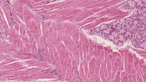
Key Characteristics of Skeletal Muscle
Voluntary movement
Multi-nucleated
Straited
Vascularized
Innervated by a plexus (network of blood vessels and nerves)
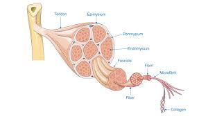
The Layers of Fascia
epimysium: surrounds the entire muscle
perimysium: surrounds a bundle of muscle fibers (fascicle)
endomysium: surrounds a single muscle fiber
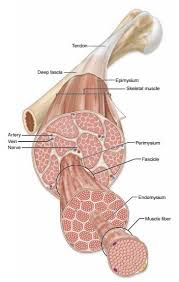
What makes up a muscle? (superficial to deep)
fascicle, muscle fiber, myofibrils, sarcomere, actin & myosin
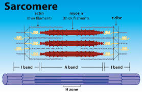
What is the structure of a sarcomere?
Z-Disc (Z-Line): Zigzag lines marking the boundaries of a sarcomere, anchoring the thin (actin) filaments.
A-Band (Anisotropic): The dark band, spanning the entire length of the thick (myosin) filaments, including overlapping thin filaments. Its length remains constant during contraction.
I-Band (Isotropic): The lighter region containing only thin (actin) filaments, bisected by the Z-disc. It shortens during contraction.
H-Zone: The pale area in the center of the A-band containing only thick (myosin) filaments (no thin filament overlap). It shrinks or disappears during contraction.
M-Line (Midline): A dark line in the very center of the H-zone, where myosin filaments are linked.
The I band and H zone shorten when a muscle contracts.
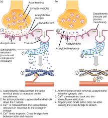
Excitation-contraction coupling
Neural Stimulation: A motor neuron releases acetylcholine (ACh) at the neuromuscular junction, generating an action potential on the muscle's sarcolemma (cell membrane).
Signal Propagation: The action potential travels along the sarcolemma and into the T-tubules (deep invaginations of the membrane).
Calcium Release: The electrical signal activates voltage-sensitive Dihydropyridine Receptors (DHPRs) in the T-tubules, which are physically linked to Ryanodine Receptors (RyRs) on the SR. This interaction pulls open the RyRs, releasing stored Ca2+ into the sarcoplasm (cytoplasm).
Cross-Bridge Cycling: The increased intracellular Ca2+ binds to troponin, causing tropomyosin to shift and uncover myosin-binding sites on actin.
Muscle Contraction: Myosin heads bind to actin, forming cross-bridges, and perform the power stroke, pulling the thin filaments towards the sarcomere's center, causing muscle shortening (contraction).
Relaxation: When the nerve signal stops, Ca2+ is actively pumped back into the SR, tropomyosin covers the binding sites, and the muscle relaxes.
Where is smooth muscle found?
walls of hollow visceral organs (stomach, bladder)
What are the key characteristics of smooth muscle
non-striated, involuntary, mono nucleated
Types of contractions produced by smooth muscle
slow and smooth, peristalsis (contraction, relax, repeat)
Where is cardiac muscle found?
only in the myocardium (wall of the heart)
The key characteristics of cardiac muscle
striated, involuntary, mononucleated
How does cardiac muscle contract?
automaticity; action potential created spontaneously by the heart
intercalated discs (gap junctions) spread action potentials throughout heart, causing rhythmic contractions of the heart