Anatomy and Physiology Dr. Brown Exam 3
1/210
There's no tags or description
Looks like no tags are added yet.
Name | Mastery | Learn | Test | Matching | Spaced |
|---|
No study sessions yet.
211 Terms
Sodium Potassium Pump
moves sodium ions (Na+) out of the cell and potassium ions (K+) into the cell, regulating ion concentration of both sides of the cell membrane
Voltage-gated ion channel
a channel that responds to changes in the electrical properties of the membrane in which it is embedded
ligand-gated ion channel
opens because a signaling molecule, a ligand, bonds to the extracellular region of the channel
Stimulus gated ion channel
when a neurotransmitter or a sensory stimulus binds to a receptor protein to signal channel opening
Resting membrane potential
when ions are distributed across the membrane, the difference in charge is -70 mV
Polarized
a charged particle with oppositely charged ends
polarized membrane
a charged membrane having a positive outside and a negative inside (unequal charges)
Depolarized
change in a cell membrane potential from rest toward zero
repolarization
return of the membrane potential to its normally negative voltage at the end of the action potential
threshold
membrane voltage at which an action potential is initiated
threshold voltage
-70mv to -55mV
Hyperpolarization
returns membrane volage to the resting value when repolarization continues past the resting membrane potential
action potential
change in voltage of a cell membrane in response to a stimulus that results in transmission of an electrical signal; unique to neurons and muscle fibers
synapse
narrow junction across which a chemical signal passes from neuron to the next, initiating a new electrical signal in the target cell
Excitability
ability to undergo neural stimulation
Irritability
ability to respond to a stimulus
Contractility
ability to shorten (contract) forcibly
Extensibility
ability to lengthen (extend)
Endomysium
loose, and well-hydrated connective tissue covering each muscle fiber in a skeletal muscle
Perimysium
connective tissue that bundles skeletal muscle fibers into fascicles within a skeletal muscle
Epimysium
outer layer of connective tissue around a skeletal muscle
Tendon
Connects muscle to bone
Fascicle
bundle of muscle fibers within a skeletal muscle
Origin
end of a skeletal muscle that is attached to another structure (usually a bone) in a fixed position
Insertion
end of a skeletal muscle that is attached to the structure (usually a bone) that is moved when the muscle contracts
Myofiber
skeletal muscle cell
Myofibril
long, cylindrical organelle that runs parallel within the muscle fiber and contains the sarcomeres
Myocyte
muscle cell
Sarcomere
longitudinally, repeating functional unit of skeletal muscle, with all of the contractile and associated proteins involved in contraction
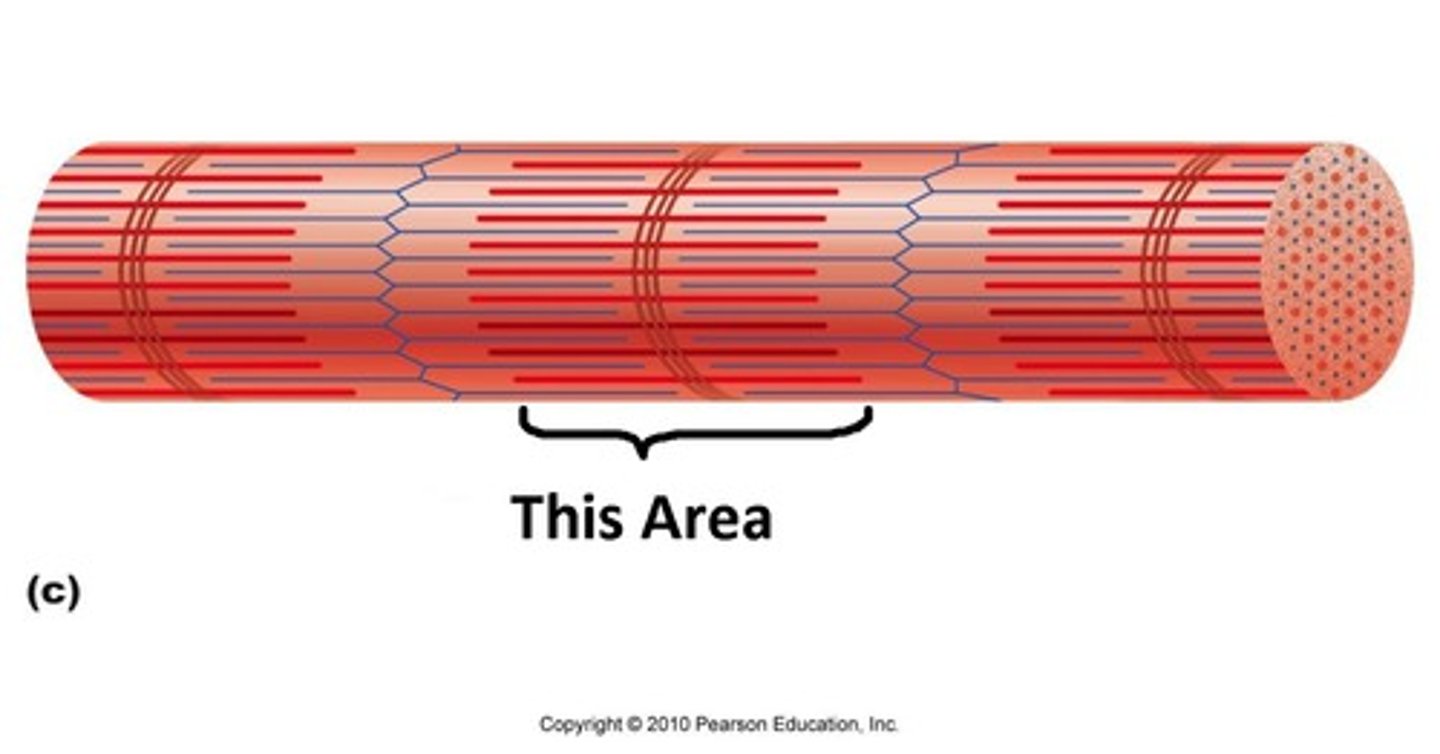
A band
when a sarcomere contracts, it stays the same length
A band
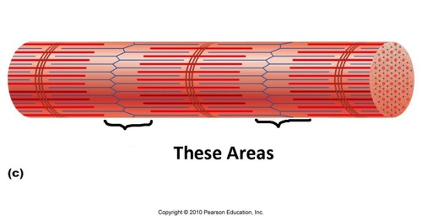
I band
when a sarcomere contracts, it becomes smaller
I band
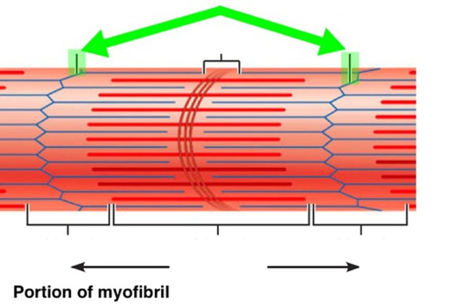
Z line
when a sarcomere contracts, these move closer together
Z-line
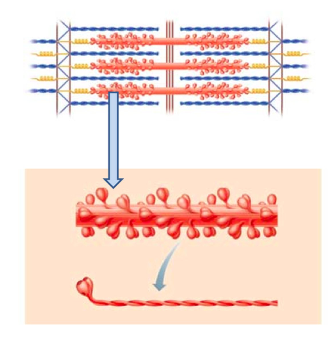
Thick filament
the thick myosin strands and their multiple heads projecting from the center of the sarcomere toward, but not all to way to, the Z-discs
Thick filament
Role of thick filament in sarcomere contraction
pulls on thin filaments to bring z-lines closer together
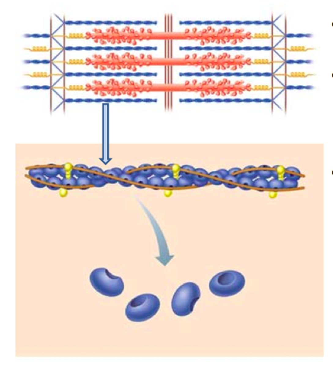
Thin filament
thin strands of actin and its troponin-tropomyosin complex projecting from the Z-discs toward the center of the sarcomere
Thin filament
Role of thin filament in sarcomere contraction
pulled by the myosin heads to slide past the thick filaments toward the center of the sarcomere.
Sarcolemma
plasma membrane of a skeletal muscle fiber
T-tubule
projection of the sarcolemma into the interior of the cell
Sarcoplasmic reticulum
specialized smooth endoplasmic reticulum, which stores, releases, and retrieves Ca++
Myosin
protein that makes up most of the thick cylindrical myofilament within a sarcomere muscle fiber
Actin
protein that makes up most of the thin myofilaments in a sarcomere muscle fiber
Troponin
regulatory protein that binds to actin, tropomyosin, and calcium
Tropomyosin
regulatory protein that covers myosin-binding sites to prevent actin from binding to myosin
Motor end plate
sarcolemma of muscle fiber at the neuromuscular junction, with receptors for the neurotransmitter acetylcholine
Neuromuscular junction
synapse between the axon terminal of a motor neuron and the section of the membrane of a muscle fiber with receptors for the acetylcholine released by the terminal
Acetylcholine
neurotransmitter that binds at a motor end-plate to trigger depolarization
Cardiac muscle
striated muscle found in the heart; joined to one another at intercalated discs and under the regulation of pacemaker cells, which contract as one unit to pump blood through the circulatory system. Is under involuntary control.
Intercalated disks
part of the sarcolemma that connects cardiac tissue, and contains gap junctions and desmosomes
Functional syncitium
The wave of contraction that allows the heart to work as a unit- begins with the pacemaker cells.
Smooth muscle
nonstriated, mononucleated muscle in the skin that is associated with hair follicles; assists in moving materials in the walls of internal organs, blood vessels, and internal passageways
Tetany
a symptom that involves involuntary muscle contractions and overly stimulated peripheral nerves
membrane potential is established and maintained
Proteins in the membrane or leakage channels allow Na+ to slowly move into the cell or K+ to slowly move out, and the Na+/K+ pump restores them.
mechanism of action potential generation
starts at -70mV with a Na+ channel opening, and Na+ atoms will flood inside the cell causing the cell to become less negative (depolarization), as membrane potential reaches +30mV, a K+ channel will open allowing K+ to flood out of the cell in a process called repolarization- this pushes the membrane potential below -70mV which will result in a period hyperpolarization.
three connective tissues that coordinate a muscle's activity
endomysium- covers each muscle fiber (cell), perimysium- covers fascicles/bundled groups, epimysium- covers the entire muscle
characteristics of muscle tissue
excitability (irritability), contractility, extensibility
step 1 of the molecular events of contraction
Attachment: myosin head (along with ADP and P molecules) binds with actin on a thin filament (other molecules include tropomyosin, Ca2+, and troponin)
step 2 of the molecular events of contraction
Power stroke: myosin head bends releasing ADP and P molecules
step 3 of the molecular events of contraction
Detachment: ATP enters cycle allowing myosin head to be released from actin
step 4 of the molecular events of contraction
Reactivation: myosin moves itself back to the cocked position utilizing energy from the transfer of ATP to ADP and P
Characteristics of cardiac muscle
tubular-shaped, found only in the heart, striated, involuntary control, less SR, myocytes are connected by intercalated discs, doesn't get fatigued, syncytium
syncytium
cells working together like a single functional cell
Characteristics of skeletal muscle
tubular-shaped, striated, voluntary, more SR, gets fatigued
Characteristics of smooth muscle
spindle-shaped, unstriated, involuntary, overlaps tapering cells, interconnected cells, thick and thin filaments crisscross cell, cell "scrunches" during contraction, doesn't contain sarcomeres
step 1 of sliding filament theory
an electrical impulse travels down a nerve fiber
what does an electrical impulse travel down in the sliding filament theory?
a nerve fiber
step 2 of the sliding filament theory
the nerve impulse reaches the end of the nerve and causes it to release acetylcholine (Ach)
Sliding Filament Theory: when the nerve impulse reaches the end of a nerve, what is released?
acetylcholine (Ach)
step 3 of the sliding filament theory
Ach binds to receptors on the muscle cell membrane and causes the electrical impulse to be transmitted to the muscle cell
Sliding filament theory: what happens when Ach binds to receptors on the muscle cell membrane?
the electrical impulse is transmitted to the muscle cell
step 4 of the sliding filament theory
the electrical impulse inside the muscle cell causes the release of calcium ions from the endoplasmic reticulum
the release of calcium ions from the ER
Sliding filament theory: the electrical impulse inside the cell causes...
step 5 of the sliding filament theory
calcium ions bind to troponin causing it to rotate
sliding filament theory: what do calcium ions bond to? what does this cause?
troponin, causes it to rotate
step 6 of the sliding filament theory
rotation of troponin move tropomyosin off the myosin binding site on actin
sliding filament theory: what does the rotation of troponin move?
tropomyosin off the myosin binding site on actin
step 7 of the sliding filament theory
the myosin head binds the myosin binding domain of actin
sliding filament theory: what does the myosin head bind?
the myosin binding domain of actin
step 8 of the sliding filament theory
myosin bends in two places, releasing ADP and pulling on the thin filament
sliding filament theory: what happens when myosin bends in 2 places?
ADP is released and myosin pulls on the thin filament
step 9 of the sliding filament theory
the Z-lines are pulled closer together and the A-band shrinks
sliding filament theory: what happens when the Z-lines are pulled closer together?
the A-bands shrink
step 10 of the sliding filament theory
the myofibril gets shorter (contracts)
step 1 of the NMJ
Action potential (electrical impulse) travels down nerve fiber
step 2 of the NMJ
Acetylcholine (Ach) is released from end of nerve
step 3 of the NMJ
Ach binds to receptor protein on muscle cell
step 4 of the NMJ
action potential is generated inside muscle cell
step 5 of the NMJ
Ca2+ is released from the sarcoplasmic reticulum
what is the effect of calcium on the thin filament?
when calcium binds to troponin, troponin rotates tropomyosin to reveal myosin binding sites on actin
step 1 of the molecular events of the contraction cycle
attachment
myosin head binds to the binding site on actin
what happens during attachment
step 2 of the molecular events of the contraction cycle
power stroke
Myosin head bends and ADP and phosphate are released.
what happens during the power stroke
step 3 of the molecular events of the contraction cycle
detachment
ATP enters the cycle allowing myosin to detach from actin
what happens during detachment
step 4 of the molecular events of the contraction cycle
reactivation